
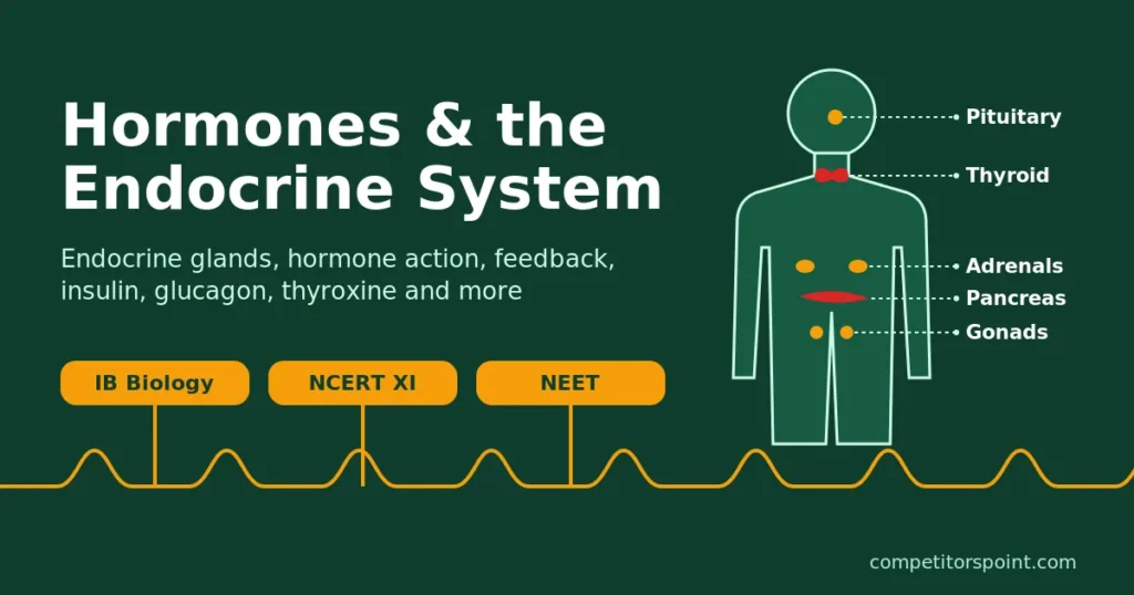
Hormones and the Endocrine System
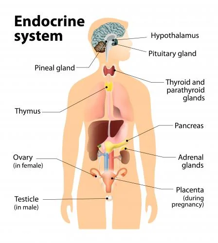
The endocrine system is the set of ductless glands and hormone-secreting tissues that release hormones directly into the blood. Hormones reach every part of the body but act only on target cells with matching receptors, regulating growth, metabolism, blood glucose, water balance and reproduction. Most are controlled by negative feedback, and the hypothalamus links the endocrine and nervous systems.
The human endocrine system for IB Biology and NEET: endocrine glands and hormones, the hypothalamus and pituitary, thyroid, parathyroid and adrenal glands, the pancreas and blood glucose control by insulin and glucagon, growth and metabolism, feedback mechanisms, how hormones act, and diabetes and other endocrine disorders. IB-only, NEET-only and beyond-the-syllabus content is marked, and each section maps the endocrine system to the IB and NCERT syllabus.
1 Endocrine glands and hormones
Syllabus link: NCERT XI 19.1 Endocrine glands and hormones; IB C3.1.3 (SL) hormonal and nervous signalling; IB C2.1.3 to C2.1.5 HL
The nervous system and the endocrine system jointly coordinate and regulate the physiological functions of the body. The neural system provides point-to-point, rapid coordination among organs, but it is short-lived. Nerve fibres do not reach every cell, and many cellular functions need continuous regulation. Hormones do that job. This chapter explains how the human endocrine system works, gland by gland, for IB Biology and NEET.
Practice MCQ
Which of the two systems of the body jointly coordinate and regulate the physiological functions in the body?
- a) The nervous system and the muscular system
- b) The nervous system and the endocrine system
- c) The respiratory system and the endocrine system
- d) The nervous system and the digestive system
Show answer
(b) Neural coordination is fast but short-lived and reaches only innervated cells; the endocrine system gives slower, longer-lasting, body-wide control.
Nervous system vs endocrine system
The nervous system and the endocrine system both carry messages between organs, but they do it in very different ways. IB asks you to distinguish them; NEET expects the same basic contrast. For a plain-language overview, see the MedlinePlus guide to the endocrine system.
Swipe the table sideways to see every column.
| Feature | Nervous system | Endocrine system |
|---|---|---|
| Signal | Electrical impulse along a neuron, then a neurotransmitter across a synapse | Chemical (hormone) secreted into the blood |
| Route | Along nerve fibres to specific cells | In the blood to every cell; only cells with the right receptor respond |
| Speed | Very fast (milliseconds) | Slower (seconds to hours) |
| Duration of effect | Short; stops when impulses stop | Longer lasting (minutes to days) |
| Area affected | Localised: one muscle or gland | Often widespread: many tissues at once |
| Examples | Pain reflex, contraction of a skeletal muscle | Insulin lowering blood glucose, GH and growth, thyroxin and metabolic rate |
Glands of the endocrine system: endocrine vs exocrine
An exocrine gland has a duct. Its epithelial cells secrete into the duct, which carries the secretion to a body surface or into a cavity (e.g. salivary glands into the mouth, sweat glands onto the skin).
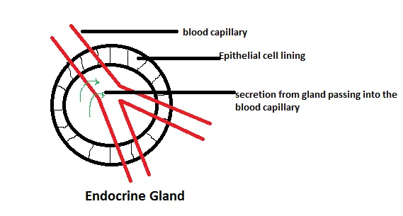
Endocrine glands are ductless. Their secretory epithelial cells release hormones into the surrounding tissue fluid, from where they enter the blood capillaries that run through the gland and travel to the rest of the body.
Practice MCQ
Which of the following statements is correct for endocrine glands?
- a) Endocrine glands are ductless glands
- b) Endocrine glands pour their secretions into ducts
- c) Both a and b
- d) Endocrine glands secrete digestive enzymes
Show answer
(a) Endocrine glands are ductless, so they secrete hormones directly into the blood capillaries.
What is a hormone? The messengers of the endocrine system
NCERT defines hormones as non-nutrient chemicals which act as intercellular messengers and are produced in trace amounts. “Non-nutrient” means they are not used as food or as a source of energy or raw material; it does not mean they cannot be swallowed. Thyroxine tablets, oral steroids and contraceptive pills all work when taken by mouth, although protein hormones such as insulin are digested in the gut and must be injected.
The word hormone comes from the Greek hormaein, “to excite or set in motion”. Many hormones are inhibitory, though; the name simply dates from when the first ones were discovered. Invertebrates have simple endocrine systems with few hormones, while vertebrates use a large number of chemicals as hormones.
Practice MCQ
Which of the following statements about hormones is correct?
- a) A hormone is a chemical messenger that acts as an intercellular messenger
- b) Hormones are non-nutrient chemicals: they are not used as food or energy
- c) Hormones are produced in trace amounts
- d) All of the above
Show answer
(d) This is the NCERT definition. Note that ‘non-nutrient’ does not mean hormones cannot be taken orally: thyroxine and steroid tablets work by mouth.
Practice MCQ
Which of the following scientists discovered the first hormone?
- a) Bayliss and Starling
- b) Skoog and Miller
- c) Ernst Mayr
- d) None of these
Show answer
(a) William Bayliss and Ernest Starling discovered secretin in 1902 and introduced the idea of a hormone.
Practice MCQ
Which hormone was the first to be discovered, by Bayliss and Starling?
- a) Cortisol
- b) Testosterone
- c) Estrogen
- d) hCG
- e) Secretin
Show answer
(e) Secretin is secreted by the epithelial lining of the duodenum and stimulates the pancreas to release bicarbonate-rich juice.
Chemical nature of hormones
Hormones of the endocrine system belong to four chemical groups. The chemical group of a hormone decides whether it can cross the plasma membrane, and so how it acts (section 9). Peptide hormones are made on the rough ER, packaged by the Golgi apparatus into vesicles and released by exocytosis; steroid hormones are made from cholesterol.
Swipe the table sideways to see every column.
| Group | Examples | Soluble in | Receptor |
|---|---|---|---|
| Peptides, polypeptides and proteins | Insulin, glucagon, pituitary hormones, hypothalamic hormones | Water | On the plasma membrane |
| Steroids | Cortisol, aldosterone, testosterone, estradiol, progesterone | Lipid | Inside the cell (cytoplasm or nucleus) |
| Iodothyronines | Thyroid hormones (T3, T4) | Lipid | Inside the cell (nucleus) |
| Amino-acid derivatives (amines) | Adrenaline (epinephrine), noradrenaline, melatonin | Mostly water (catecholamines) | On the plasma membrane |
IB only HL Signalling chemicals: local and distant effects
- Hormones, neurotransmitters, cytokines and calcium ions are all signalling chemicals in animals (C2.1.3).
- Hormones are chemically diverse: amines, proteins and steroids (C2.1.4).
- Hormones of the endocrine system have distant effects, carried in the blood; neurotransmitters have localised effects, diffusing only across a synaptic gap (C2.1.5).
2 Hypothalamus and pituitary gland
Syllabus link: NCERT XI 19.2.1 The hypothalamus, 19.2.2 The pituitary gland; IB C3.1.13 (SL) control of the endocrine system by the hypothalamus and pituitary gland
The hypothalamus links the nervous system to the endocrine system. It monitors the blood (temperature, osmotic concentration, hormone levels) and controls the pituitary gland, which in turn controls several other endocrine glands, so the pair sit at the top of the human endocrine system.
The hypothalamus
- It is the basal part (floor) of the diencephalon of the forebrain.
- It contains several groups of neurosecretory cells called nuclei: neurons that secrete hormones (neurohormones).
- Its hormones pass to the pituitary gland and regulate the synthesis and secretion of pituitary hormones.
- It is also part of the limbic system and controls the autonomic nervous system, body temperature, thirst and hunger.
Practice MCQ
Which of the following is true for the hypothalamus?
- a) It is the floor (basal part) of the diencephalon of the forebrain
- b) It contains groups of neurosecretory cells called nuclei
- c) Hormones secreted by the hypothalamus pass to the pituitary gland
- d) Hypothalamic hormones regulate the synthesis and secretion of pituitary hormones
- e) All of the above
Show answer
(e)
A stalk (the infundibulum) connects the hypothalamus to the pituitary gland, which sits in a bony cavity of the sphenoid bone called the sella turcica. The hypothalamus reaches the two lobes of the pituitary in two different ways:
- Anterior pituitary: neurosecretory cells release releasing and inhibiting hormones into a capillary bed in the hypothalamus. Blood from the superior hypophyseal arteries (branches of the internal carotid artery) carries them down the hypophyseal portal vein to a second capillary bed in the anterior pituitary.
- Posterior pituitary: the axons of some neurosecretory cells run down the stalk into the posterior pituitary (neurohypophysis) and release their hormones there, straight into the blood.
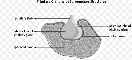
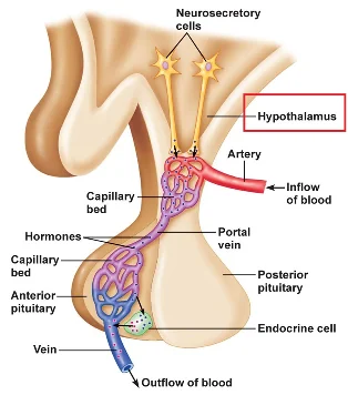
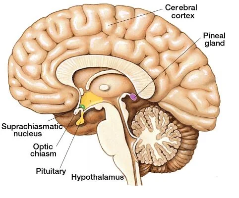
A portal vein is a vein that begins in one capillary bed and ends in a second capillary bed instead of going straight to the heart. The hypophyseal portal system lets hypothalamic hormones reach the anterior pituitary in high concentration, without being diluted in the general circulation.
Beyond the syllabus Other portal systems
The hepatic portal system carries blood from the stomach, intestines, pancreas and spleen (through the splenic, superior mesenteric and inferior mesenteric veins) to the liver before it returns to the heart; see absorption of digested food. A renal portal system (blood from the hind limbs passing through the kidneys) is well developed in fishes and amphibians, reduced in reptiles and birds, and absent in mammals.
Hypothalamic releasing and inhibiting hormones
Hypothalamic hormones are either releasing hormones, which stimulate the pituitary to secrete a hormone, or inhibiting hormones, which stop it. NCERT gives GnRH and somatostatin as examples; NEET books list seven:
- GHRH (growth hormone-releasing hormone): stimulates the anterior pituitary to release growth hormone.
- GHIH (growth hormone-inhibiting hormone), also called somatostatin: inhibits the release of growth hormone.
- TRH (thyrotropin-releasing hormone): stimulates the anterior pituitary to release TSH (thyroid-stimulating hormone).
- GnRH (gonadotropin-releasing hormone): stimulates the anterior pituitary to release the gonadotropins FSH and LH.
- CRH (corticotropin-releasing hormone): stimulates the anterior pituitary to release ACTH (adrenocorticotropic hormone).
- PRH (prolactin-releasing hormone): stimulates the anterior pituitary to release prolactin.
- PIH (prolactin-inhibiting hormone), which is dopamine: inhibits the release of prolactin.
🧠 Mnemonic: GO-GO-GO TO CONTROL PITUITARY PERFORMANCE
GHRH, GHIH, GnRH, TRH, CRH, PRH, PIH: the seven hypothalamic hormones that control the pituitary. The two inhibiting ones are GHIH and PIH; the other five are releasing hormones.
Note: PIH is dopamine. A separate human PRH has not been clearly identified (TRH also stimulates prolactin), but it is listed in NEET books. IB does not need this list.
Practice MCQ
How many releasing and inhibiting hormones are produced by the hypothalamus (in the list above)?
- a) 5 releasing hormones and 2 inhibiting hormones
- b) 4 releasing hormones and 3 inhibiting hormones
- c) 3 releasing hormones and 4 inhibiting hormones
- d) 6 releasing hormones and 1 inhibiting hormone
Show answer
(a) GHRH, TRH, GnRH, CRH and PRH are releasing hormones; GHIH (somatostatin) and PIH (dopamine) are inhibiting hormones.
Posterior pituitary hormones: made in the hypothalamus
Two hormones are made in the hypothalamus, carried down the axons and only stored and released by the posterior pituitary (neurohypophysis):
- Oxytocin, made mainly by the paraventricular nuclei: contracts the smooth muscle of the uterus during childbirth and causes milk ejection from the mammary glands. A synthetic form (Pitocin) is used to induce labour.
- Vasopressin (ADH, antidiuretic hormone), made mainly by the supraoptic nuclei: increases water reabsorption in the late DCT and collecting duct of the kidney. Too little ADH causes diabetes insipidus: large volumes of dilute urine and great thirst. See Human Excretory System: ADH, RAAS and ANF for how ADH and aquaporins work.
Both nuclei make some of each hormone; “mainly” is the accurate word.
Practice MCQ
Which of the following hormones are released from the posterior pituitary?
- a) Oxytocin
- b) Vasopressin
- c) Both a and b
- d) ACTH
Show answer
(c) Both are made by neurosecretory cells of the hypothalamus; the posterior pituitary only stores and releases them. ACTH comes from the anterior pituitary.
The pituitary gland: master gland of the endocrine system
- Also called the hypophysis; a small, pea-sized gland in the sella turcica.
- Called the master gland because its hormones control several other endocrine glands, though it is itself controlled by the hypothalamus.
- Divided into the adenohypophysis and the neurohypophysis (pars nervosa). The adenohypophysis has two parts: pars distalis (anterior pituitary) and pars intermedia, which in adult humans is almost merged with the pars distalis.
- It is ectodermal in origin, from two sources: the anterior lobe grows up from the roof of the embryo’s mouth (Rathke’s pouch), and the posterior lobe grows down from the brain (the floor of the diencephalon).
- It secretes trophic (tropic) hormones, which stimulate other endocrine glands, and non-trophic hormones, which act directly on non-endocrine tissues.
Practice MCQ
Which of the following statements is true for the pituitary gland?
- a) It sits in a cavity of the sphenoid bone called the sella turcica
- b) It is called the master gland because it regulates other glands
- c) It is ectodermal in origin: the anterior lobe from Rathke’s pouch and the posterior lobe from the brain
- d) It secretes both trophic and non-trophic hormones
- e) All of the above
Show answer
(e)
IB only How much detail do you need?
IB C3.1.13 asks for a general understanding of how the hypothalamus and pituitary control the endocrine system: the hypothalamus detects changes and controls the pituitary, which releases hormones that control other glands and organs. You are not required to know the differences between the mechanisms of the anterior and posterior pituitary, or the lists of releasing hormones.
Trophic hormones of the anterior pituitary
These act on other endocrine glands:
- TSH (thyroid-stimulating hormone, thyrotropin): stimulates the thyroid gland to secrete T3 and T4.
- FSH (follicle-stimulating hormone): in females, stimulates growth and development of ovarian follicles; in males, with androgens, regulates spermatogenesis.
- LH (luteinising hormone): in females, induces ovulation and maintains the corpus luteum; in males, stimulates the Leydig cells to secrete androgens.
- ACTH (adrenocorticotropic hormone): stimulates the adrenal cortex to secrete glucocorticoids.
🧠 Mnemonic: FLAT
FSH, LH, ACTH, TSH: the four trophic hormones of the anterior pituitary.
Practice MCQ
Which of the following pituitary hormones is trophic in nature?
- a) Growth hormone (GH)
- b) Prolactin (PRL)
- c) Melanocyte-stimulating hormone (MSH)
- d) Thyroid-stimulating hormone (TSH)
- e) All of the above
Show answer
(d) TSH acts on another endocrine gland, the thyroid.
Practice MCQ
Which of the following pituitary hormones is non-trophic in nature?
- a) Thyroid-stimulating hormone
- b) Follicle-stimulating hormone
- c) Adrenocorticotropic hormone
- d) Melanocyte-stimulating hormone
Show answer
(d) MSH acts on melanocytes in the skin, not on an endocrine gland.
Non-trophic hormones of the anterior and intermediate lobes
These act directly on non-endocrine tissues:
- GH (growth hormone, somatotropin): increases protein synthesis and raises blood glucose. Much of its growth effect works through an intermediate: GH makes the liver release somatomedins, chiefly IGF-1 (insulin-like growth factor 1). IGF-1 is anabolic: it binds to cells of bone, cartilage, skeletal muscle and other tissues and promotes cell division and protein synthesis, so the body grows. GH also acts directly on fat cells, promoting lipolysis (fat breakdown), which supplies energy between meals and during fasting. GH is still secreted in adults, where it maintains and repairs tissues.
- Prolactin (PRL): stimulates the growth of the mammary glands and milk production. Prolactin levels rise steadily during pregnancy, but high progesterone and estrogen hold back milk secretion until after birth. Its release is stimulated by PRH and inhibited by PIH (dopamine).
- MSH (melanocyte-stimulating hormone): the only hormone of the pars intermedia (NCERT). It acts on melanocytes in the skin and regulates pigmentation.
🧠 Mnemonic: GPM
GH, Prolactin, MSH: the non-trophic hormones of the anterior and intermediate lobes. (Oxytocin and ADH from the posterior lobe are also non-trophic.)
Growth hormone disorders
- Pituitary dwarfism: too little GH in childhood; stunted but proportionate growth.
- Gigantism: too much GH in childhood, before the growth plates (epiphyses) close; abnormal increase in height.
- Acromegaly: too much GH in adults, especially in middle age. The growth plates have already closed, so bones cannot lengthen; instead they thicken and enlarge, especially the hands, feet, jaw and face, causing severe disfigurement.
Practice MCQ
Acromegaly arises from hypersecretion of GH in adults. Its main features are:
- a) Enlarged hands and feet
- b) Prognathism (protruding lower jaw)
- c) Kyphosis (hunched back)
- d) Coarse facial features and thick skin
- e) All of the above
Show answer
(e) Bones thicken rather than lengthen because the growth plates have closed.
3 Thyroid, parathyroid and adrenal glands
Syllabus link: NCERT XI 19.2.4 Thyroid, 19.2.5 Parathyroid, 19.2.7 Adrenal gland; IB C3.1.12 (SL) adrenaline; IB D3.3.5 (SL) thyroxin
These three glands of the endocrine system control metabolic rate, blood calcium, salt and water balance, the stress response and the response to emergencies.
Thyroid gland
- The largest gland of the human endocrine system; butterfly-shaped.
- Two lobes lie on either side of the trachea, just below the larynx, joined by a thin flap of tissue called the isthmus.
- Each lobe is made of many follicles, the structural and functional units, richly supplied with blood vessels.
- Follicles are lined by cuboidal epithelium and filled with a gelatinous colloid (stored thyroglobulin).
- Follicular cells secrete T3 (triiodothyronine) and T4 (tetraiodothyronine or thyroxine), both made by iodination of the amino acid tyrosine.
- Parafollicular (C) cells, between the follicles, secrete thyrocalcitonin (TCT), which lowers blood Ca2+.
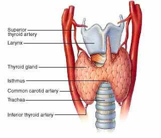
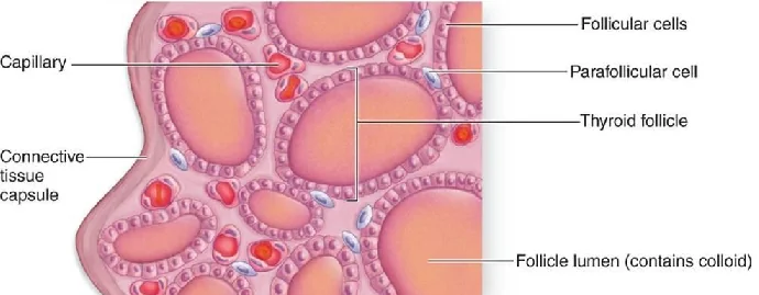
Secretion is controlled by the hypothalamus and pituitary: TRH (hypothalamus) → TSH (anterior pituitary) → T3 and T4 (thyroid). When T3 and T4 rise in the blood they feed back negatively on both the hypothalamus and the pituitary, reducing TRH and TSH (see the TRH–TSH feedback diagram in section 7).
Functions of T3 and T4
The thyroid releases much more T4 than T3, but T3 is the more active form: most T4 is converted to T3 in the tissues. Thyroid hormones act on almost every cell.
- Regulate the basal metabolic rate (BMR), the minimum energy use needed to stay alive. They increase cell respiration of carbohydrates, fats and amino acids, so energy and heat production rise.
- Increase protein synthesis; essential for normal physical and mental growth.
- Support the process of red blood cell formation.
- Control the metabolism of carbohydrates, proteins and fats.
- Help maintain water and electrolyte balance.
- Needed for a normal menstrual cycle.
- In amphibians, needed for metamorphosis of the tadpole into an adult frog.
Hypothyroidism: too little T3 and T4
- Cretinism: hypothyroidism during pregnancy or in infancy causes defective development of the baby: stunted growth, mental retardation and low IQ, abnormal skin, a protruding belly and thick tongue, and deaf-mutism.
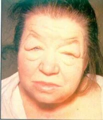
- Myxoedema Beyond the syllabus: hypothyroidism in adults. Puffy appearance, low BMR, weight gain, lethargy and sensitivity to cold; in women the menstrual cycle may become irregular. Treated with daily thyroxine tablets.
- Hashimoto’s thyroiditis Beyond the syllabus: an autoimmune disease in which the immune system attacks and damages the thyroid, so it cannot make enough hormone.
- Simple goitre: enlargement of the thyroid caused by iodine deficiency in the diet. It is common in hilly regions where rain washes iodine out of the soil, so local crops contain little iodine. With too little iodine, little T3/T4 is made; negative feedback weakens, so TSH stays high and keeps stimulating the gland, which enlarges. Iodised salt prevents it.
Hyperthyroidism: too much T3 and T4
- BMR rises; the person loses weight, feels nervous and cannot tolerate heat.
- Caused by thyroid tumours or nodules, or by Graves’ disease.
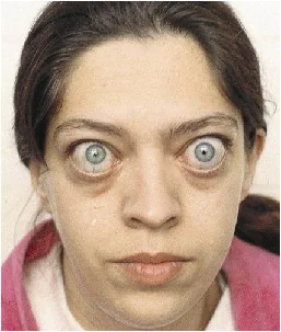
- Graves’ disease (exophthalmic goitre): an autoimmune disease. Antibodies bind to and stimulate the TSH receptor, so the thyroid keeps secreting even when TSH is low. The gland enlarges and the eyeballs protrude because the tissues behind the eyes swell.
- Beyond the syllabus Treatment includes antithyroid drugs such as thiourea-type drugs (carbimazole, propylthiouracil), which block the synthesis of thyroid hormones.
🧠 Mnemonic: Chief Minister has Goiter
Cretinism, Myxoedema, Hashimoto’s thyroiditis, simple Goitre: disorders linked to too little thyroid hormone (simple goitre may have normal hormone levels). Too much thyroid hormone: Graves’ disease, whose enlarged gland is called exophthalmic goitre.
Thyrocalcitonin (TCT)
- A peptide hormone of 32 amino acids, secreted by the parafollicular cells.
- Released when blood calcium is high (hypercalcaemia).
- Lowers blood Ca2+ mainly by inhibiting osteoclasts, the large multinucleate cells that break down bone, and by increasing Ca2+ excretion by the kidneys.
- No disorder of calcitonin deficiency is known.
Parathyroid glands
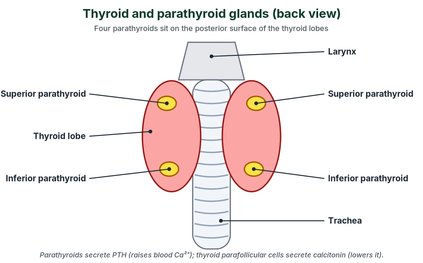
- Humans have four parathyroid glands on the back surface of the thyroid, one pair on each lobe.
- They secrete parathyroid hormone (PTH, parathormone), a peptide hormone, when blood Ca2+ falls (hypocalcaemia). Its action is antagonistic to calcitonin.
- PTH raises blood Ca2+: it stimulates bone resorption (osteoclasts dissolve bone mineral), increases Ca2+ reabsorption in the kidney tubules, and stimulates the kidney to make calcitriol (active vitamin D, 1,25-dihydroxycholecalciferol) from inactive vitamin D. Calcitriol then increases Ca2+ absorption from digested food in the gut.
- PTH is therefore a hypercalcaemic hormone.
Too little PTH: hypocalcaemic tetany
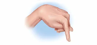
Calcium ions on the outside of nerve cell membranes help keep voltage-gated Na+ channels closed at rest. When blood Ca2+ falls, these channels open much more easily, so nerves and muscles become over-excitable and fire on their own. The result is tetany: twitching, cramps and sustained spasms of skeletal muscles, typically of the hands and feet (carpal spasm). Blood Na+ and K+ do not rise; the cause is the low Ca2+.
Too much PTH: hyperparathyroidism Beyond the syllabus
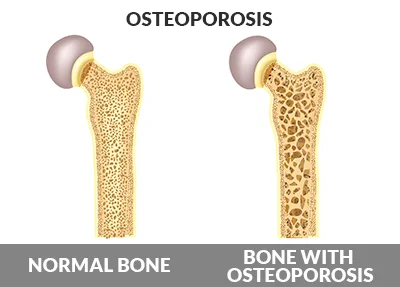
Osteoclast activity rises and bones lose calcium (demineralisation), becoming fragile and fracturing easily; in severe cases the bone matrix is replaced by fibrous tissue and cysts (osteitis fibrosa cystica). The high blood Ca2+ can also cause kidney stones.
Adrenal glands
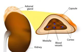
- A pair of glands, one on top of each kidney; also called suprarenal glands.
- Two distinct regions: the central adrenal medulla (from neural crest, ectoderm) and the outer adrenal cortex (from mesoderm).
Adrenal medulla: adrenaline for vigorous activity
The adrenal medulla secretes adrenaline (epinephrine) and noradrenaline (norepinephrine), together called catecholamines; about 80% is adrenaline. They are secreted rapidly in response to stress and emergencies, so they are called the emergency hormones or hormones of fight or flight (fright, fight and flight).
Adrenaline prepares the body for vigorous activity (IB C3.1.12):
Swipe the table sideways to see every column.
| Target | Effect of adrenaline | Why it helps vigorous activity |
|---|---|---|
| Heart | Faster heart rate and stronger contractions (more stroke volume) | Higher cardiac output delivers more O2 and glucose to muscles |
| Airways | Bronchioles dilate; breathing rate rises | More air reaches the alveoli, so more O2 in and CO2 out |
| Blood vessels | Vessels to skin and gut constrict; vessels to skeletal muscle, heart and liver dilate | Blood is redirected to the organs doing the work |
| Liver and muscle | Glycogen broken down to glucose (glycogenolysis) | More glucose for respiration in the muscles |
| Adipose tissue | Lipolysis releases fatty acids | An extra fuel for respiration |
| Eyes, skin | Pupils dilate; sweating; hair stands up (piloerection) | Alertness; cooling during effort |
| Gut | Peristalsis and digestive secretions decrease | Energy and blood are saved for muscles |
Noradrenaline’s main effect is vasoconstriction throughout the body, which raises blood pressure. The sympathetic nervous system releases noradrenaline from its nerve endings and works together with the adrenal medulla (the sympathetico-adrenal system), a clear example of the nervous system and endocrine system working together; the parasympathetic system has the opposite effects at rest.
Beyond the syllabus Disorders of the adrenal medulla
No common deficiency disorder is known; loss of the medulla is largely compensated by the sympathetic nervous system. A rare tumour of the chromaffin (pheochromocytes) cells, pheochromocytoma, usually benign, secretes excess catecholamines and causes very high blood pressure, sweating and palpitations.
Adrenal cortex
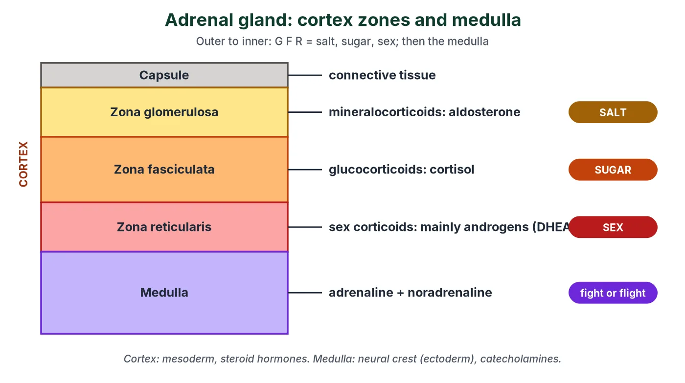
The cortex has three zones, from the outside in: zona glomerulosa, zona fasciculata and zona reticularis. Together they make more than 20 hormones, all steroids (corticoids), grouped by function:
- Zona glomerulosa: mineralocorticoids (mainly aldosterone): salt and water balance.
- Zona fasciculata: glucocorticoids (mainly cortisol): glucose metabolism and stress.
- Zona reticularis: sex corticoids, mainly weak androgens.
🧠 Mnemonic: 3S = salt, sugar, sex + GFR
Outside to inside, the zones are Glomerulosa, Fasciculata, Reticularis, and they make salt (mineralocorticoids), sugar (glucocorticoids) and sex (sex corticoids) hormones in that order. “GFR” here is not the kidney’s glomerular filtration rate.
Mineralocorticoids: aldosterone
- Made in the zona glomerulosa; the main one is aldosterone.
- Released when blood pressure or blood volume falls, when blood Na+ is low or blood K+ is high, and when the renin-angiotensin-aldosterone system (RAAS) is activated.
- Acts on the late DCT and collecting duct: increases Na+ reabsorption (water follows) and K+ and phosphate excretion.
- So it maintains electrolytes, body fluid volume, osmotic pressure and blood pressure.
RAAS in four steps
- Low blood pressure or low Na+ is sensed by the juxtaglomerular apparatus (JG cells and macula densa); sympathetic nerves also stimulate it.
- JG cells release renin, which converts angiotensinogen (a plasma protein from the liver) into angiotensin I.
- Angiotensin-converting enzyme (ACE), mainly in the lungs, converts angiotensin I to angiotensin II, a powerful vasoconstrictor.
- Angiotensin II stimulates the adrenal cortex to release aldosterone; more Na+ and water are reabsorbed, and blood volume and pressure rise. (About 99% of filtered Na+ is reabsorbed anyway; aldosterone fine-tunes the last few per cent.)
The full story continues with ANF (atrial natriuretic factor), the heart’s check on RAAS.
Glucocorticoids: cortisol
The glucocorticoids are cortisol, cortisone and corticosterone; cortisol is the most important. They are released in response to ACTH, especially during stress.
- Raise blood glucose mainly by gluconeogenesis (making new glucose from amino acids and glycerol), with lipolysis and proteolysis supplying the raw materials. They also inhibit the uptake and use of amino acids by cells.
- Glucose-sparing effect: cortisol makes most cells use fatty acids and amino acids for respiration instead of glucose, saving glucose for the brain, which depends on it. (In the liver cortisol actually promotes glycogen storage; glycogen breakdown is the job of glucagon and adrenaline.)
- Anti-inflammatory: blocks the effects of histamine and stabilises lysosome membranes, limiting tissue damage; also anti-allergic.
- Suppresses the immune response (fewer lymphocytes) and stimulates RBC production.
- Helps the body cope with long-term stress; maintains the cardiovascular system and kidney function.
- In large doses inhibits growth and increases bone resorption.
Sex corticoids
- Made in the zona reticularis in response to ACTH.
- Mainly weak androgens such as DHEA (dehydroepiandrosterone); only a little estrogen is formed, mostly by conversion in other tissues.
- Role: growth of axial (armpit), pubic and facial hair during puberty (NCERT).
Beyond the syllabus Excess sex corticoids
In boys, excess adrenal androgens before puberty cause early development of male features without the testes maturing (precocious pseudopuberty). In females they cause adrenal virilism: male secondary sexual characters such as facial hair and a deep voice.
Disorders of the adrenal cortex
- Addison’s disease (NEET list): failure of the adrenal cortex, so both cortisol and aldosterone are too low. Low cortisol alters carbohydrate metabolism and causes acute weakness, fatigue and low blood glucose; low aldosterone causes low blood Na+, high K+, dehydration and low blood pressure. The skin shows a bronze pigmentation, because the pituitary makes extra ACTH, which also stimulates melanocytes.
- Conn’s syndrome Beyond the syllabus: too much aldosterone, usually from a tumour. Na+ and water are retained, causing high blood pressure; K+ is lost, causing muscle weakness and cramps.
- Cushing’s syndrome Beyond the syllabus: too much cortisol, from a cortex tumour, excess ACTH or long-term steroid drugs. Features: high blood glucose, fat redistributed to the abdomen (pendulous belly), face (moon face) and upper back (buffalo hump), purple stretch marks (striae), excess hair (hirsutism), amenorrhoea and slow wound healing.
🧠 Mnemonic: MAC
Mineralocorticoid-linked disorders: Addison’s (aldosterone and cortisol too low) and Conn’s (aldosterone too high). Remember that Addison’s is a failure of the whole cortex, not of aldosterone alone.
🧠 Mnemonic: Glucose Causes Hyperglycemia
Excess Glucocorticoid → Cushing’s syndrome → Hyperglycaemia. Too little cortisol is part of Addison’s disease.
4 Pineal, thymus, gonads and other hormone sources
Syllabus link: NCERT XI 19.2.3 Pineal gland, 19.2.6 Thymus, 19.2.9 Testis, 19.2.10 Ovary, 19.3 Hormones of heart, kidney and gastrointestinal tract; IB C3.1.11 (SL) melatonin
The human endocrine system also includes the pineal gland, the thymus and the gonads, plus hormone-secreting cells in the heart, kidney and gut. All are in the NEET syllabus, and the pineal gland’s melatonin is an IB SL statement.
Pineal gland and melatonin
- Also called the epiphysis cerebri; a small pine-cone-shaped gland on the dorsal side of the forebrain (roof of the diencephalon, behind the third ventricle).
- Secretes melatonin, an amine hormone made from the amino acid tryptophan (through serotonin).
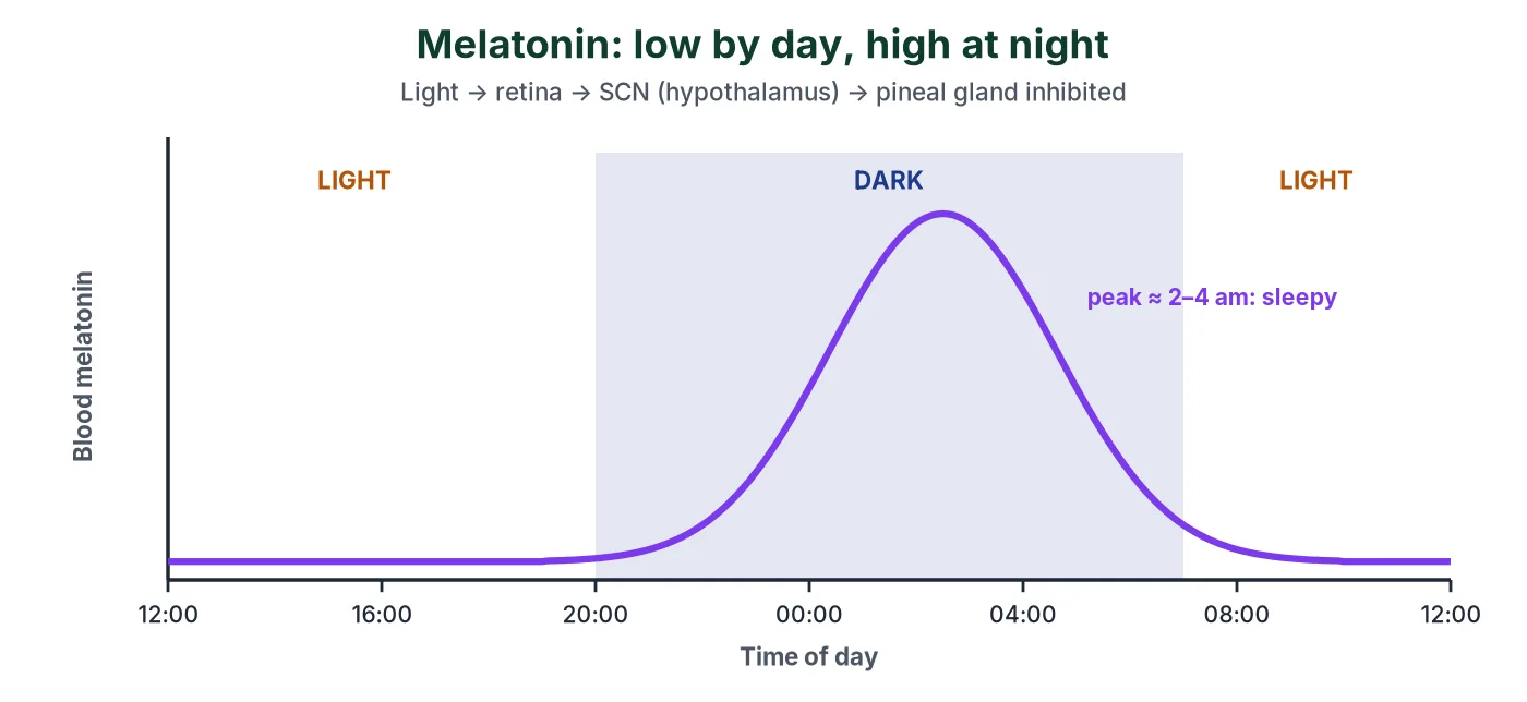
- Melatonin regulates the 24-hour (diurnal) rhythm of the body: the sleep-wake cycle and body temperature. It also influences metabolism, pigmentation, the menstrual cycle and our defence capability (NCERT).
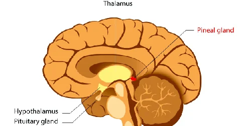
How melatonin sets the sleep cycle (IB C3.1.11): light falling on the retina sends nerve impulses to the suprachiasmatic nucleus (SCN) of the hypothalamus, the body’s master clock. From the SCN, a pathway through the spinal cord and sympathetic nerves reaches the pineal gland and inhibits melatonin secretion. So melatonin is low in daylight and we stay awake. As light fades in the evening the inhibition stops, melatonin rises, peaks in the middle of the night and makes us drowsy; it falls again towards morning.
IB only Applications of melatonin
- Jet lag: after flying across time zones the SCN is out of step with local daylight; melatonin taken at local bedtime can help reset the cycle.
- Shift work and screen light at night suppress melatonin and disturb sleep.
Beyond the syllabus Melatonin and seasonal breeding
In animals that breed in a particular season, the length of the nightly melatonin pulse tells the body the time of year. In long-day breeders such as hamsters, long winter nights (more melatonin) switch the gonads off; in short-day breeders such as sheep, the longer melatonin signal of autumn switches breeding on.
Thymus
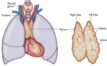
- A lobular gland between the lungs, behind the sternum and on the front of the aorta (in the mediastinum).
- Large in childhood; it shrinks after puberty and in old age is largely replaced by fat.
- Secretes peptide hormones, thymosins, which help T lymphocytes differentiate (cell-mediated immunity) and also promote antibody production (humoral immunity).
- Because the thymus degenerates in old age, thymosin production falls and immune responses weaken (NCERT).
Beyond the syllabus How the thymus ‘trains’ T cells
Developing T cells that react strongly against the body’s own antigens are removed in the thymus by apoptosis (negative selection) and the debris is cleared by macrophages. Failure of this process can lead to autoimmune disease. Hassall’s corpuscles are whorls of keratinised epithelial cells in the thymic medulla; they are not the cells that remove T cells.
Testes
- A pair of testes in the scrotum. They are both gametogenic and endocrine glands (sometimes called heterocrine).
- Leydig (interstitial) cells between the seminiferous tubules secrete androgens, mainly testosterone, an anabolic steroid. Sertoli cells in the tubules secrete inhibin, which inhibits FSH secretion.
- Beyond the syllabus Misuse of anabolic steroids (synthetic testosterone) causes acne and, through negative feedback that reduces LH and FSH, a fall in sperm count.
Practice MCQ
Leydig cells in the intertubular spaces produce androgens, mainly testosterone. Androgens are responsible for:
- a) Development, maturation and functions of the male accessory sex organs: epididymis, vas deferens, seminal vesicles, prostate gland and urethra
- b) Muscular growth, growth of facial and axillary hair, aggressiveness and low pitch of voice
- c) A major stimulatory role in spermatogenesis
- d) Anabolic (synthetic) effects on protein and carbohydrate metabolism
- e) All of the above
Show answer
(e)
Ovaries
- A pair of ovaries in the abdomen; they produce ova and hormones.
- Growing ovarian follicles secrete estrogen (mainly estradiol).
- After ovulation, the ruptured follicle becomes the corpus luteum, which secretes mainly progesterone.
- Late in pregnancy the ovary also secretes relaxin, which softens the ligaments of the pelvis and the cervix for birth (NCERT XII, Human Reproduction).
Practice MCQ
The ovary produces estrogen and progesterone. Which of the following is a function of estrogen?
- a) It stimulates growth and activities of the female secondary sex organs
- b) It stimulates the development of growing ovarian follicles
- c) It causes the appearance of female secondary sex characters, such as a high-pitched voice, and mammary gland development
- d) It regulates female sexual behaviour
- e) All of the above
Show answer
(e)
Practice MCQ
Which of the following is a function of progesterone?
- a) It supports pregnancy
- b) It acts on the mammary glands and stimulates the formation of alveoli
- c) It stimulates milk secretion
- d) All of the above
Show answer
(d) This is the NCERT answer. In reality it is prolactin that drives milk production; progesterone prepares the alveoli and its high level in pregnancy holds back milk until after birth.
IB only Reproductive hormones in the IB guide
Swipe the table sideways to see every column.
| Statement | Hormones | Key idea |
|---|---|---|
| D3.1.5 (SL) | FSH, LH, oestradiol, progesterone | Control the ovarian and uterine cycles by negative and positive feedback (LH surge) |
| D3.1.7 (SL) | Hormone drugs | IVF: normal secretion is suppressed, then high doses of FSH cause superovulation |
| D3.1.13 HL | GnRH, LH, FSH, sex steroids | Rising GnRH from the hypothalamus starts puberty |
| D3.1.17 HL | hCG | Pregnancy tests detect hCG with monoclonal antibodies |
| D3.1.19 HL | Progesterone, oxytocin | Progesterone maintains pregnancy; falling progesterone and positive feedback on oxytocin drive childbirth |
Hormones of the heart, kidney and gut: the diffuse endocrine system
Some tissues that are not endocrine glands also secrete hormones (NCERT 19.3), so the endocrine system is wider than its glands.
Swipe the table sideways to see every column.
| Source | Hormone | Action |
|---|---|---|
| Atrial wall of the heart | ANF (atrial natriuretic factor) | Released when the atria are stretched by high blood volume or pressure. Causes vasodilation and more Na+ and water excretion, lowering blood pressure; opposes RAAS |
| Juxtaglomerular cells of the kidney (NCERT) | Erythropoietin (EPO) | Stimulates RBC formation in the bone marrow; secreted when O2 supply falls (hypoxia) |
| Kidney JG cells | Renin (an enzyme) | Starts RAAS |
| Stomach (pyloric) mucosa | Gastrin | Stimulates gastric glands to secrete HCl and pepsinogen |
| Duodenal epithelium | Secretin | Stimulates the pancreas to secrete water and bicarbonate |
| Duodenal epithelium | CCK (cholecystokinin) | Contracts the gall bladder to release bile; stimulates pancreatic enzyme secretion |
| Duodenal epithelium | GIP (gastric inhibitory peptide) | Inhibits gastric secretion and motility |
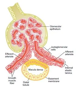
Beyond NCERT: most EPO actually comes from interstitial cells around the kidney tubules, but NCERT and NEET credit the juxtaglomerular cells. More on the gut hormones: Digestion and Absorption: hormonal control. More on ANF and renin: the renin-angiotensin-aldosterone system.
5 Pancreas and regulation of blood glucose
Syllabus link: NCERT XI 19.2.8 Pancreas; IB D3.3.1 to D3.3.3 (SL) homeostasis and regulation of blood glucose
Blood glucose is a homeostatic variable: it is kept close to a set point of about 5 mmol L−1 (90 mg dL−1) despite meals, fasting and exercise. Too high damages blood vessels and nerves; too low starves the brain. The pancreas controls it by negative feedback, the best-known example of the endocrine system in homeostasis.
The pancreas: a mixed gland
- The pancreas is both exocrine (pancreatic juice with enzymes, into the duodenum) and endocrine.
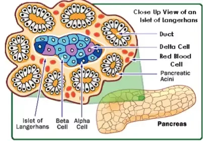
- The endocrine part is the islets of Langerhans: about 1 to 2 million clusters of cells, only 1 to 2% of the pancreatic tissue. See pancreatic juice and the digestive glands for the exocrine part.
- α (alpha) cells secrete glucagon; β (beta) cells secrete insulin.
- Beyond the syllabus δ (delta) cells secrete somatostatin, which inhibits the release of both insulin and glucagon; PP cells secrete pancreatic polypeptide, which inhibits the secretion of pancreatic (exocrine) juice.
The blood glucose control loop
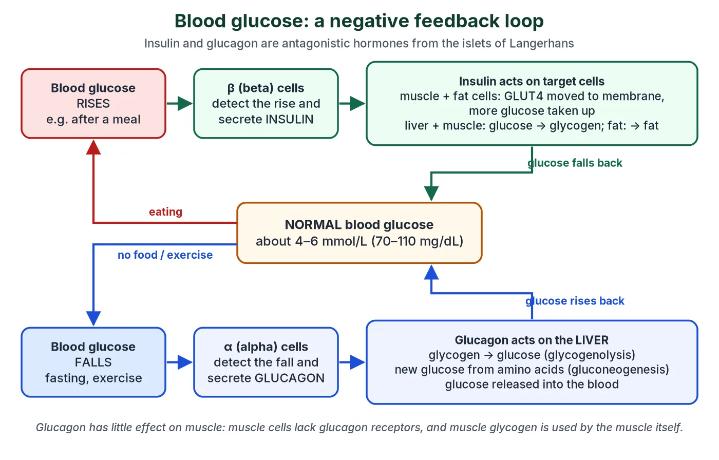
- The islet cells themselves are the receptors: they detect the glucose concentration of the blood flowing through the pancreas.
- If glucose rises, β cells secrete insulin into the blood. Its targets are liver cells, skeletal muscle cells and adipose (fat) cells, which take up glucose and store it as glycogen or fat.
- If glucose falls, α cells secrete glucagon. Its main target is the liver, which breaks down glycogen and makes new glucose, then releases glucose into the blood.
- In each case the response cancels the change: negative feedback returns blood glucose to the set point.
- Insulin and glucagon are antagonistic hormones: they have opposite effects on the same variable.
Try it: what happens to blood glucose…
After a meal Blood glucose rising
Glucose absorbed from the gut raises blood glucose. β cells detect the rise and secrete insulin; α cells secrete less glucagon. Muscle and fat cells move GLUT4 transporters into their membranes and take up glucose; liver and muscle convert it to glycogen (glycogenesis); fat cells convert it to fat. Blood glucose falls back to normal.
Overnight fast Blood glucose falling
Between meals and overnight, glucose is used up and blood glucose starts to fall. α cells detect the fall and secrete glucagon; insulin falls. Glucagon acts on liver cells: glycogen is broken down to glucose (glycogenolysis) and new glucose is made from amino acids and glycerol (gluconeogenesis). The liver releases glucose and blood glucose rises back to normal.
A sprint or an exam Stress or vigorous activity
Muscles take up glucose fast. Adrenaline from the adrenal medulla, with glucagon, triggers glycogenolysis in the liver and in the muscles themselves, while insulin secretion is held down. In longer stress, cortisol adds gluconeogenesis. Blood glucose is kept up so the muscles and brain never run short.
Don’t mix these up
- Glucose: the sugar in the blood. Glycogen: the storage polysaccharide in liver and muscle. Glucagon: the hormone that makes the liver break glycogen down.
- Gluconeogenesis = making new glucose; glycogenolysis = splitting glycogen; glycogenesis = making glycogen.
- Glucagon acts mainly on the liver; muscle cells have no glucagon receptors and keep their glycogen for their own use.
6 Insulin and glucagon
Syllabus link: NCERT XI 19.2.8 Pancreas; IB D3.3.3 (SL) effects on target cells; IB C2.1.11 HL insulin receptor with tyrosine kinase activity
Insulin and glucagon are the two endocrine system hormones that matter most for blood glucose. Both are peptide hormones from the islets of Langerhans, carried in the blood plasma to their target cells.
Swipe the table sideways to see every column.
| Insulin | Glucagon | |
|---|---|---|
| Secreted by | β cells | α cells |
| Chemical nature | Peptide hormone of 51 amino acids in two chains | Peptide hormone (29 amino acids) |
| Secreted when | Blood glucose is high (hyperglycaemia) | Blood glucose is low (hypoglycaemia) |
| Main targets | Liver, skeletal muscle, adipose tissue | Liver (hepatocytes) |
| Effects | Increases glucose uptake and use by cells (for example in glycolysis) (GLUT4 in muscle and fat); glucose → glycogen (glycogenesis) in liver and muscle; glucose → fat in adipocytes; increases amino acid uptake and protein synthesis | Glycogen → glucose (glycogenolysis) in the liver; gluconeogenesis; reduces cellular glucose uptake and use |
| Effect on blood glucose | Lowers it: hypoglycaemic hormone | Raises it: hyperglycaemic hormone |
IB only HL Cell signalling: how insulin and adrenaline act
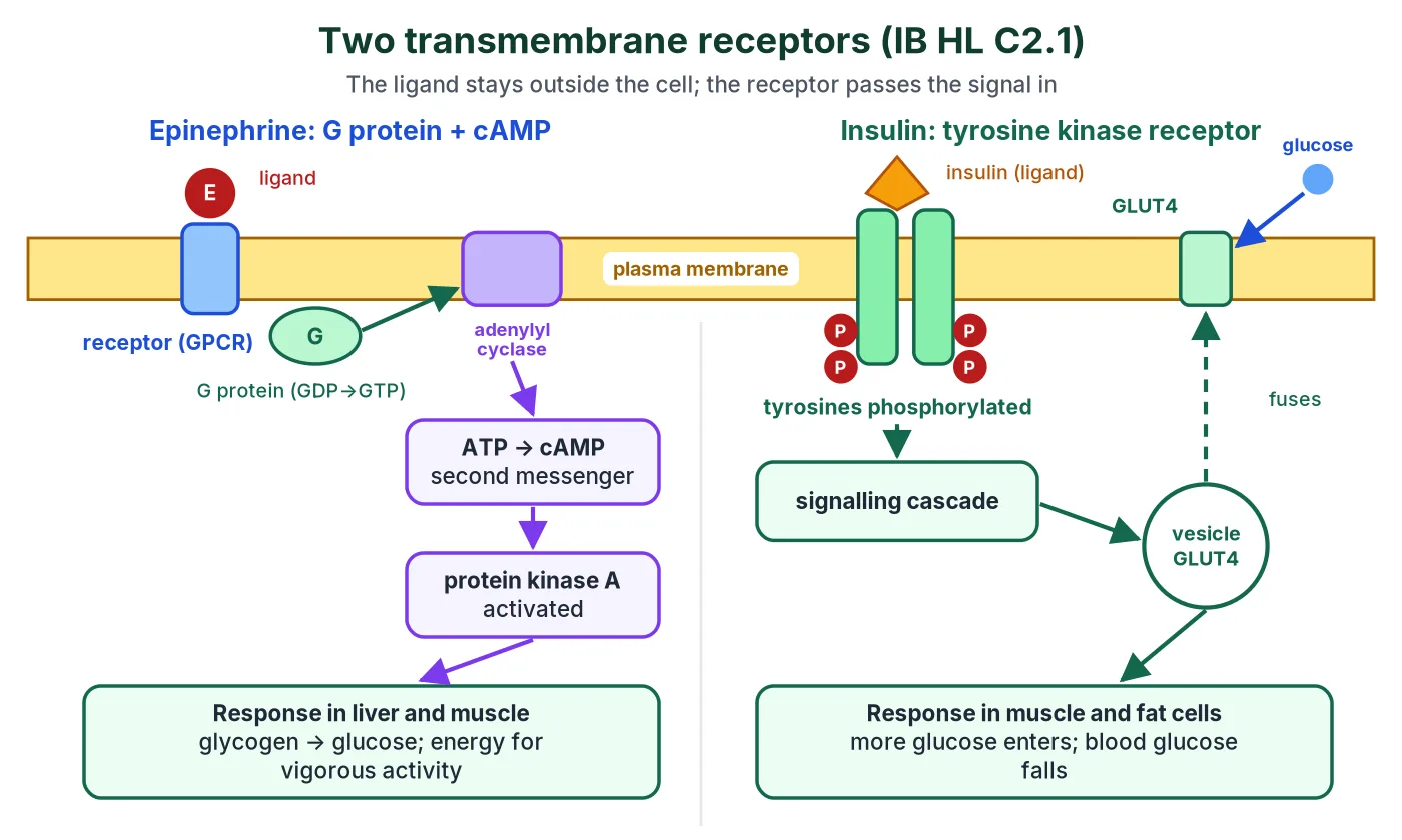
In the endocrine system, a signalling chemical is called a ligand; its receptor is a protein with a binding site specific to that ligand. Binding starts a signal transduction pathway inside the cell.
- Transmembrane receptors span the plasma membrane: their outside has hydrophilic amino acids and the part crossing the membrane has hydrophobic ones. They bind hydrophilic ligands (peptides, adrenaline) that cannot enter the cell.
- Intracellular receptors in the cytoplasm or nucleus bind hydrophobic ligands (steroids) that diffuse through the membrane (section 9).
- Adrenaline binds a G protein-coupled receptor. The activated G protein switches on adenylyl cyclase, which makes cyclic AMP (cAMP), the second messenger; cAMP activates protein kinase A, leading, in liver and muscle, to glycogen breakdown.
- Insulin binds a receptor with tyrosine kinase activity. Tyrosines on the inside of the receptor become phosphorylated, a chain of reactions follows, and vesicles carrying GLUT4 glucose transporters move to and fuse with the plasma membrane, so more glucose enters the cell.
7 Hormonal control of growth and metabolism
Syllabus link: NCERT XI 19.2 (GH, thyroid hormones, glucocorticoids); IB D3.3.5 and D3.3.6 (SL) thermoregulation with thyroxin
The endocrine system controls growth and metabolism through several hormones acting together. GH and thyroxine are the main growth hormones; insulin, glucagon, adrenaline and cortisol decide whether fuel is stored or used.
Swipe the table sideways to see every column.
| Hormone | Source | Effect on growth and metabolism |
|---|---|---|
| Growth hormone (GH) | Anterior pituitary | Growth of bones and muscles through IGF-1 from the liver; protein synthesis; lipolysis; raises blood glucose |
| Thyroxine (T4) and T3 | Thyroid | Sets the basal metabolic rate and heat production; needed for growth and brain development |
| Insulin | Pancreas β cells | Anabolic: glucose uptake and storage, fat synthesis, protein synthesis |
| Glucagon, adrenaline | Pancreas α cells, adrenal medulla | Mobilise stored fuel: glycogenolysis, lipolysis |
| Cortisol | Adrenal cortex | Gluconeogenesis, glucose sparing, protein breakdown during long-term stress |
| Testosterone, estrogen | Testes, ovaries | Growth spurt and secondary sexual characters at puberty; testosterone is anabolic (muscle) |
Thyroxine, metabolic rate and body temperature
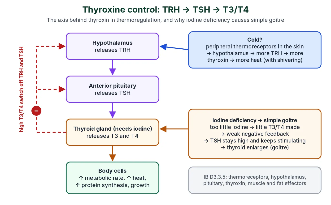
Body temperature is another homeostatic variable controlled by negative feedback (IB D3.3.5); here the nervous system and the endocrine system act together. Peripheral thermoreceptors in the skin and receptors in the hypothalamus detect temperature changes. When the body is cold, the hypothalamus increases TRH, the pituitary releases more TSH and the thyroid releases more thyroxin. Thyroxin raises the metabolic rate of cells, so more heat is released by respiration. Other effectors work at the same time:
Swipe the table sideways to see every column.
| When the body is too cold | When the body is too hot |
|---|---|
| Vasoconstriction of skin arterioles: less heat lost | Vasodilation of skin arterioles: more heat lost |
| Shivering: skeletal muscles contract rapidly, releasing heat | Sweating: evaporation of sweat cools the skin |
| Uncoupled respiration in brown adipose tissue: energy released as heat instead of ATP | Less thyroxin; lower metabolic rate |
| Hair erection (piloerection): traps a layer of air | Hairs lie flat |
IB D3.3.6 asks for these human thermoregulation mechanisms; they are covered again in the Homeostasis chapter (card 9 of Human Physiology).
8 Hormonal feedback mechanisms
Syllabus link: IB D3.3.2 (SL) negative feedback loops; IB D3.1.5 (SL) positive and negative feedback in the menstrual cycle; IB C2.1.14 HL; NCERT XI 19.2 (feedback control of hormone secretion)
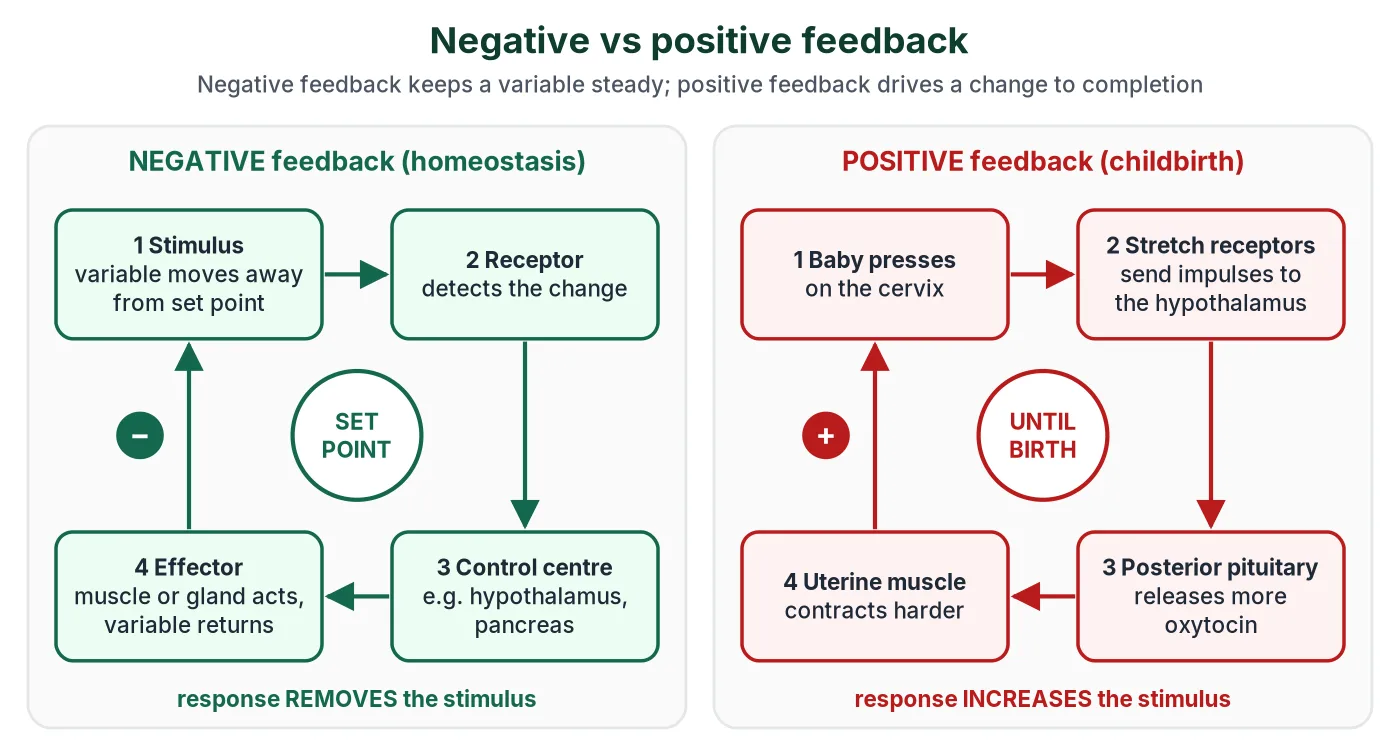
Feedback is how the endocrine system controls itself. Most hormone secretion is controlled by feedback: the result of a hormone’s action changes how much of the hormone is secreted.
Negative feedback in the endocrine system
In negative feedback the response reverses the change that caused it. If a variable rises above its set point, the response lowers it; if it falls below, the response raises it. This keeps homeostatic variables within narrow limits.
Swipe the table sideways to see every column.
| Variable | Detected by | Hormone response | Result |
|---|---|---|---|
| Blood glucose | α and β cells of islets | High: insulin. Low: glucagon | Glucose back to about 5 mmol L−1 |
| Blood Ca2+ | Parathyroid and thyroid C cells | Low: PTH. High: calcitonin | Ca2+ back to normal |
| Thyroxine level | Hypothalamus and anterior pituitary | High T3/T4 reduce TRH and TSH | Thyroxine stays steady |
| Blood osmotic concentration | Osmoreceptors in hypothalamus | High: more ADH. Low: less ADH | Water reabsorption adjusted |
| Body temperature | Thermoreceptors, hypothalamus | Cold: more TSH and thyroxin | Heat production rises |
Many hypothalamus-pituitary axes work by long-loop negative feedback: the final hormone (thyroxine, cortisol, testosterone, estradiol) inhibits both the hypothalamus and the anterior pituitary. This is the commonest control pattern in the human endocrine system.
Positive feedback
In positive feedback the response increases the change, so it grows until some outside event ends it. The endocrine system uses it for changes that need to be completed quickly, not for homeostasis.
- Childbirth: stretching of the cervix → more oxytocin → stronger uterine contractions → more stretching, until the baby is born.
- The LH surge: before ovulation, high oestradiol switches from negative to positive feedback on the hypothalamus and pituitary, causing a surge of LH that triggers ovulation.
- Milk ejection: suckling → oxytocin → milk let-down → more suckling.
Why homeostasis uses negative feedback
- Negative feedback returns a variable to the set point from values above or below it, so the internal environment stays stable.
- Positive feedback would push a variable further from its set point, so it is only safe for one-off events that end on their own.
Antagonistic and synergistic hormones
Hormones of the endocrine system often act in pairs or teams.
- Antagonistic hormones have opposite effects on the same variable: insulin and glucagon (blood glucose), calcitonin and PTH (blood Ca2+).
- Synergistic hormones work together to produce a bigger combined effect: estrogen, progesterone, prolactin and oxytocin together prepare the mammary glands and produce and eject milk; glucagon, adrenaline and cortisol together raise blood glucose.
Beyond the syllabus Nervous and hormonal antagonism at the heart
Heart rate is raised by adrenaline and noradrenaline (through β-receptors, a G protein and more cAMP) and lowered by acetylcholine from the vagus nerve. Acetylcholine acts through muscarinic receptors linked to an inhibitory G protein: cAMP falls and K+ channels open. Acetylcholine is a neurotransmitter here, not a hormone.
9 Mechanism of hormone action
Syllabus link: NCERT XI 19.4 Mechanism of hormone action (elementary idea); IB C2.1.6 to C2.1.13 HL
Hormones of the endocrine system travel in the blood to every cell, but only target cells respond, because only they have receptors for that hormone. A hormone binding to its receptor forms a hormone-receptor complex, and each receptor is specific to one hormone. The hormone is the first messenger.
- Membrane-bound receptors are on the plasma membrane of target cells. They bind water-soluble hormones (peptides, proteins, catecholamines), which cannot cross the lipid bilayer.
- Intracellular receptors are inside the cell, mostly in the nucleus (nuclear receptors). They bind lipid-soluble hormones: steroids (corticoids, testosterone, estrogen, progesterone) and iodothyronines (T3, T4), which diffuse through the membrane.
Lipid-soluble hormones: steroids and thyroid hormones
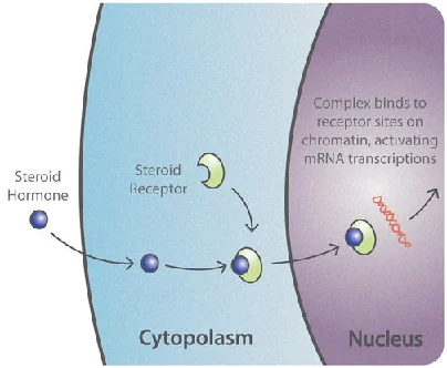
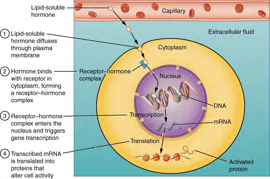
- The hormone diffuses through the plasma membrane into the target cell.
- It binds a receptor protein in the cytoplasm or nucleus to form a hormone-receptor complex.
- The complex binds a specific DNA sequence in the nucleus.
- Transcription of particular genes is switched on (or off), making mRNA.
- The mRNA is translated into new proteins, such as enzymes or membrane channels, which change the cell’s activity.
Because they work through gene expression, these hormones act more slowly but their effects last longer, which is typical of the endocrine system.
Water-soluble hormones: second messengers
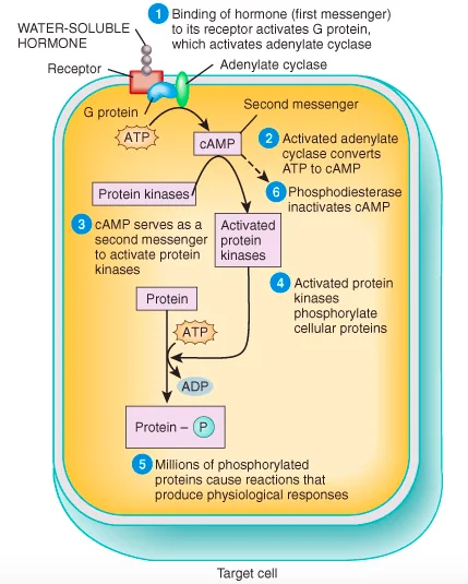
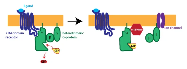
These hormones do not enter the cell. The receptor passes the signal inside:
- The hormone binds its membrane receptor.
- The receptor activates a G protein on the inside of the membrane; the G protein has α, β and γ subunits, and the α subunit swaps its GDP for GTP.
- The activated G protein activates the enzyme adenylate cyclase.
- Adenylate cyclase converts ATP into cyclic AMP (cAMP) + pyrophosphate (PPi).
- cAMP is the second messenger. It activates protein kinase, which was present in an inactive form.
- The protein kinase phosphorylates other enzymes, setting off the reactions that make up the cell’s response. One hormone molecule can lead to millions of product molecules (amplification).
- Common second messengers: cAMP, cGMP, IP3 (inositol trisphosphate), DAG (diacylglycerol) and Ca2+. IP3 and DAG are made by splitting the membrane lipid PIP2, which is their precursor, not a messenger itself.
- Insulin is different: its receptor is itself an enzyme (a receptor tyrosine kinase), not a G protein-coupled receptor (see the IB box in section 6).
IB only HL Steroid hormones and their target cells
- Oestradiol, progesterone and testosterone bind intracellular receptors; the activated receptor binds specific DNA sequences and promotes transcription (C2.1.12).
- Oestradiol acts on cells in the hypothalamus that secrete GnRH; progesterone acts on cells of the endometrium, keeping it ready for implantation (C2.1.13).
10 Diabetes mellitus and selected endocrine disorders
Syllabus link: NCERT XI 19.2 (hypo- and hyperactivity disorders: dwarfism, acromegaly, cretinism, goitre, exophthalmic goitre, diabetes, Addison’s disease); IB D3.3.4 (SL) type 1 and type 2 diabetes
When the endocrine system secretes too much (hyper-) or too little (hypo-) of a hormone, a disorder results. NEET lists seven to know in brief; IB asks only about diabetes, in more depth. For more on these conditions, see the MedlinePlus overview of endocrine diseases.
Endocrine system disorders for NEET at a glance
Swipe the table sideways to see every column.
| Disorder | Gland and hormone | Cause | Main features |
|---|---|---|---|
| Pituitary dwarfism | Anterior pituitary, GH | Too little GH in childhood | Short but proportionate body |
| Gigantism | Anterior pituitary, GH | Too much GH in childhood | Excessive height |
| Acromegaly | Anterior pituitary, GH | Too much GH in adults | Thickened bones of hands, feet, jaw and face |
| Diabetes insipidus | Hypothalamus / posterior pituitary, ADH | Too little ADH | Large volumes of dilute urine; thirst |
| Cretinism | Thyroid, T3/T4 | Too little in fetus or infant | Stunted growth, mental retardation, deaf-mutism |
| Simple goitre | Thyroid | Iodine deficiency | Enlarged thyroid |
| Exophthalmic goitre (Graves’) | Thyroid, T3/T4 | Too much (autoimmune) | Enlarged thyroid, protruding eyes, high BMR, weight loss |
| Tetany | Parathyroid, PTH | Too little PTH, low blood Ca2+ | Muscle spasms and cramps |
| Addison’s disease | Adrenal cortex, cortisol and aldosterone | Too little | Weakness, fatigue, low BP, bronzed skin |
| Diabetes mellitus | Pancreas, insulin | Too little insulin or insulin resistance | High blood glucose, glycosuria, ketone bodies |
Diabetes mellitus: the commonest endocrine system disorder
Diabetes mellitus is prolonged hyperglycaemia caused by too little insulin or by cells failing to respond to it. Without insulin action, cells take up little glucose, the liver keeps releasing glucose, and fat and protein are broken down for energy.
- Glycosuria: blood glucose rises above the renal threshold (about 180 mg dL−1), so the kidney cannot reabsorb all the filtered glucose and it appears in the urine.
- Polyuria and thirst: glucose in the filtrate holds water by osmosis, so a large volume of urine is produced and the person is always thirsty.
- Ketone bodies: fat is broken down so fast that the liver makes ketone bodies (e.g. acetoacetic acid, acetone). They make the blood acidic (ketoacidosis), especially in type 1, and can be life-threatening (NCERT: ‘harmful compounds known as ketone bodies’).
- Weight loss, tiredness and, in the long term, damage to blood vessels, eyes, kidneys and nerves.
Read more in the NIDDK (US National Institutes of Health) guide to diabetes.
Swipe the table sideways to see every column.
| Type 1 diabetes | Type 2 diabetes | |
|---|---|---|
| Older name | IDDM (insulin-dependent) | NIDDM (non-insulin-dependent) |
| Cause | Autoimmune destruction of β cells: the immune system (mainly T cells) attacks them, so little or no insulin is made | Target cells become resistant to insulin (receptors respond less); insulin may be normal or high at first, but β cells may later fail |
| Usual onset | Childhood or early adulthood; often sudden | Usually adults over 40, but increasingly in young people; develops slowly |
| Proportion of cases | About 5 to 10% | About 90% |
| Risk factors | Family history and genes; a possible viral trigger | Obesity (especially abdominal fat), physical inactivity, diet high in sugar and refined carbohydrate, age, family history, some ethnic groups, diabetes in a previous pregnancy |
| Prevention | No known way to prevent it | Healthy body weight, regular exercise, a diet high in fibre and low in sugar and saturated fat |
| Treatment | Insulin (recombinant human insulin) by injection or pump, several times a day, matched to meals and blood glucose tests; replacing β cells with insulin-secreting cells grown from stem cells is being researched | Diet, exercise and weight loss first; then oral drugs such as metformin; insulin injections when these are not enough |
Common mistakes
- Type 2 diabetics can and often do use insulin; diet and exercise are the first step, not the only one.
- An insulinoma is a tumour of β cells. The condition it causes, from too much insulin, is hypoglycaemia (low blood glucose).
- Diabetes insipidus is an ADH problem (dilute urine, no glucose); diabetes mellitus is an insulin problem (glucose in urine).
Summary: the human endocrine system in eight points
- The human endocrine system is made of ductless glands that secrete hormones into the blood; only target cells with the right receptors respond.
- The hypothalamus links the nervous system and the endocrine system and controls the pituitary, which controls the thyroid, adrenal cortex and gonads.
- The thyroid sets the metabolic rate, PTH and calcitonin control blood calcium, and the adrenal glands handle stress, salt balance and emergencies.
- Insulin and glucagon from the islets of Langerhans keep blood glucose near its set point: the best example of negative feedback in the endocrine system.
- GH, thyroxine, cortisol, insulin and the sex hormones together control growth and metabolism.
- Negative feedback keeps the endocrine system stable; positive feedback (oxytocin in childbirth, the LH surge) drives one-off events to completion.
- Water-soluble hormones act through membrane receptors and second messengers; steroid and thyroid hormones act through intracellular receptors and gene expression.
- Too much or too little of a hormone causes disorders of the endocrine system, the commonest being diabetes mellitus.
11 Common doubts about hormones and the endocrine system
These are the doubts students raise most often about the human endocrine system, with short answers.
If hormones reach every cell in the body, why do only some cells respond?
Only target cells have the right receptor. Insulin can only affect a cell with insulin receptors, just as a radio signal is only heard by a radio tuned to that station. This is also why one hormone can have different effects in different tissues: the receptors and the pathways behind them differ. It is how the endocrine system can broadcast to the whole body yet act selectively.
Why is the pituitary called the master gland if the hypothalamus controls it?
Because its trophic hormones (TSH, ACTH, FSH, LH) control the thyroid, adrenal cortex and gonads. But the pituitary is itself under the command of the hypothalamus through releasing and inhibiting hormones, and of feedback from its target glands. ‘Master gland’ is a traditional name; the hypothalamus is really the boss of the endocrine system.
Are oxytocin and ADH pituitary hormones or hypothalamic hormones?
Both answers appear in books. They are made by neurosecretory cells in the hypothalamus and released from the posterior pituitary. In an MCQ that asks which hormones are ‘released from’ or ‘stored in’ the posterior pituitary, the answer is oxytocin and ADH; if it asks where they are synthesised, the answer is the hypothalamus.
Glucose, glycogen, glucagon: which is which?
Glucose is the sugar in the blood. Glycogen is the polysaccharide store of glucose in liver and muscle. Glucagon is the hormone from α cells that tells the liver to break glycogen down. A useful trick: glucagon is what you need when glucose is gone.
If type 2 diabetics make insulin, why do some of them need insulin injections?
In type 2 diabetes the cells are resistant, so more insulin is needed for the same effect. At first the β cells make extra, but over years they often wear out and secretion falls. When diet, exercise and tablets can no longer keep blood glucose down, extra insulin overcomes the resistance. The MedlinePlus diabetes page explains the treatment options.
Why does diabetes make you pass so much urine and feel thirsty?
When blood glucose goes above the renal threshold (about 180 mg dL−1), the kidney tubules cannot reabsorb all the filtered glucose. The glucose left in the filtrate holds water by osmosis, so much more urine is produced (polyuria). The body loses water and the person feels very thirsty.
Is adrenaline a hormone or a neurotransmitter?
Adrenaline (epinephrine) secreted by the adrenal medulla into the blood is a hormone. Its close relative noradrenaline is mainly the neurotransmitter of sympathetic nerve endings, and is also secreted in small amounts as a hormone. The same kind of chemical can be a hormone or a neurotransmitter depending on how it is delivered: through the blood or across a synapse. The nervous system and the endocrine system share several of these chemicals.
Why does a lack of iodine make the thyroid bigger, when it makes less hormone?
The thyroid needs iodine to make T3 and T4. With too little iodine, hormone levels fall, so the hypothalamus and pituitary get less negative feedback and secrete more TRH and TSH. TSH keeps stimulating the thyroid to grow and work harder, and it enlarges into a goitre, but without iodine it still cannot make enough hormone. It is a neat example of endocrine system feedback going wrong.
Why does homeostasis rely on negative rather than positive feedback?
In the endocrine system, negative feedback opposes any change, so it brings a variable back towards its set point whether it has risen or fallen. Positive feedback reinforces a change, so the variable would keep moving away from the set point. That is useful only for events that must finish quickly, such as childbirth or ovulation.
Can hormones be taken as tablets if they are ‘non-nutrient’?
Yes, many can. ‘Non-nutrient’ only means the hormones of the endocrine system are not used for energy or building material. Steroid and thyroid hormones survive digestion and are absorbed, so thyroxine, cortisol-type drugs and contraceptive pills work by mouth. Protein hormones such as insulin are digested by proteases in the gut, which is why insulin has to be injected.
12 Key terms
Many one-mark questions on the endocrine system test a definition.
Swipe the table sideways to see every column.
| Term | Meaning |
|---|---|
| Hormone | A non-nutrient chemical messenger of the endocrine system, made in trace amounts and carried in the blood to target cells. |
| Endocrine gland | A ductless gland of the endocrine system that secretes hormones into the blood. |
| Target cell | A cell with receptors for a particular hormone. |
| Ligand | The signalling chemical that binds to a receptor. |
| Neurosecretory cell | A neuron that secretes a hormone (e.g. in hypothalamic nuclei). |
| Portal vein | A vein that links two capillary beds, e.g. hypothalamus to anterior pituitary. |
| Trophic hormone | A hormone that stimulates another endocrine gland (TSH, ACTH, FSH, LH). |
| Negative feedback | A response that reverses the change that caused it. |
| Positive feedback | A response that increases the change that caused it. |
| Set point | The normal value around which a variable is kept. |
| Glycogenesis / glycogenolysis | Making glycogen from glucose / breaking glycogen down to glucose. |
| Gluconeogenesis | Making new glucose from non-carbohydrates such as amino acids and glycerol. |
| GLUT4 | The glucose transporter that insulin moves into the membrane of muscle and fat cells. |
| Second messenger | A molecule inside the cell (cAMP, IP3, Ca2+) that relays the signal from a membrane receptor. |
| Glycosuria | Glucose in the urine. |
| Catecholamines | Adrenaline and noradrenaline. |
13 FAQ
Short answers to the questions we are asked most about hormones and the endocrine system.
What is the endocrine system?
The human endocrine system is the set of ductless glands and tissues that secrete hormones into the blood: the hypothalamus, pituitary, pineal, thyroid, parathyroids, thymus, adrenals, pancreatic islets and gonads, plus hormone-secreting cells in the heart, kidney and gut.
How is the endocrine system different from the nervous system?
The endocrine system sends hormones in the blood: its effects are slower, last longer and are often widespread. The nervous system sends impulses along neurons: fast, short-lived and aimed at particular cells.
Which is the largest gland of the endocrine system?
The thyroid gland. (The pancreas is larger overall, but most of it is exocrine.)
Which hormone lowers blood glucose?
Insulin, from the β cells of the islets of Langerhans. Glucagon, adrenaline, cortisol and GH all raise it.
Which chapter is this for NEET?
NCERT Class XI Biology, Chapter 19 ‘Chemical Coordination and Integration’ (Chapter 22 in older editions), in Unit 5 Human Physiology, which covers the human endocrine system. See the official NEET UG 2026 syllabus (NTA).
Which IB topics does this page cover?
IB Biology covers the endocrine system in C3.1 Integration of body systems (adrenaline, melatonin, hypothalamus and pituitary), D3.3 Homeostasis (blood glucose, diabetes, thermoregulation) and, for HL, C2.1 Chemical signalling. IB-only and NEET-only boxes are marked.
14 Quick check
Tap each question to check what you know about the human endocrine system.
Test yourself on hormones and the endocrine system
1. What links the nervous system to the endocrine system?
The hypothalamus, which controls the pituitary gland.
2. Which gland of the endocrine system is called the master gland?
The pituitary gland.
3. Who discovered the first hormone, and which was it?
Bayliss and Starling; secretin (1902).
4. Where are oxytocin and ADH made?
In neurosecretory cells of the hypothalamus; they are released from the posterior pituitary.
5. Name the four trophic hormones of the anterior pituitary.
FSH, LH, ACTH and TSH (FLAT).
6. What does too much GH in adults cause?
Acromegaly.
7. Which cells of the thyroid secrete calcitonin?
Parafollicular (C) cells.
8. Which hormone raises blood calcium?
Parathyroid hormone (PTH).
9. Which zone of the adrenal cortex makes aldosterone?
Zona glomerulosa.
10. Which hormone does the pineal gland secrete, and when?
Melatonin, mainly at night.
11. Which cells secrete glucagon?
α cells of the islets of Langerhans.
12. What does insulin move into the membranes of muscle and fat cells?
GLUT4 glucose transporters.
13. What is the second messenger made by adenylate cyclase?
Cyclic AMP (cAMP).
14. Name one example of positive feedback.
Oxytocin and uterine contractions during childbirth (or the LH surge).
15. What is the main cause of type 1 diabetes?
Autoimmune destruction of the β cells.
16. What causes simple goitre?
Iodine deficiency.
Related chapters and practice
The endocrine system works with almost every other body system. Continue with these chapters and tests:
- Digestion and Absorption: the gut’s own part of the endocrine system (gastrin, secretin, CCK, GIP).
- Human Excretory System: how ADH, aldosterone and ANF act on the nephron.
- Cell Membrane: glycoproteins and cell recognition: the receptor proteins that hormones bind.
- Membrane Transport (IB Biology): diffusion, osmosis and active transport behind glucose uptake.
- Gas Exchange: control of ventilation: breathing and the blood gases.
- NEET sample test papers: try the XI Ch 19 Chemical Coordination and Integration test.
- NCERT Class XI Biology chapters: the NCERT chapter on chemical coordination and its neighbours.
- IB Biology topics: every IB topic on this site, including C2.1 and D3.3.
- all Human Physiology chapters: the full map of body systems, from nutrition to the endocrine system.
© competitorspoint.com · Biology notes. Labelled flow diagrams are original to competitorspoint.com.