
Cell Structure: Prokaryotic, Plant and Animal Cells
Cell structure is the arrangement of the parts that make up a cell. Every cell has a plasma membrane, cytoplasm, ribosomes and DNA. Prokaryotic cells, such as bacteria, have no nucleus and no membrane-bound organelles, while eukaryotic cells, such as plant and animal cells, have a nucleus and membrane-bound organelles such as mitochondria.
Cell structure from the bacterium to the plant and animal cell: every organelle, prokaryotic and eukaryotic cells compared, the atypical cells IB asks about and the NCERT facts NEET tests, with labelled diagrams and a quiz.
1 What is cell structure?
Syllabus link: NCERT XI Chapter 8 Cell: The Unit of Life; IB A2.2 Cell structure
NCERT = statement or value as given in NCERT Class XI (use in NEET/CBSE answers) IB = IB Biology (first assessment 2025) link
Cell structure means the parts a cell is built from and how each part is arranged to do a job. Every cell has a plasma membrane, cytoplasm, ribosomes and DNA, but the way these are organised splits all life into two great groups: prokaryotic and eukaryotic cells. Inside eukaryotic cells we then find a set of compartments, the cell organelles, each with its own structure and function. Read more on the cell on Wikipedia.
This chapter is card 2 of our Cell Biology series. Cell theory, cell size, magnification basics, surface area to volume ratio and unicellular organisms are already covered in Introduction to cells, so here we go straight to what is inside the cell: bacterial, plant and animal cells, every organelle named in NCERT Chapter 8, the atypical cells that IB loves to ask about, and a set of specialised cells. The boundary has its own page on the cell membrane.

Key points
- All cells share four things: a plasma membrane, cytoplasm, ribosomes and DNA as genetic material.
- Prokaryotic cells have no nucleus and no membrane-bound organelles; eukaryotic cells have both.
- Cell structure and function are linked: the shape and organelles of a cell match the work it does.
2 Prokaryotic cells
Syllabus link: NCERT XI 8.4 Prokaryotic cells; IB A2.2 prokaryote cell structure
The word prokaryote means “before the nucleus”, and it describes the simplest cell structure. NCERT Prokaryotic cells are represented by bacteria, blue-green algae (cyanobacteria), mycoplasma and PPLO (pleuro-pneumonia like organisms). They are generally smaller than eukaryotic cells and multiply more rapidly. Mycoplasmas, the smallest known cells, are only about 0.3 µm long, while typical bacteria are 3-5 µm.
All prokaryotes share the same basic cell structure:
- No well-defined nucleus. The genetic material is a single, naked, circular DNA molecule lying in the cytoplasm in a region called the nucleoid. It has no envelope and no histones.
- Plasmids: many bacteria also carry small circles of DNA outside the genomic DNA. Plasmids give extra characters such as antibiotic resistance.
- No membrane-bound organelles such as mitochondria, ER, Golgi or chloroplasts. The only organelle common to all prokaryotes is the ribosome.
- 70S ribosomes, made of 50S and 30S subunits.
- A cell wall outside the plasma membrane in almost all of them (mycoplasma is the exception, with no wall at all).
- Inclusion bodies: reserve material lying free in the cytoplasm, not bound by membranes.
Shapes: bacillus (rod), coccus (sphere), vibrio (comma) and spirillum (spiral). Prokaryotes divide by binary fission, not by mitosis, because there is no spindle and no nucleus to divide.
Exam tips
- Prokaryotes DO have ribosomes, a membrane and DNA: say no membrane-bound organelles, not no organelles.
- Naked DNA means the DNA is not associated with histone proteins (IB wording).
- Mycoplasma has no cell wall, so penicillin (which blocks wall synthesis) does not kill it.
3 Bacterial cell structure in detail
Syllabus link: NCERT XI 8.4.1 Cell envelope and its modifications; IB A2.2 labelling a prokaryotic cell
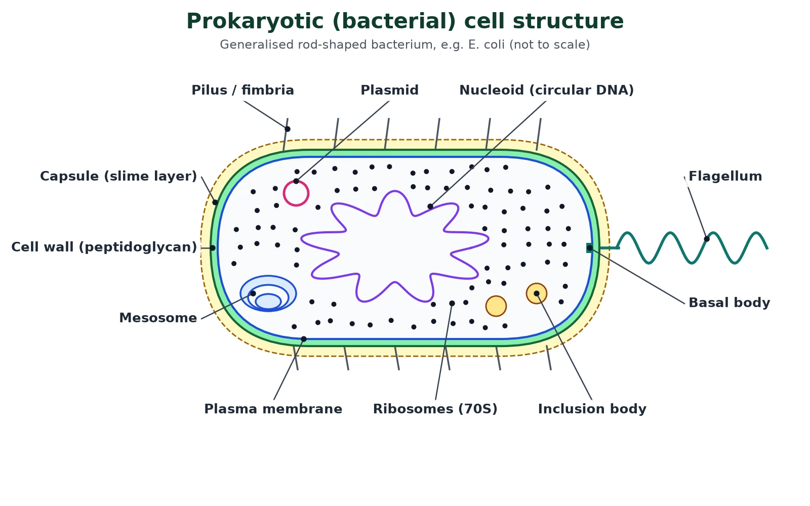
A bacterium is the model of prokaryotic cell structure in both syllabuses. Learn this cell structure diagram well: IB papers often ask you to draw and label it, and NEET asks about each layer.
The cell envelope: three layers
NCERT Most bacterial cells have a chemically complex cell envelope made of three tightly bound layers: the outer glycocalyx, then the cell wall, then the plasma membrane. Together they act as one protective unit.
- Glycocalyx: differs in composition and thickness between bacteria. A loose sheath is a slime layer; a thick, tough one is a capsule. It protects against drying out and phagocytosis.
- Cell wall: made of peptidoglycan (murein). It gives shape and stops the cell bursting.
- Plasma membrane: selectively permeable and structurally similar to the eukaryotic membrane.
Bacteria are split into two groups by the way their envelope reacts to the stain developed by Gram: those that take up and keep the stain are Gram positive, and those that do not are Gram negative. Gram positive walls are thick peptidoglycan; Gram negative walls are thin with an outer lipid membrane.
Mesosomes and chromatophores
NCERT A mesosome is a special membranous structure formed by infolding of the plasma membrane, in the form of vesicles, tubules and lamellae. It helps in cell wall formation, DNA replication and distribution of the DNA to daughter cells, respiration, secretion, and increases the surface area of the membrane and its enzyme content. In cyanobacteria, other membranous extensions called chromatophores carry the photosynthetic pigments.
Flagella, pili and fimbriae
Motile bacteria have thin filamentous flagella growing out of the cell wall. A bacterial flagellum has three parts: filament (the longest part), hook and basal body. It rotates like a propeller. It does not have the 9+2 arrangement of microtubules found in eukaryotic flagella. Pili are elongated tubular structures of a special protein; fimbriae are small bristle-like fibres that help bacteria attach to rocks in streams and to host tissues. Neither pili nor fimbriae help in movement.
Ribosomes and polysomes
Bacterial ribosomes are attached to the plasma membrane or free in the cytoplasm. They are about 15-20 nm in size, 70S, with a 50S and a 30S subunit. Several ribosomes on one mRNA form a polysome, which makes many copies of the protein at once.
4 Eukaryotic cells
Syllabus link: NCERT XI 8.5 Eukaryotic cells; IB A2.2 eukaryote cell structure
Eukaryotic cell structure is found in all protists, plants, animals and fungi. NCERT In eukaryotic cells there is extensive compartmentalisation of the cytoplasm through membrane-bound organelles, and a well-organised nucleus with a nuclear envelope. They also have complex locomotory and cytoskeletal structures, and their genetic material is organised into chromosomes.
Compartments are the defining idea of eukaryotic cell structure. They matter because each one can keep its own conditions: lysosomes stay acidic for their enzymes, mitochondria build up a proton gradient, and toxic or reactive chemicals are kept away from the rest of the cytoplasm.
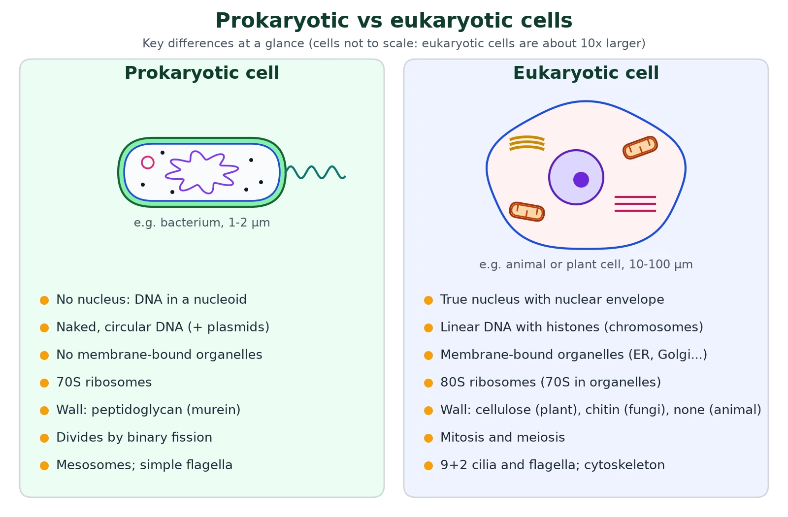
Prokaryotic and eukaryotic cells compared: cell structure table
Swipe the table sideways to see every column.
| Feature | Prokaryotic cell | Eukaryotic cell |
|---|---|---|
| Examples | Bacteria, cyanobacteria, mycoplasma, PPLO | Protists, fungi, plants, animals |
| Typical size | 1-5 µm (mycoplasma 0.3 µm) | 10-100 µm |
| Nucleus | Absent; DNA in a nucleoid | True nucleus with a double nuclear envelope and nucleolus |
| DNA | Single, circular, naked (no histones); plasmids common | Linear, wrapped round histones as chromosomes; no plasmids (except some, e.g. yeast) |
| Membrane-bound organelles | Absent | Present: mitochondria, ER, Golgi, lysosomes, plastids, vacuoles |
| Ribosomes | 70S (50S + 30S) | 80S (60S + 40S) in cytoplasm; 70S in mitochondria and chloroplasts |
| Cell wall | Usually present; peptidoglycan | Cellulose in plants, chitin in fungi, absent in animals |
| Cytoskeleton | No true cytoskeleton (NCERT level) | Microtubules, microfilaments, intermediate filaments |
| Flagella | Simple, made of flagellin, rotate | 9+2 microtubules, covered by membrane, whip-like beat |
| Cell division | Binary fission | Mitosis and meiosis with a spindle |
Exam tips
- IB marks differences as matched pairs: ‘prokaryotes have 70S ribosomes whereas eukaryotes have 80S’.
- Mitochondria and chloroplasts having their own circular DNA and 70S ribosomes is evidence for the endosymbiotic origin of eukaryotes.
5 Plant cells: plant cell structure
Syllabus link: NCERT XI 8.5 (plant cell, Figure 8.3a); IB A2.2 differences between plant, animal and fungal cells
The cell structure of a typical plant cell, such as a leaf mesophyll cell, is box-shaped because it is held in by a rigid wall. Three structures mark it out from an animal cell: a cellulose cell wall, plastids (including chloroplasts) and a large central vacuole.
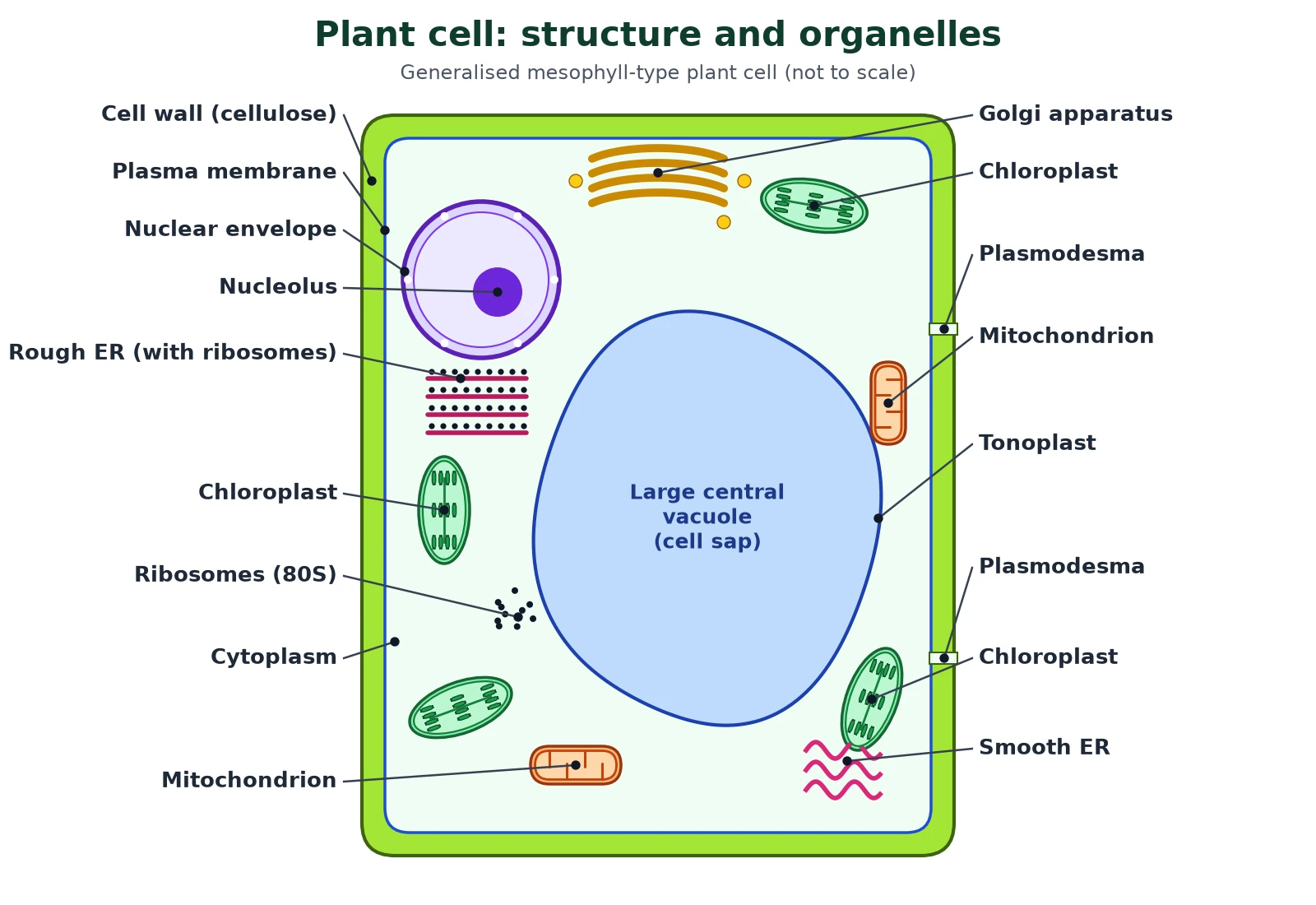
- Cell wall: outside the plasma membrane; mainly cellulose, with hemicellulose and pectins. Cells are linked through plasmodesmata, cytoplasmic strands crossing the walls.
- Large central vacuole: bounded by the tonoplast and filled with cell sap. NCERT notes it can occupy up to 90 per cent of the cell volume.
- Plastids: chloroplasts in green cells, chromoplasts in coloured petals and fruits, leucoplasts in storage tissue.
- Centrioles absent in almost all plant cells; the spindle forms without them.
- Stores starch, not glycogen.
6 Animal cells: animal cell structure
Syllabus link: NCERT XI 8.5 (animal cell, Figure 8.3b); IB A2.2 differences between plant, animal and fungal cells
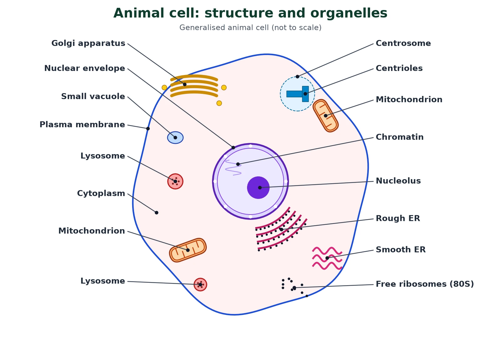
In animal cell structure there is no cell wall, so it is held in shape by its cytoskeleton and can be round, flat, long or irregular. It has a centrosome with two centrioles, only small vacuoles, and stores glycogen.
Plant and animal cell compared
Swipe the table sideways to see every column.
| Feature | Plant cell | Animal cell |
|---|---|---|
| Cell wall | Present, cellulose | Absent |
| Shape | Fixed, often rectangular | Variable, often rounded or irregular |
| Plastids | Present (chloroplasts, chromoplasts, leucoplasts) | Absent |
| Vacuole | One large central vacuole with tonoplast | Small and temporary, if present |
| Centrioles | Absent in almost all plant cells | Present, in the centrosome |
| Lysosomes | Rare (the vacuole does much of this work) | Common |
| Stored carbohydrate | Starch | Glycogen |
| Cytokinesis | Cell plate forms from the centre | Cleavage furrow from the outside |
IB Fungal cells
IB also asks you to compare fungi. Fungal cells have a cell wall made of chitin (not cellulose), no plastids, may have a large vacuole, have no centrioles in most species and store carbohydrate as glycogen.
7 Cell organelles: structure and function
Syllabus link: NCERT XI 8.5.1-8.5.11; IB A2.2 eukaryote cell structure and organelle functions
Now we take each of the cell organelles in turn. For every one, learn three things: membrane (none, single or double), structure and function. The table at the end of this section is a one-page revision summary of cell structure and function.
Cell wall
NCERT The cell wall is a non-living rigid structure that forms an outer covering for the plasma membrane of fungi and plants. It gives shape, protects against damage and infection, and blocks unwanted macromolecules. Plant walls are mainly cellulose with hemicellulose, pectins and proteins (algae add galactans, mannans and calcium carbonate). A growing cell has a primary wall; a secondary wall is laid down inside it as the cell matures, and the calcium pectate middle lamella glues neighbouring cells together.
Nucleus
NCERT The nucleus was first described by Robert Brown in 1831, and Flemming later named the material stained by basic dyes chromatin. The interphase nucleus contains chromatin, the nuclear matrix (nucleoplasm) and one or more nucleoli. The nuclear envelope has two parallel membranes with a 10-50 nm perinuclear space between them. The envelope is broken at intervals by nuclear pores, formed where the two membranes fuse, which let RNA and protein move in both directions.
The nucleolus is a part of nuclear cell structure that is not bound by a membrane. It is the site of active rRNA synthesis, so protein-making cells have larger and more nucleoli.

During cell division the chromatin condenses into chromosomes. Each has a primary constriction, the centromere, carrying disc-shaped kinetochores. By the position of the centromere, NCERT classes chromosomes as metacentric (middle), sub-metacentric (slightly off-centre), acrocentric (very near one end) and telocentric (at the end). Division itself is covered in cell cycle and cell division.
Endoplasmic reticulum (ER)
The ER is a network of tubules and flattened sacs (cisternae) that divides the cytoplasm into luminal and extraluminal compartments. Rough ER carries ribosomes on its outer surface, is continuous with the outer nuclear membrane and is extensive in cells that make and secrete protein. Smooth ER has no ribosomes and is the major site of lipid synthesis; in animal cells, lipid-like steroid hormones are made in the SER.
Ribosomes
NCERT Ribosomes are present in every cell structure, prokaryotic or eukaryotic. They were first seen with the electron microscope as dense granules by George Palade (1953). They are made of RNA and protein and are not surrounded by any membrane. Eukaryotic ribosomes are 80S (60S + 40S) and prokaryotic ribosomes are 70S (50S + 30S). S (Svedberg unit) is the sedimentation coefficient, an indirect measure of size and density. (S values are not additive.)
Golgi apparatus
Camillo Golgi (1898) first saw these densely stained reticular structures near the nucleus. The Golgi has a distinctive cell structure: a stack of flat, disc-shaped cisternae, 0.5-1.0 µm in diameter, arranged near the nucleus with a convex cis (forming) face and a concave trans (maturing) face. ER vesicles fuse with the cis face and modified products leave from the trans face. The Golgi packages materials to go to targets inside the cell or to be secreted, and it is the main site where glycoproteins and glycolipids are formed.
Lysosomes
Lysosomes, common in animal cell structure, are single-membrane vesicles budded off from the Golgi. They are rich in almost all types of hydrolytic enzymes (lipases, proteases, carbohydrases), which work best at an acidic pH and can digest carbohydrates, proteins, lipids and nucleic acids. They digest engulfed material and worn-out organelles (autophagy).
Vacuoles
A vacuole is a membrane-bound space in the cytoplasm containing water, sap, excretory products and other material not needed by the cell. Its single membrane is the tonoplast, which pumps ions and other materials into the vacuole against their concentration gradient, so their concentration in the vacuole is much higher than in the cytoplasm. In Amoeba the contractile vacuole removes excess water; protists also form food vacuoles. Water enters by osmosis (see membrane transport).
Mitochondria
The cell structure of a mitochondrion is only clear with the electron microscope; under a light microscope mitochondria are hard to see unless specially stained. NCERT Each is sausage-shaped or cylindrical, 0.2-1.0 µm (average 0.5 µm) in diameter and 1.0-4.1 µm long. It has two membranes: a smooth outer membrane and an inner membrane folded into cristae that increase its surface area. The two membranes enclose the intermembrane space and the inner one encloses the dense matrix.
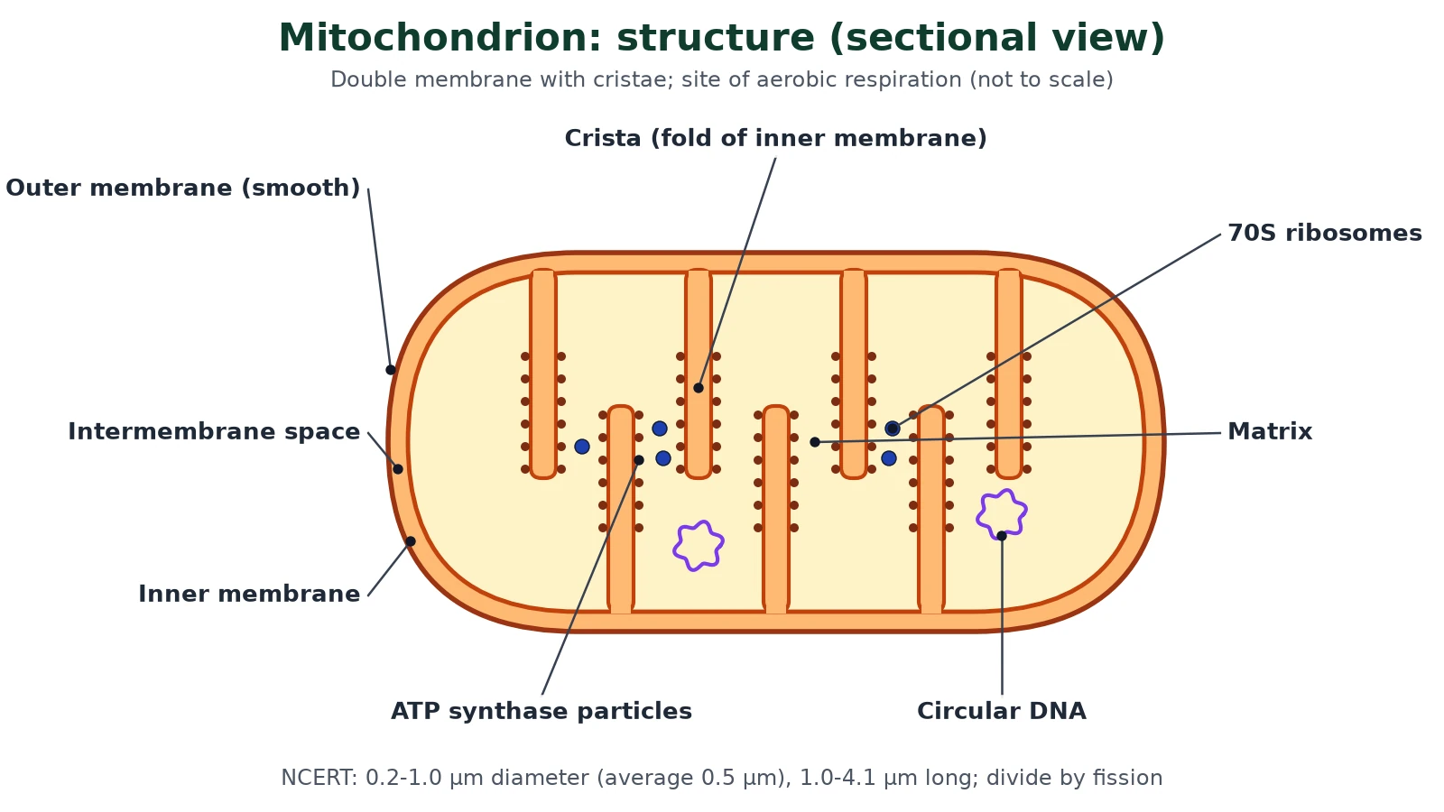
Mitochondria are the sites of aerobic respiration and make energy in the form of ATP, so they are called the “power houses” of the cell. The matrix holds circular DNA, a few RNA molecules and 70S ribosomes. Mitochondria divide by fission.
Plastids
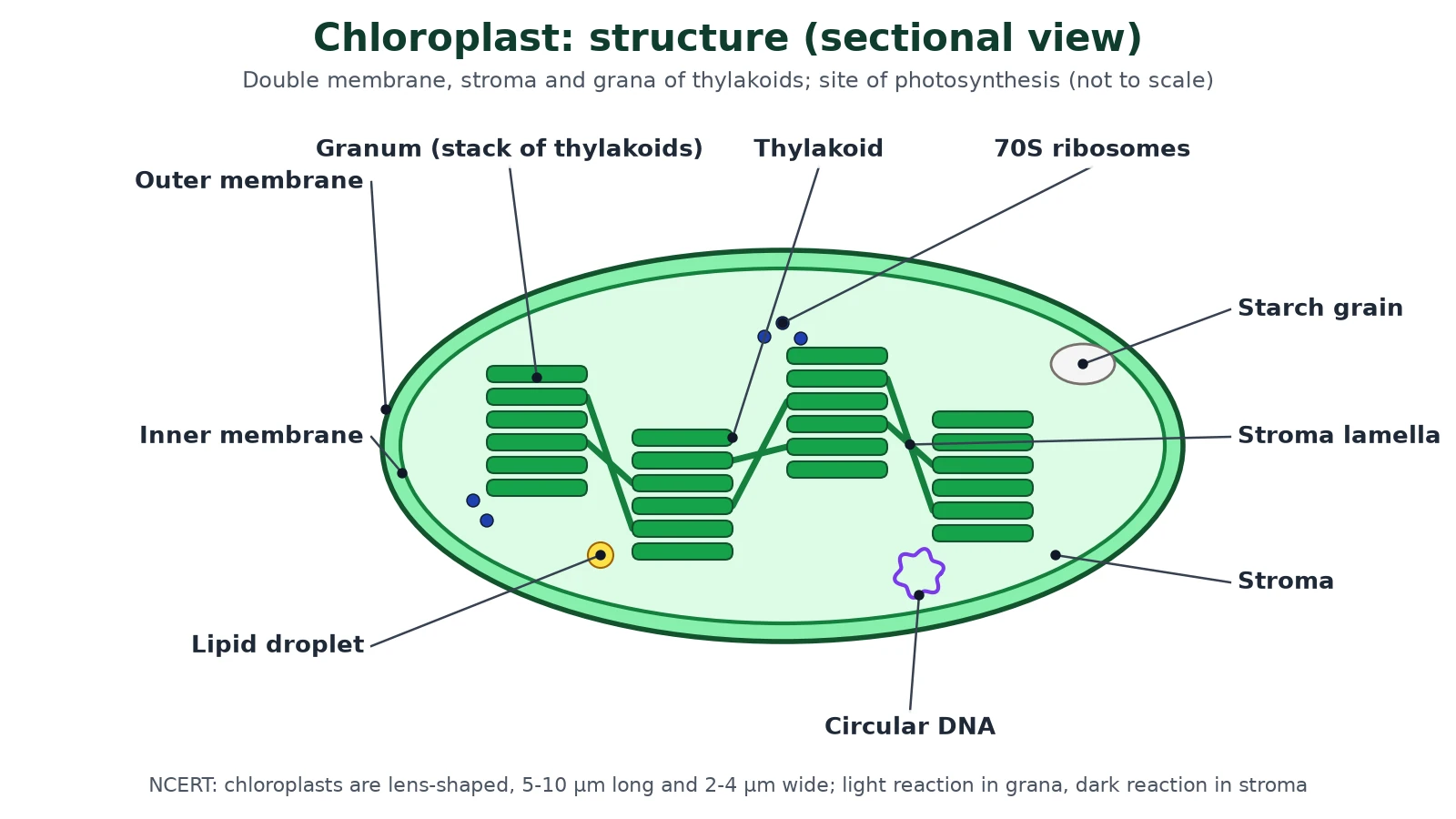
NCERT Plastids are found in all plant cells and in euglenoids. Depending on their pigments they are chloroplasts (chlorophyll and carotenoids, for photosynthesis), chromoplasts (fat-soluble carotenoids such as carotene and xanthophylls, giving yellow, orange or red colours) and leucoplasts (colourless storage plastids). Leucoplasts are of three kinds: amyloplasts store starch (as in potato), elaioplasts store oils and fats, and aleuroplasts store proteins.
In higher plants, chloroplasts are mostly lens-shaped, oval, spherical, discoid or ribbon-like, 5-10 µm long and 2-4 µm wide. There is one per cell in Chlamydomonas and 20-40 per mesophyll cell. They have a double membrane; the inner one is less permeable. The space inside the inner membrane is the stroma, in which flattened sacs called thylakoids are stacked like coins into grana. Flat membranous tubules called stroma lamellae connect the thylakoids of different grana. The stroma holds enzymes, circular DNA and 70S ribosomes; chlorophyll is in the thylakoids.
Cytoskeleton
The cytoskeleton is the internal framework of cell structure: an elaborate network of protein filaments in the cytoplasm made of microtubules (tubulin), microfilaments (actin) and intermediate filaments. It gives support and shape and drives motility, organelle movement and cell division.
Centrosome and centrioles
The centrosome usually contains two cylindrical centrioles lying at right angles to each other, surrounded by amorphous pericentriolar material. Each centriole has a cartwheel organisation of nine evenly spaced peripheral triplets of tubulin microtubules. A central protein hub is joined to the triplets by radial spokes. Centrioles form the basal bodies of cilia and flagella and the spindle apparatus during cell division in animal cells.

Cilia and flagella
Cilia and flagella are hair-like outgrowths of the plasma membrane. Cilia are short and beat like oars; flagella are longer and move the whole cell. In eukaryotes both have a core called the axoneme with nine doublets of radially arranged peripheral microtubules and a pair of central microtubules: the 9+2 array. The central tubules sit in a central sheath joined to each peripheral doublet by a radial spoke. Both grow from a basal body with the 9+0 triplet pattern.
Microbodies
NCERT Many membrane-bound minute vesicles called microbodies, containing various enzymes, are found in both plant and animal cells. Peroxisomes and glyoxysomes are examples.
NCERT One-line summary: membranes of organelles
- Double membrane: nucleus (nuclear envelope), mitochondria, chloroplasts and other plastids.
- Single membrane: ER, Golgi, lysosomes, vacuoles, microbodies.
- No membrane: ribosomes, centrioles, nucleolus, cytoskeleton.
Cell structure and function: revision table
Swipe the table sideways to see every column.
| Organelle | Structure | Main function |
|---|---|---|
| Nucleus | Double envelope with pores; chromatin; nucleolus | Holds DNA; controls the cell; rRNA made in the nucleolus |
| Rough ER | Cisternae studded with ribosomes | Synthesis and transport of proteins for secretion or membranes |
| Golgi apparatus | Stack of cisternae with cis and trans faces | Modifies and packages proteins; makes glycoproteins, glycolipids and lysosomes |
| Mitochondrion | Double membrane, cristae, matrix, 70S ribosomes, DNA | Aerobic respiration; ATP production |
| Chloroplast | Double membrane, grana of thylakoids, stroma | Photosynthesis; starch storage |
| Cilia and flagella | 9+2 axoneme | Movement of the cell or fluid |
| Cell wall | Cellulose (plants), chitin (fungi), peptidoglycan (bacteria) | Shape, support, protection |
8 Specialized cells: how cell structure fits function
Syllabus link: IB A2.2 atypical cell structure; IB B2.3 Cell specialization; NCERT XI 8.3 cell shapes
In a multicellular organism cells differentiate so that their cell structure matches one job. NCERT shows this with cell shapes: RBCs are round and biconcave, WBCs are amoeboid, columnar epithelial cells are long and narrow, and nerve cells are branched and are among the longest cells.
Atypical cells (IB)
IB The typical eukaryotic cell has one nucleus. IB asks you to explain four exceptions to normal cell structure, and to say whether each still fits cell theory.
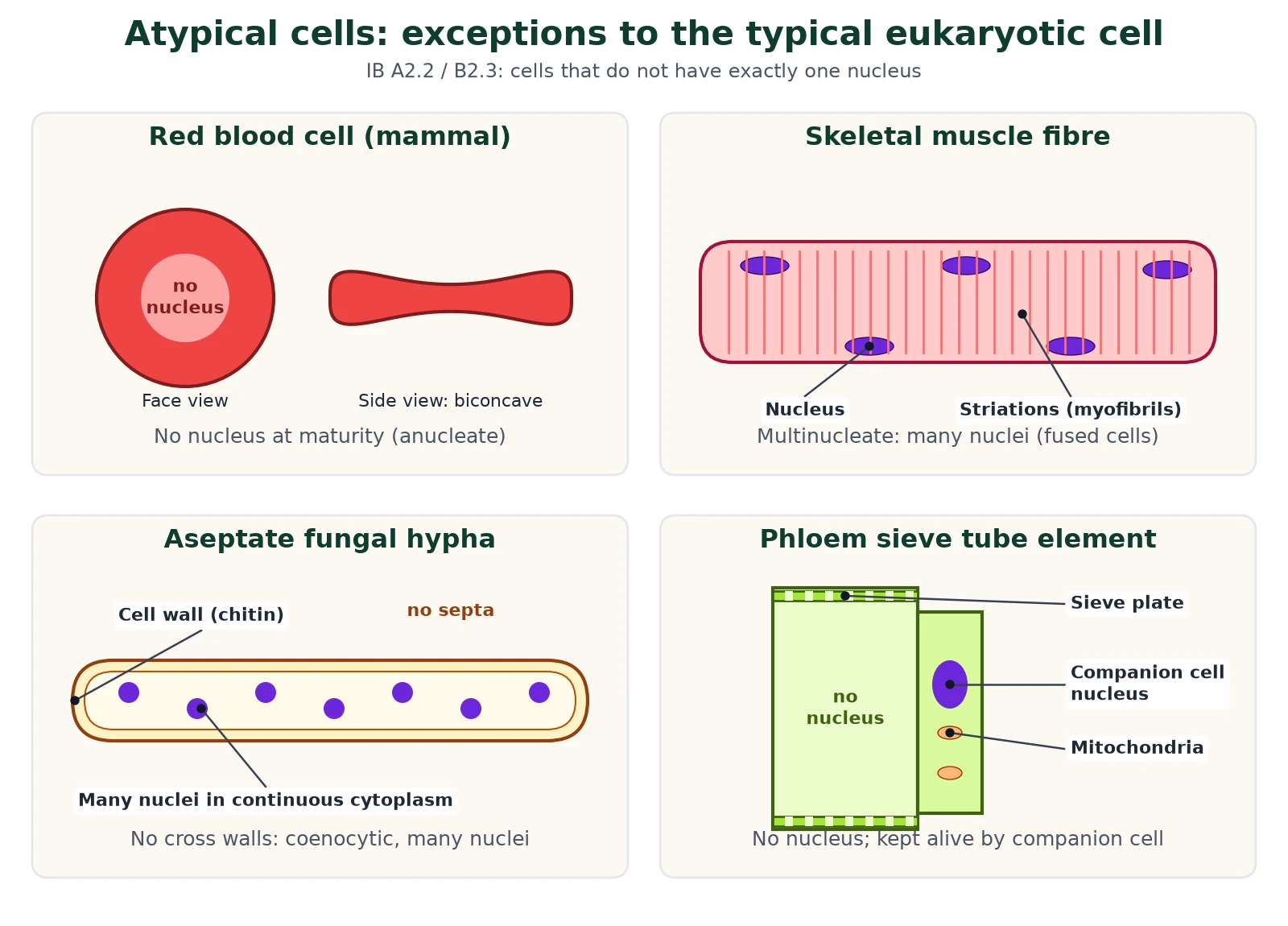
- Red blood cells (mammals): lose their nucleus (and mitochondria) as they mature. This leaves more room for haemoglobin, and the biconcave disc adds surface area. The cost: they cannot divide or repair themselves. NCERT gives the human RBC diameter as about 7.0 µm.
- Skeletal muscle fibres: very long (up to several cm) and multinucleate, because they form when many myoblast cells fuse. Many nuclei supply enough protein for the whole fibre.
- Aseptate fungal hyphae: some fungi (for example Rhizopus) have long hyphae with no septa (cross walls), so many nuclei share one continuous cytoplasm. This coenocytic cell structure lets cytoplasm stream quickly along the hypha.
- Phloem sieve tube elements: lose the nucleus and most organelles as they mature, leaving open tubes joined end to end through sieve plates, so sap flows freely. They stay alive because a companion cell next to each one, linked by many plasmodesmata, supplies proteins and ATP.
More examples of specialized cells
Other favourites: the neurone (long axon for fast impulses), the sperm (flagellum and a midpiece packed with mitochondria), the root hair cell (long outgrowth for absorption) and the pancreatic acinar cell (masses of rough ER and Golgi for enzyme secretion). How cells become specialized is explained in cell differentiation and stem cells.
Micrographs and magnification (IB skill)
IB IB expects you to identify organelles in electron micrographs, draw and annotate them, and calculate magnification or actual size. Use:
Magnification formula
- Magnification = image size / actual size (both in the same unit).
- Actual size = image size / magnification.
- 1 mm = 1000 µm; 1 µm = 1000 nm.
- Worked example: a mitochondrion is 30 mm long on a micrograph at ×15 000. Actual length = 30 000 µm / 15 000 = 2 µm, which fits the NCERT range of 1.0-4.1 µm.
- With a scale bar: measure the bar (say 20 mm for 1 µm), so magnification = 20 000 µm / 1 µm = ×20 000.
Cell structure clues for micrographs: mitochondria show cristae, chloroplasts show grana, rough ER is dotted with ribosomes and a bacterium shows only a pale nucleoid, never a nucleus.
IB vs NEET: what each exam wants on cell structure
IB Explain and compare: prokaryote vs eukaryote, plant vs animal vs fungal cells, atypical cells, drawing organelles from electron micrographs and magnification calculations. Use words like compartmentalisation, naked DNA and 70S/80S.
NCERT NEET takes most questions word for word from NCERT: dates (Brown 1831, Golgi 1898, Palade 1953), sizes, 9+2 and 9+0 patterns, leucoplast types, centromere positions and the bacterial envelope.
9 FAQs on cell structure
What is cell structure?
Cell structure is the arrangement of the parts of a cell (membrane, cytoplasm, nucleus and organelles) and how each part supports its function.
What are the main differences between prokaryotic and eukaryotic cells?
Prokaryotic cells have no nucleus, no membrane-bound organelles, naked circular DNA and 70S ribosomes. Eukaryotic cells have a nucleus, membrane-bound organelles, linear DNA with histones and 80S ribosomes.
Which organelles have a double membrane?
The nucleus, mitochondria and plastids such as chloroplasts.
Which organelles have no membrane?
Ribosomes, centrioles, the nucleolus and the cytoskeleton.
Why are mitochondria called the power house of the cell?
They are the sites of aerobic respiration and make most of the cell’s ATP.
What are the three differences between plant and animal cells?
Plant cells have a cellulose cell wall, plastids and a large central vacuole; animal cells lack these but have centrioles.
Is a red blood cell a prokaryotic cell because it has no nucleus?
No. A mature RBC is a eukaryotic cell that has lost its nucleus during development. It develops from a nucleated stem cell.
What is the 9+2 arrangement?
The axoneme of eukaryotic cilia and flagella has nine peripheral doublet microtubules around two central single microtubules. Centrioles and basal bodies instead have nine triplets and no central pair (9+0).
10 Quick check
Test yourself on cell structure
1. Which ribosomes are found in mitochondria?
70S ribosomes.
2. Name the infolding of the bacterial plasma membrane that helps in DNA replication and respiration.
Mesosome.
3. Which face of the Golgi apparatus is the maturing face?
The trans face (concave).
4. Give one reason a sieve tube element is called atypical.
It has no nucleus when mature and depends on its companion cell.
5. What is the membrane around the plant vacuole called?
Tonoplast.
6. A 2 µm bacterium appears 40 mm long in a micrograph. What is the magnification?
40 000 µm / 2 µm = ×20 000.
Ready to practise? Download the Cell Structure IB and NEET question set with markscheme (25 MCQs + 6 structured questions, 65 marks, free for signed-up members).
⬇ Download practice questions (PDF)© competitorspoint.com · Biology notes. New diagrams are original to competitorspoint.com.