
IB Biology › Cell Biology › Cell Membrane
Cell Membrane
The cell membrane is the thin, flexible boundary around every cell. It keeps the inside of the cell different from the outside, yet lets the right substances in and out. This IB Biology chapter explains what the cell membrane is made of, how the fluid mosaic model describes it, the evidence behind that model, and why its make-up controls fluidity and permeability.
Intro What is the cell membrane?
Syllabus link: B2.1.1 Lipid bilayers as the basis of cell membranes; B2.1.2 Lipid bilayers as barriers
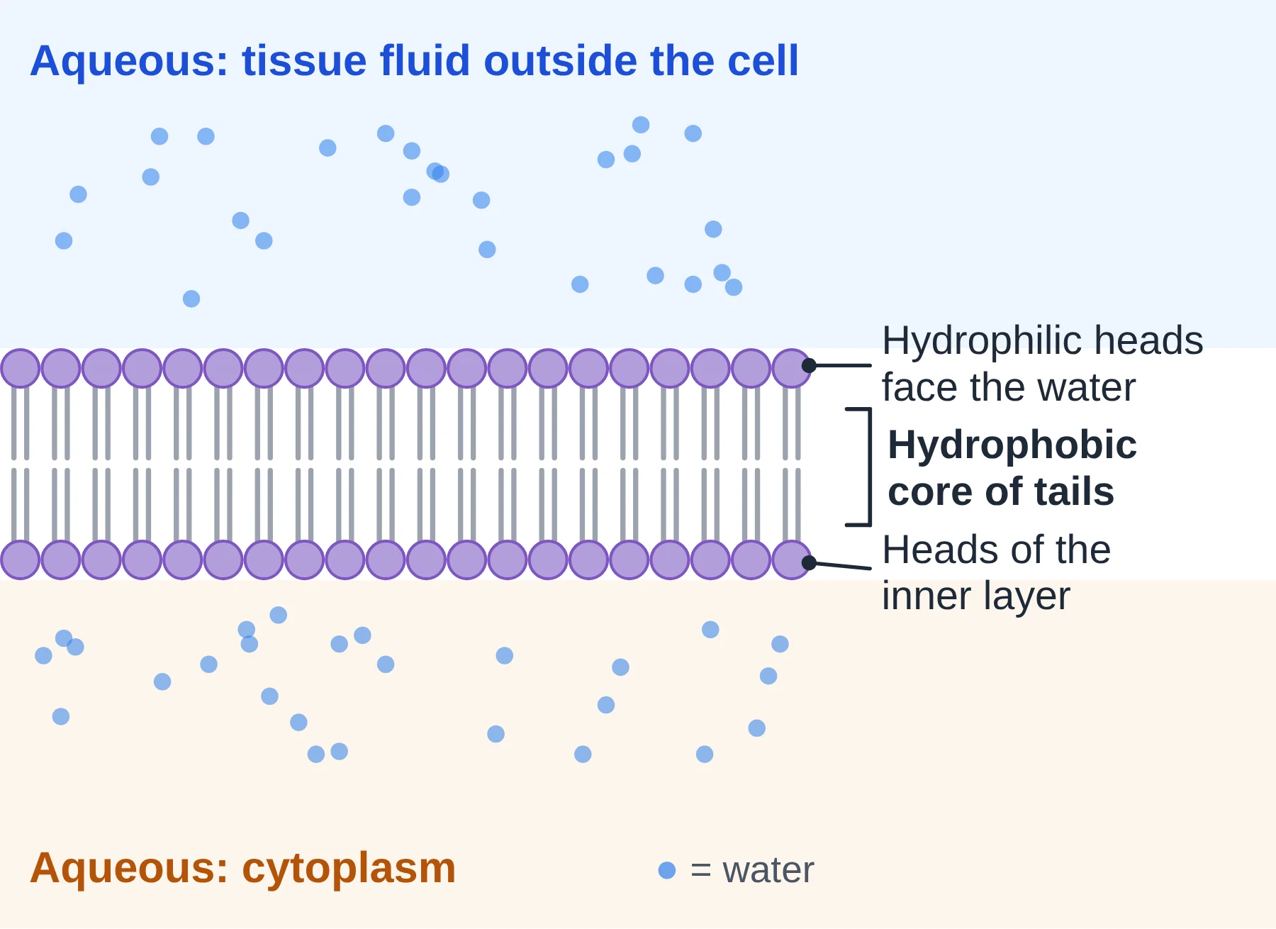
Every living cell is surrounded by a cell membrane, also called the plasma membrane. It separates the watery fluid inside the cell (the cytoplasm) from the watery fluid outside it (tissue fluid, pond water or the medium of a culture). Both sides are aqueous, and that single fact explains much of the membrane’s structure.
The membrane is a barrier, but not a wall. It lets some substances pass and stops others, so it is described as selectively permeable (or partially permeable). It also carries receptors, enzymes and recognition markers, so it is an active, working part of the cell rather than a simple bag.
Membranes are not only found at the cell surface. In eukaryotic cells, the same kind of membrane surrounds the nucleus, mitochondria, chloroplasts, the endoplasmic reticulum, the Golgi apparatus and vesicles. These internal membranes divide the cell into compartments, so that different reactions can run side by side under different conditions.
What is a cell membrane made of?
Each type of membrane has its own exact recipe, but every cell membrane is built from the same three kinds of molecule. For a typical plasma membrane, approximate proportions by mass are:
- Proteins: about 50%
- Lipids (mainly phospholipids, plus cholesterol in animal cells): about 45–50%
- Carbohydrates: a few per cent, always attached to proteins (glycoproteins) or lipids (glycolipids)
These figures vary a lot between membranes. The myelin sheath around nerve fibres is about four-fifths lipid, which suits its job as an electrical insulator, while the inner mitochondrial membrane, packed with respiratory enzymes, is about three-quarters protein. Even when protein and lipid are equal by mass, lipid molecules far outnumber protein molecules (roughly 50 to 1), because each protein is much larger than a lipid.
Key points
- The cell membrane (plasma membrane) separates two aqueous environments: cytoplasm and extracellular fluid.
- It is selectively permeable: it controls what enters and leaves.
- It is made of lipids, proteins and a small amount of carbohydrate; proportions differ with the membrane’s function.
- Internal membranes create compartments in eukaryotic cells.
Common mistakes
- Writing ‘semi-permeable’ and stopping there. In IB answers, explain what crosses and why.
- Saying only animal cells have a cell membrane. Every cell, including plant cells and bacteria, has one; the cell wall is outside it.
Test yourself (click a question to reveal the answer)
Why is it important that both surfaces of the cell membrane face an aqueous environment?
Phospholipids arrange themselves according to water: their hydrophilic heads face the water on both sides and their hydrophobic tails are hidden in the middle. Water on both sides is what makes a bilayer the stable arrangement.
The inner mitochondrial membrane is about 75% protein. Suggest why.
It carries large numbers of electron-transport proteins and ATP synthase for aerobic respiration, so a higher proportion of protein reflects its role as a site of many enzyme-catalysed reactions.
1 Phospholipids and the phospholipid bilayer
Syllabus link: B2.1.1 Lipid bilayers as the basis of cell membranes; B2.1.2 Lipid bilayers as barriers (links to B1.1 lipids)
Building blocks: glycerol and fatty acids
Many lipids are built from glycerol and fatty acids. Each fatty acid joins to one of glycerol’s three –OH groups in a condensation reaction, which forms an ester bond and releases one molecule of water.
- Glycerol + 1 fatty acid gives a monoglyceride (monoacylglycerol).
- Glycerol + 2 fatty acids gives a diglyceride.
- Glycerol + 3 fatty acids gives a triglyceride, with 3 ester bonds and 3 molecules of water released. Triglycerides are energy stores (fats and oils), not membrane lipids.
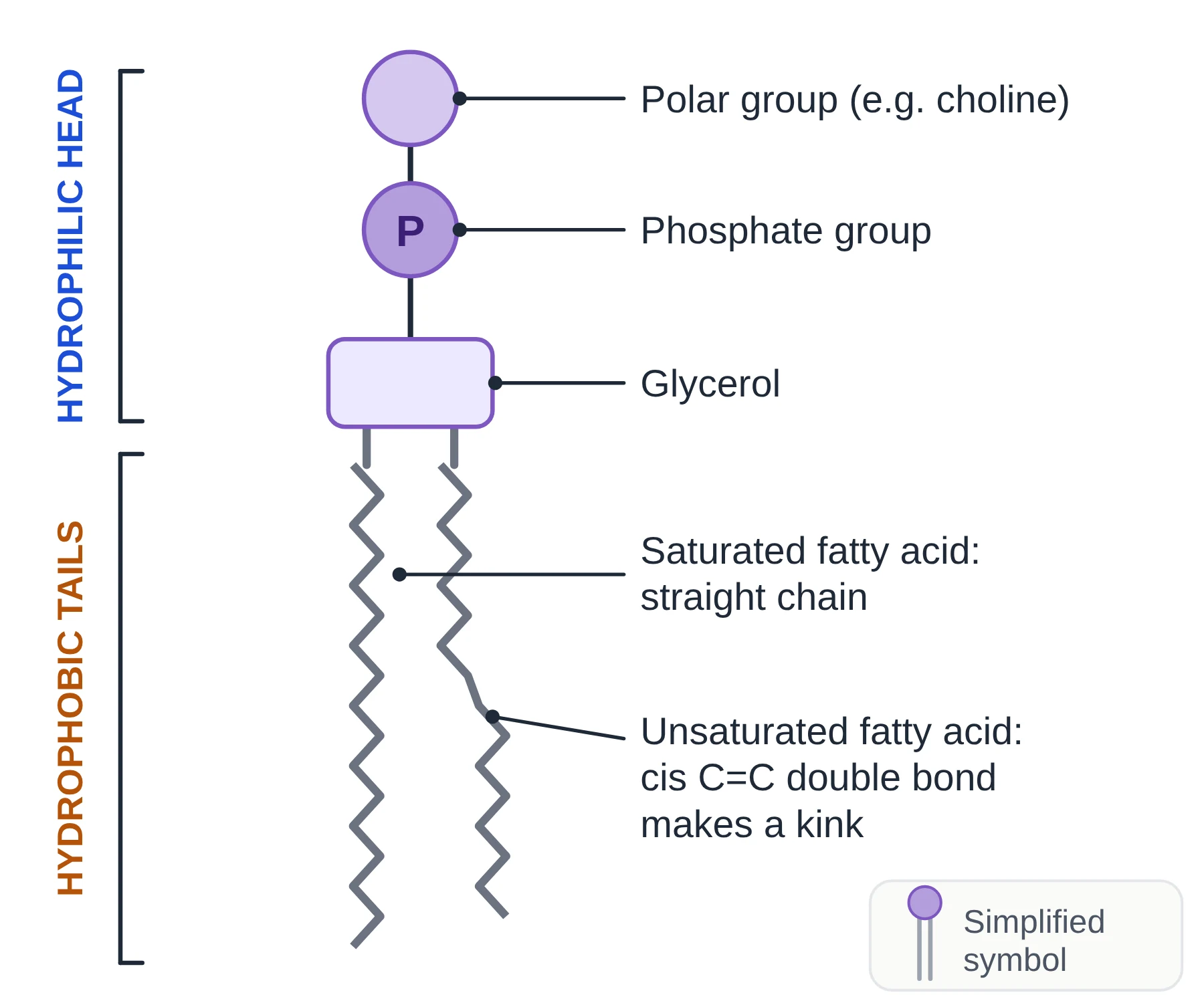
A phospholipid has glycerol linked to two fatty acids and, in the third position, a phosphate group. The phosphate is usually joined to another small polar or charged group, such as choline. A useful way to picture it is a triglyceride in which one fatty acid has been replaced by a phosphate group. In the cell, however, phospholipids are not made by removing a fatty acid from a triglyceride; they are assembled from glycerol phosphate, two fatty acids and the extra polar group.
Amphipathic molecules
A phospholipid is amphipathic: one part of the molecule is hydrophilic and another part is hydrophobic.
- The head (phosphate and its attached group) is charged or polar, so it forms hydrogen bonds and other attractions with water. It is hydrophilic.
- The two tails are long hydrocarbon chains. They are non-polar, so they cannot form hydrogen bonds with water. They are hydrophobic.
- The tails are not repelled by water in any active sense. Water molecules simply attract one another more strongly than they attract the tails, so the tails are pushed together. This is called the hydrophobic effect.
How the bilayer forms
Put phospholipids into water and they arrange themselves without any energy from the cell. The most stable arrangement is a bilayer: two layers (leaflets) of phospholipids, with the heads of each layer facing the water and the tails of both layers pointing inwards towards each other, out of contact with water.
- The bilayer is a continuous sheet. Exposed edges would put tails in contact with water, so bilayers tend to close up into spheres and will reseal themselves after small tears.
- The tails attract one another only by weak hydrophobic interactions (London dispersion forces), not by bonds that lock them in place. Individual phospholipids can therefore move sideways within their layer.
- The whole membrane is very thin: about 7–10 nm including its proteins, so it can only be seen as a structure with an electron microscope.
The bilayer as a barrier
The hydrophobic core is the part that controls permeability. Ions and polar molecules are surrounded by water molecules and are not soluble in the oily core, so the bilayer has very low permeability to them. Small and non-polar molecules, such as oxygen, dissolve in the core and pass through easily. Section 8 covers this in detail.
The two layers are not identical
The two leaflets of a membrane have different compositions: the bilayer is asymmetrical. For example, glycolipids are found only in the outer leaflet, and some phospholipids are kept mainly in the inner leaflet. Phospholipids move sideways within a layer very quickly, but they rarely ‘flip’ from one layer to the other, so this asymmetry is kept.
Key points
- A phospholipid = glycerol + 2 fatty acids + a phosphate group (often with another polar group attached).
- Phospholipids are amphipathic: hydrophilic head, hydrophobic tails.
- In water, phospholipids form a bilayer with heads outwards and tails inwards; this happens spontaneously.
- The hydrophobic core makes the bilayer a barrier to ions and polar molecules.
- The two leaflets differ in composition (asymmetry) and lipids move sideways but rarely flip across.
Common mistakes
- Calling the tails ‘water-hating’ as if they push water away. The tails are excluded because water molecules bond to each other.
- Saying the fatty acid tails do not attract each other. They do, weakly; the weakness is what keeps the membrane fluid.
- Drawing a bilayer with heads pointing inwards. Heads always face the water on both sides.
Exam tip
- When asked to explain why phospholipids form a bilayer, link three ideas: amphipathic molecule, both sides aqueous, hydrophobic tails shielded from water in the centre.
Test yourself (click a question to reveal the answer)
Use the term amphipathic to explain why phospholipids form bilayers in water.
Phospholipids are amphipathic: they have a hydrophilic head and hydrophobic tails. In water, the heads face outwards into the water on both sides while the tails point inwards, away from water, giving a bilayer.
How does a phospholipid differ from a triglyceride?
A triglyceride has three fatty acids on glycerol and is entirely non-polar. A phospholipid has two fatty acids and a phosphate group, so it has a polar head and is amphipathic.
Why does a small tear in a bilayer reseal?
Exposed edges would put hydrophobic tails in contact with water, which is unstable, so the phospholipids move to close the gap and shield the tails.
2 The fluid mosaic model
Syllabus link: B2.1.10 Fluid mosaic model of membrane structure
The model used today to describe the cell membrane is the fluid mosaic model, proposed by S. J. Singer and Garth Nicolson in 1972. Details have been added since, but its main ideas still hold.
- Fluid: phospholipids and many proteins are not fixed in place. They drift sideways within their layer, so the membrane behaves like a thin, flexible liquid film rather than a rigid skin.
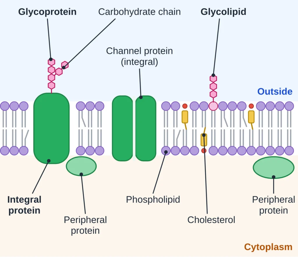
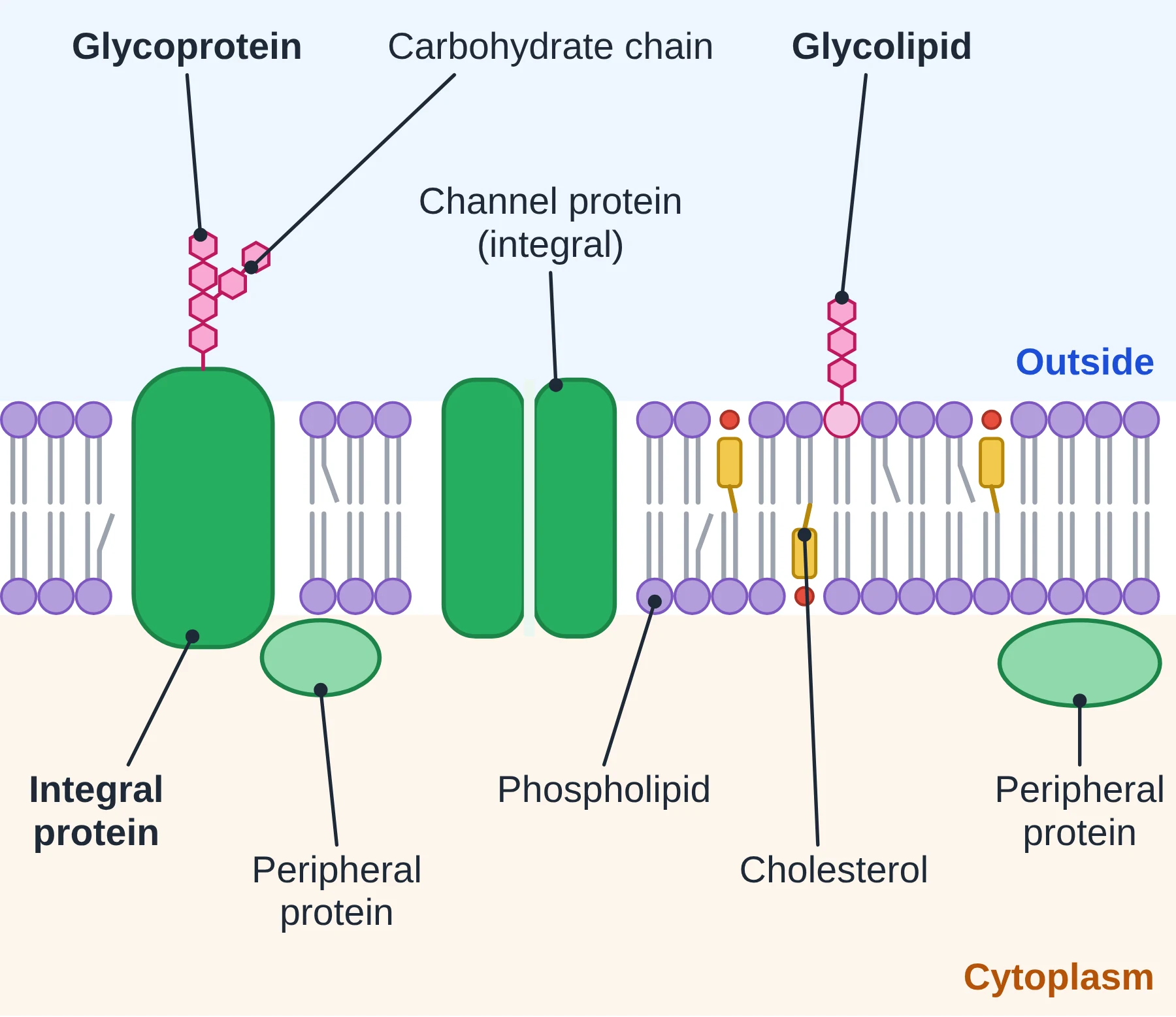
- Mosaic: proteins of many shapes and sizes are scattered through the phospholipid bilayer, like tiles of different colours in a mosaic picture. Some go right through; some sit on one surface.
Components of the model
- Phospholipid bilayer: the basic sheet; the hydrophobic core is the barrier.
- Integral proteins: embedded in the hydrophobic core; many span the whole membrane (transmembrane proteins).
- Peripheral proteins: attached to the inner or outer surface, not embedded in the core.
- Glycoproteins and glycolipids: proteins and lipids with carbohydrate chains, on the outer surface only.
- Cholesterol (animal cells): fits between phospholipids and adjusts fluidity.
Why does fluidity matter?
- The membrane can bend and change shape as cells move, divide and grow.
- Membranes can fuse and pinch off, so vesicles can form in endocytosis and join the plasma membrane in exocytosis (see the Membrane Transport chapter).
- A growing cell can add new phospholipids and enlarge its membrane.
- Proteins can move to where they are needed, and some enzymes and receptors only work when they can move and change shape within the membrane.
Drawing the fluid mosaic model
IB questions often ask for a two-dimensional drawing of the model. Include and label:
- a phospholipid bilayer, with heads and tails clearly drawn;
- an integral protein that passes through the bilayer, and a peripheral protein on one surface;
- a glycoprotein, with its carbohydrate on the outside of the membrane;
- cholesterol between the phospholipid tails;
- the hydrophilic and hydrophobic regions.
Key points
- Fluid mosaic model: Singer and Nicolson, 1972.
- Fluid = components move sideways; mosaic = many different proteins scattered in the bilayer.
- Proteins may be integral (in the core) or peripheral (on a surface).
- Carbohydrate is only on the extracellular side.
Common mistakes
- Drawing carbohydrate chains on both sides of the membrane.
- Drawing proteins as a continuous layer coating the bilayer (that is the old Davson–Danielli model).
- Drawing cholesterol in the heads or outside the bilayer. It sits among the tails, with its small polar end at head level.
Test yourself (click a question to reveal the answer)
What is meant by the word ‘mosaic’ in the fluid mosaic model?
The membrane contains many different proteins scattered through the bilayer, forming a varied pattern like the tiles of a mosaic.
Give two reasons why membrane fluidity is important to a cell.
Any two: allows vesicle formation and fusion (endocytosis, exocytosis); lets the cell change shape, divide and grow; lets proteins move and work.
3 Evidence: from Davson–Danielli to Singer–Nicolson
Syllabus link: Nature of Science: models are tested against evidence and replaced (background to B2.1.10)
The fluid mosaic model was not the first model of membrane structure. Its history is a good example of how scientists test a model, find evidence that does not fit, and replace it with a better one (falsification).
The Davson–Danielli model (1935)
Hugh Davson and James Danielli suggested that a phospholipid bilayer is sandwiched between two continuous layers of protein, one on each surface. This explained why membranes are such good barriers and why they seemed to be coated with protein.
In the 1950s, electron micrographs of membranes showed a three-layered ‘railway track’ pattern: dark, light, dark. At first this was taken as support for the sandwich (dark = protein, light = lipid). We now know the dark lines are mainly the phosphate heads, which take up the heavy-metal stain, and the light line is the hydrophobic core.
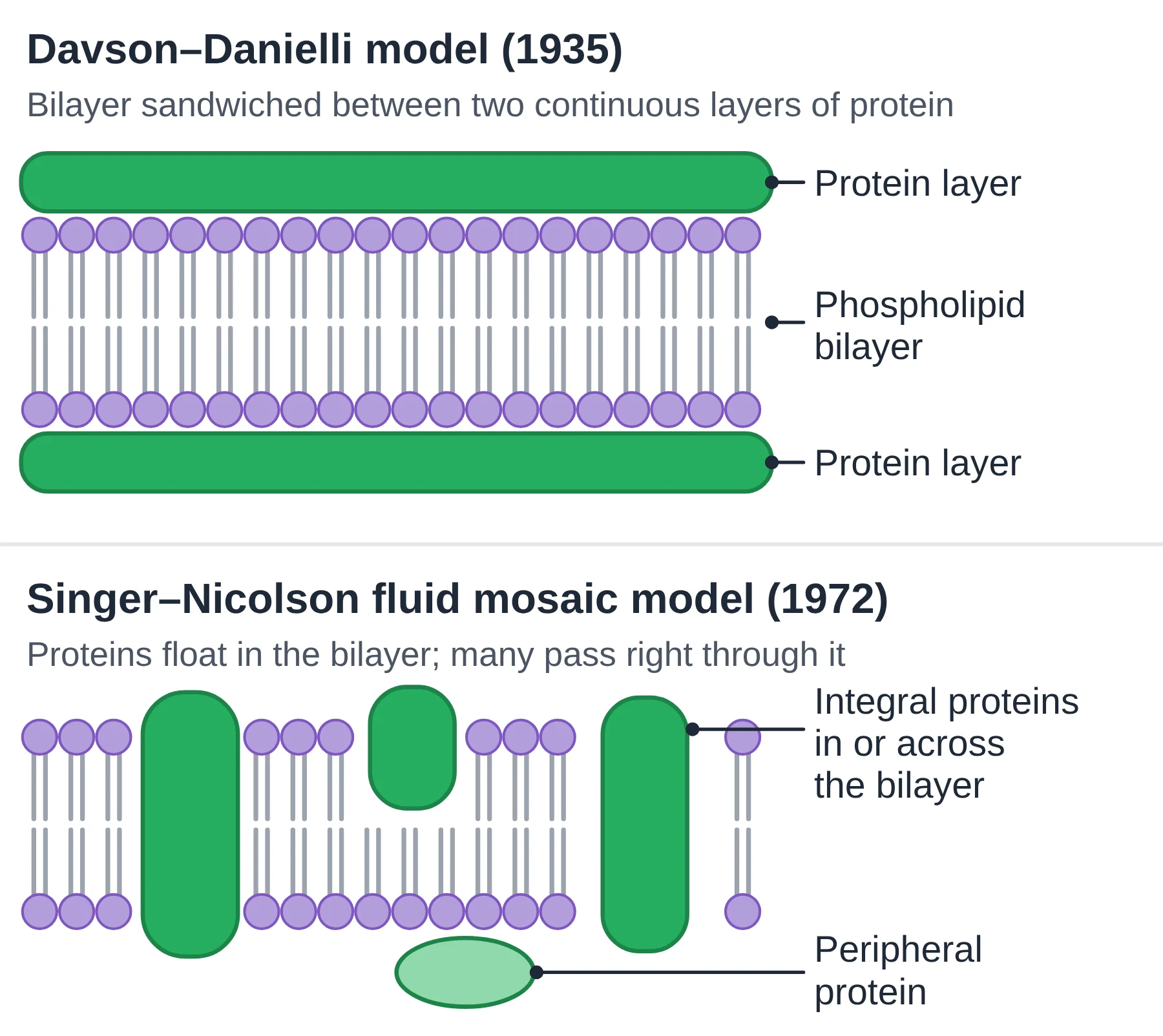
Evidence that falsified the sandwich model
- Freeze-fracture electron microscopy showed particles inside the bilayer (see below). Proteins must pass into or through the hydrophobic core, not just coat the surface.
- Membrane proteins are varied and amphipathic. When extracted, they turned out to differ greatly in size and shape, and many had hydrophobic regions. A hydrophobic surface would not sit stably in contact with water as a continuous outer coat, but fits well inside the hydrophobic core.
- Membranes differ. Different membranes have very different protein-to-lipid ratios and functions. A single uniform sandwich could not explain this.
- Membrane proteins move, as shown by the fluorescent antibody cell fusion experiment. A fixed protein coat would not allow this.
Freeze-fracture
In freeze-fracture, cells are frozen very rapidly and then split with a cold knife. The fracture tends to run along the weakest plane, which is the centre of the bilayer between the two layers of tails. A replica of the exposed surface is made and examined with the electron microscope.
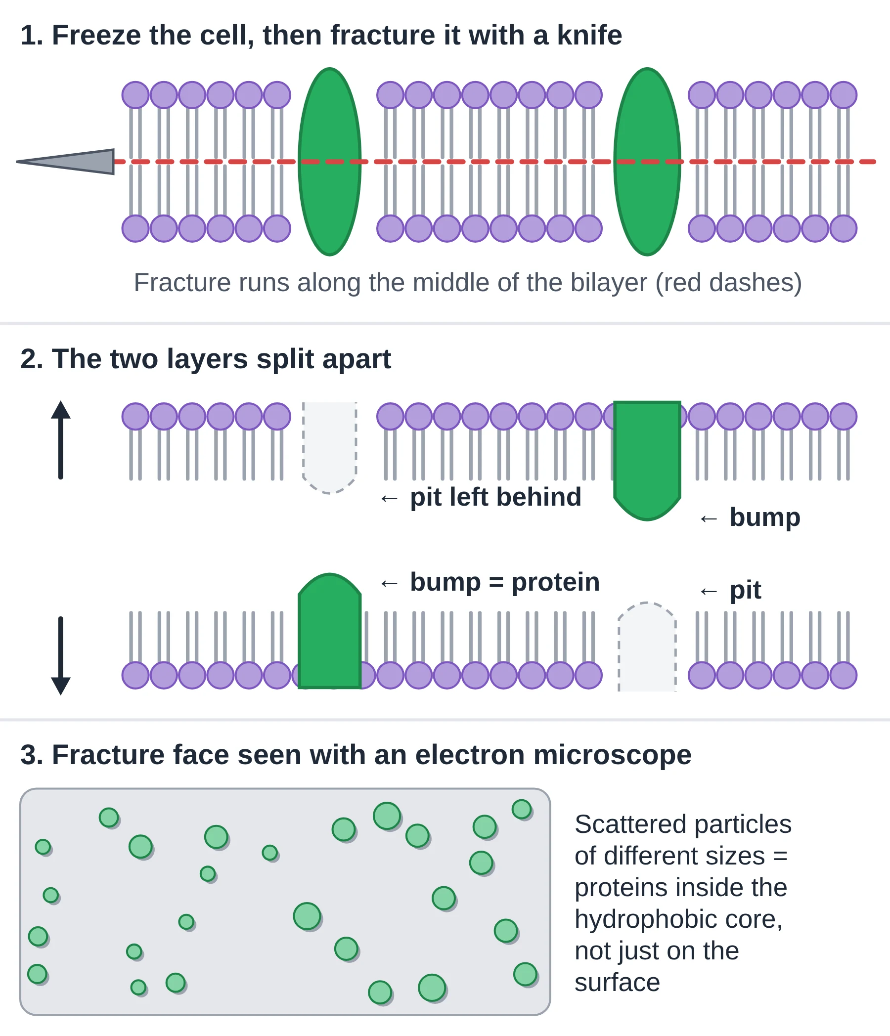
The fracture faces are covered with small particles of different sizes, with matching pits on the opposite face. These are proteins that were embedded in the bilayer. The Davson–Danielli model predicts smooth fracture faces, so this observation does not fit it.
The cell fusion experiment (Frye and Edidin, 1970)
- Mouse cells and human cells were grown in culture.
- Antibodies that bind mouse membrane proteins were tagged with a green fluorescent marker; antibodies for human membrane proteins were tagged with a red marker.
- The two cell types were fused (a virus was used to make the membranes join), producing hybrid cells with one membrane.
- Straight after fusion, green-labelled proteins were on one half of the hybrid cell and red-labelled proteins on the other.
- After about 40 minutes at 37 °C the two colours were completely mixed over the whole surface.
- At low temperatures (below about 15 °C) very little mixing happened.
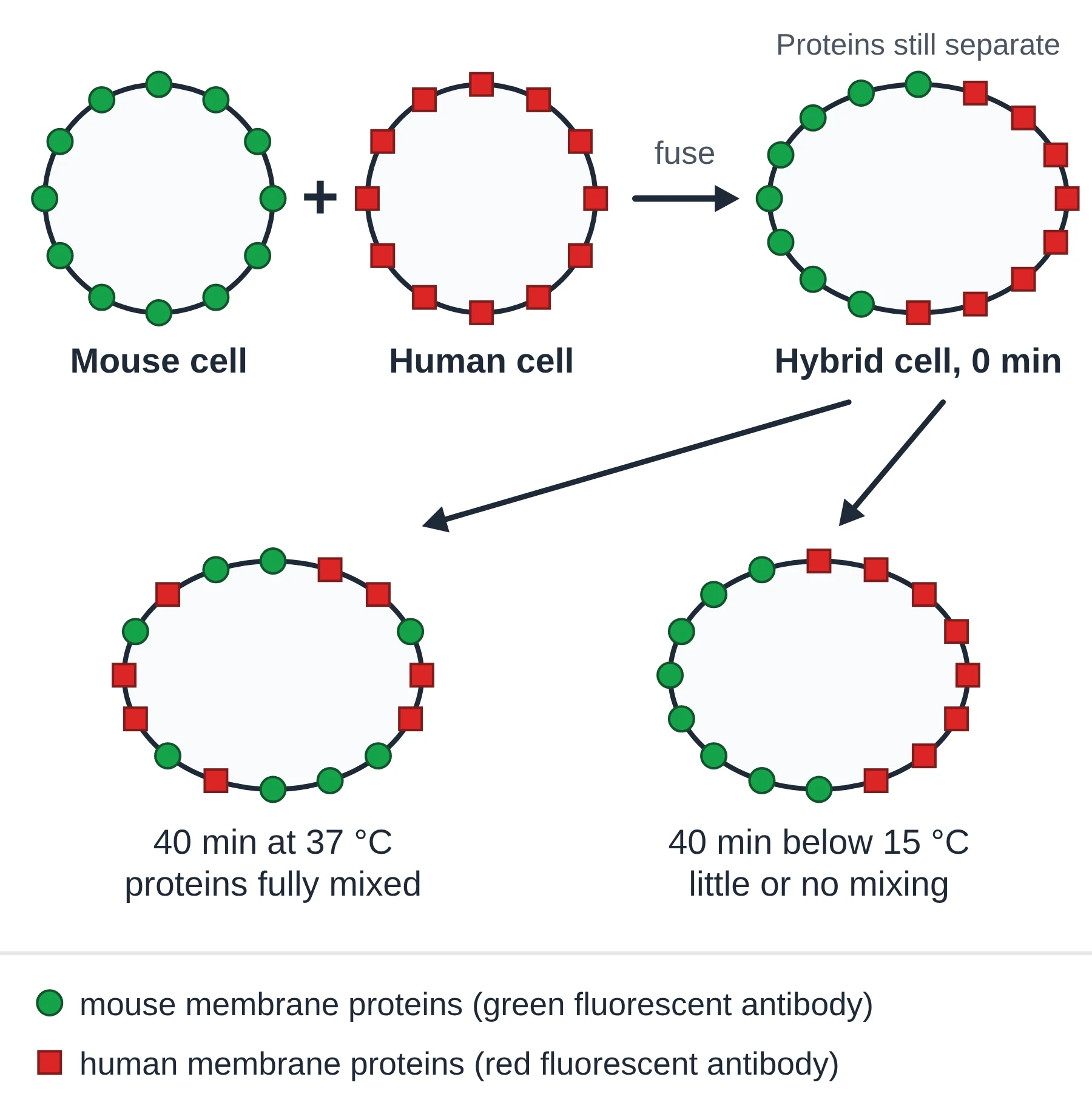
Conclusion: membrane proteins can diffuse sideways within the membrane, so the membrane is fluid. Slower mixing at low temperature fits this, because molecules move less when they have less kinetic energy.
| Feature | Davson–Danielli (1935) | Singer–Nicolson (1972) |
|---|---|---|
| Position of proteins | Two continuous layers coating both surfaces | Scattered; integral proteins in or through the core, peripheral proteins on a surface |
| Protein movement | Proteins form a fixed coat | Proteins and lipids move sideways (fluid) |
| Variety between membranes | All membranes alike | Different membranes have different proteins |
| Fit with freeze-fracture | Predicts smooth fracture faces: does not fit | Particles in core expected: fits |
| Status | Falsified | Accepted (refined since) |
Key points
- Davson–Danielli (1935): protein–lipid–protein sandwich.
- Evidence against it: freeze-fracture particles, amphipathic proteins of varied size, variation between membranes, protein movement.
- Frye and Edidin’s cell fusion showed proteins move sideways; movement slowed at low temperature.
- Replacing an old model with a better one when evidence disagrees is falsification.
Common mistakes
- Saying the cell fusion experiment shows that phospholipids move. It tracked proteins (phospholipid movement is shown by other methods).
- Writing that freeze-fracture splits the membrane between the bilayer and the proteins. It splits the bilayer down its middle.
Test yourself (click a question to reveal the answer)
Explain how freeze-fracture images provided evidence against the Davson–Danielli model.
The fracture splits the bilayer along its hydrophobic centre. The faces showed particles (proteins) embedded in the core, whereas the Davson–Danielli model has proteins only on the surfaces and predicts smooth faces.
In the cell fusion experiment, why was mixing much slower at low temperature?
At lower temperature molecules have less kinetic energy and the membrane is less fluid, so proteins diffuse sideways more slowly.
4 Membrane proteins
Syllabus link: B2.1.4 Integral and peripheral proteins in membranes; AHL B2.1.17 Adhesion of cells to form tissues
Phospholipids make the barrier; proteins give each membrane its particular jobs. That is why membranes with different functions carry different proteins.
Integral and peripheral proteins
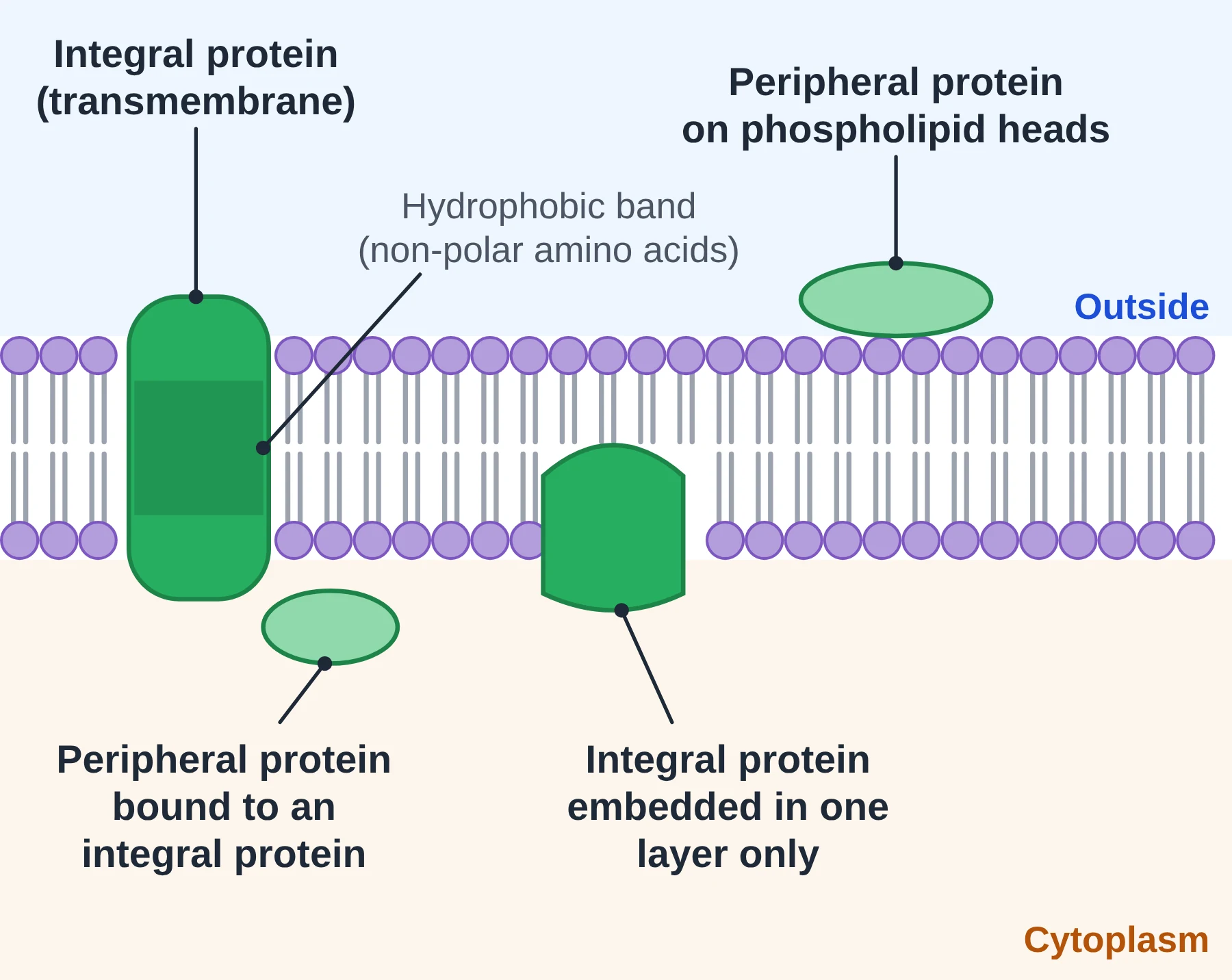
- Integral proteins are embedded in the hydrophobic core of the bilayer. Most are transmembrane: they pass all the way through and have parts exposed on both sides. A few are embedded in one layer only. Integral proteins are amphipathic: the region in contact with the tails has mostly non-polar (hydrophobic) amino acids, often coiled as an α-helix, while the parts in contact with water have polar amino acids.
- Peripheral proteins are not embedded in the core. They are attached to the inner or outer surface, held by weak, non-covalent attractions to the phospholipid heads or to an integral protein. Because they are held weakly, they can often be removed without breaking up the bilayer (for example with a salt solution). Integral proteins can only be removed by destroying the bilayer, for example with detergent.
Strictly, ‘integral’ and ‘transmembrane’ are not the same: all transmembrane proteins are integral, but not every integral protein crosses the whole membrane.
Functions of membrane proteins
Membrane proteins carry out at least six general roles:
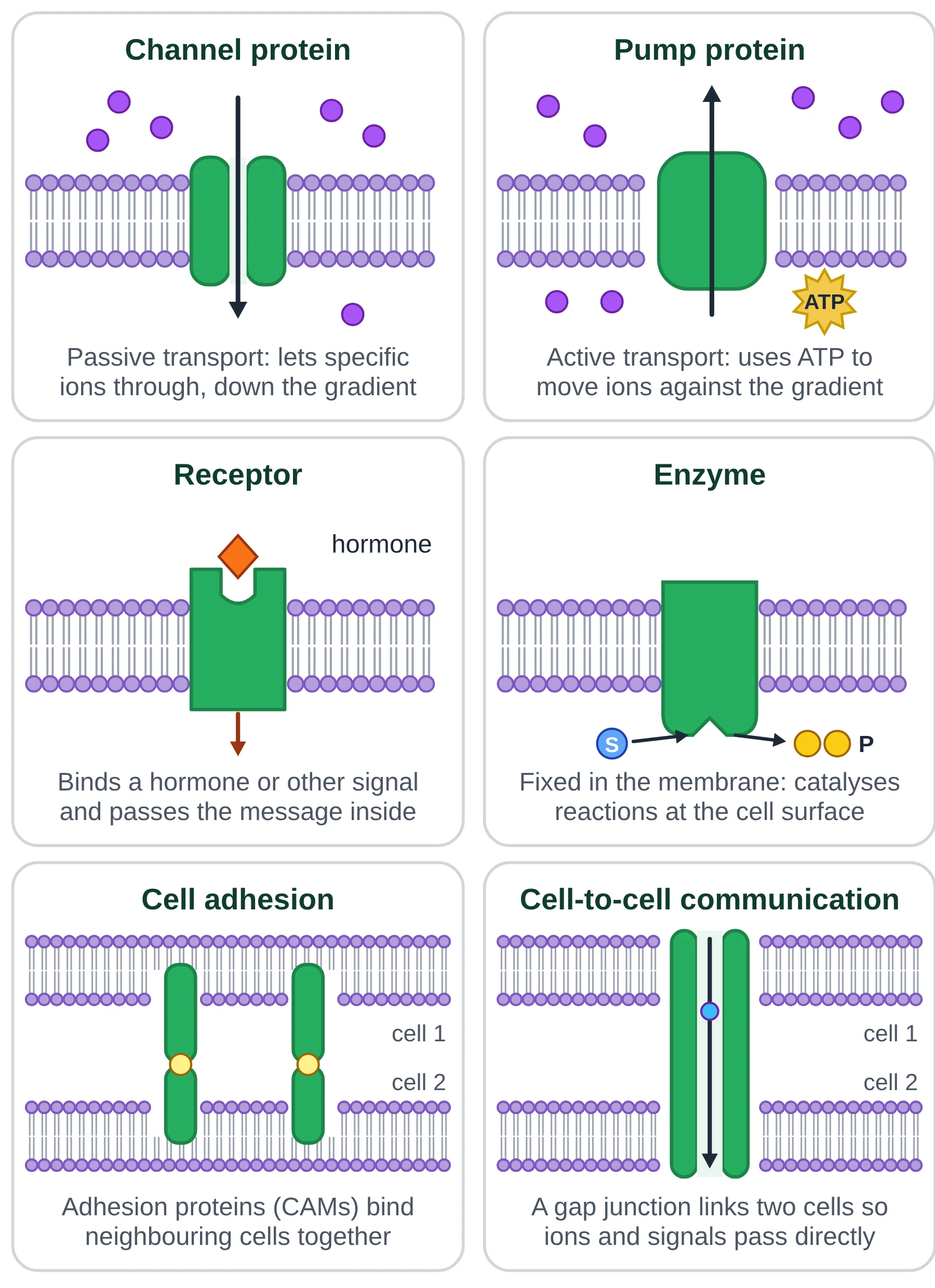
- Channels for passive transport. Channel proteins have a hydrophilic pore that lets specific ions or water (aquaporins) cross by facilitated diffusion. Carrier proteins also allow facilitated diffusion by changing shape.
- Pumps for active transport. Pump proteins use energy from ATP to move particles against their concentration gradient, for example the sodium–potassium pump.
- Receptors (hormone binding sites). A receptor has a binding site of specific shape for one signal molecule, such as insulin. Binding changes the receptor’s shape and sets off a response inside the cell.
- Enzymes (immobilised enzymes). Some enzymes are fixed in membranes, for example digestive enzymes on the surface of intestinal epithelial cells and ATP synthase in mitochondria. Fixing enzymes side by side can speed up a sequence of reactions.
- Cell adhesion. Cell-adhesion molecules (CAMs) bind to matching molecules on neighbouring cells or on the extracellular matrix, holding cells together in tissues.
- Cell-to-cell communication. Proteins allow cells to exchange signals: receptors detect chemical messages, and in animal tissues gap junctions form protein channels that connect the cytoplasm of two neighbouring cells so ions and small molecules pass directly.
Glycoproteins also act in cell recognition: their carbohydrate chains work as identity tags (Section 5).
AHL Cell-adhesion molecules and tissues
AHL Cells in tissues are held together by cell-adhesion molecules (CAMs), which are integral membrane proteins. Part of each CAM is anchored in the membrane, and part projects outside the cell to bind a CAM on a neighbouring cell (or a component of the extracellular matrix).
- Different forms of CAM are used to build different types of cell–cell junction. For example, some junctions seal neighbouring cells tightly so fluid cannot leak between them, while others anchor cells strongly so that a tissue resists being pulled apart.
- Many CAMs bind only to the same type of CAM, so a cell sticks most strongly to cells of the same kind. This helps cells sort themselves into the right tissues during development.
- Some CAMs need calcium ions in order to bind; others do not.
You do not need detailed knowledge of individual CAMs or junction types for IB; it is enough to know that CAMs are membrane proteins and that different CAMs form different junctions.
Key points
- Integral proteins are embedded in the hydrophobic core (most are transmembrane); peripheral proteins are attached to a surface.
- Integral proteins have a hydrophobic region where they contact the tails.
- Six functions: channels, pumps, receptors, enzymes, adhesion and cell-to-cell communication (plus recognition by glycoproteins).
- AHL CAMs are membrane proteins that hold cells together; different CAMs form different junctions.
Common mistakes
- Saying peripheral proteins are covalently bonded to the membrane. They are held by weak, non-covalent attractions.
- Saying all integral proteins are channels. Receptors, pumps, carriers, many enzymes and CAMs are integral too.
- Saying gap junctions or tight junctions are ‘formed by cell adhesion’. Junctions are built from specific proteins; adhesion proteins hold cells together, and gap junction proteins form channels between cells.
Exam tip
- ‘Distinguish’ questions want a contrast in one sentence: integral proteins are embedded in the hydrophobic core, whereas peripheral proteins are attached to the surface only.
Test yourself (click a question to reveal the answer)
State one structural feature of an integral protein that allows it to sit in the bilayer.
A region with non-polar (hydrophobic) amino acids, often an α-helix, that is in contact with the hydrophobic fatty acid tails.
Insulin cannot enter cells, yet it changes their activity. Explain how a membrane protein makes this possible.
Insulin binds to a specific receptor protein in the plasma membrane. Binding changes the receptor’s shape, which triggers a response inside the cell.
AHL What are CAMs and why do tissues need them?
Cell-adhesion molecules: membrane proteins that bind neighbouring cells (or the matrix) together. Without them cells would not stay joined into tissues, and different CAMs form different junctions.
5 Glycoproteins, glycolipids and the glycocalyx
Syllabus link: B2.1.9 Structure and function of glycoproteins and glycolipids
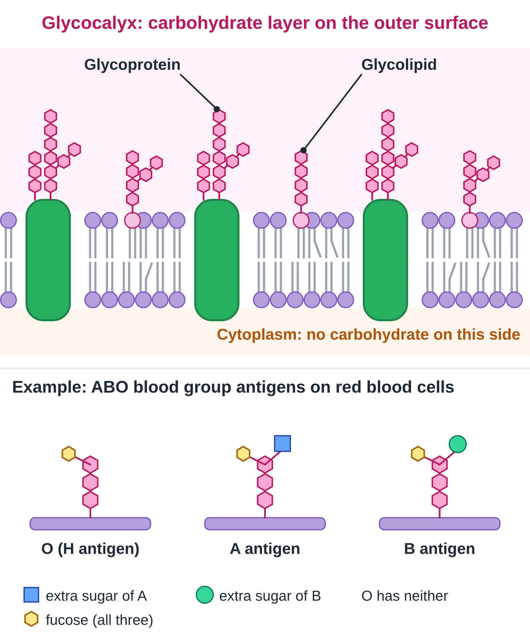
Some membrane proteins and lipids have short, often branched carbohydrate chains attached to them. A protein with carbohydrate is a glycoprotein; a lipid with carbohydrate is a glycolipid.
- The carbohydrate is always on the outer (extracellular) side of the plasma membrane, never facing the cytoplasm.
- The chains are made of a few to many sugar units, and their exact sequence and branching vary hugely. This variety lets them act as specific markers.
- Together, the carbohydrate parts of glycoproteins and glycolipids form a sugar-rich coat over the cell surface called the glycocalyx.
Functions of glycoproteins and glycolipids
- Cell recognition. The carbohydrate chains act as identity tags. The immune system uses them to recognise ‘self’ cells and to detect foreign cells, such as bacteria or cells from another person.
- Cell adhesion. They help cells attach to each other and bind into tissues.
- Receptors. Some glycoproteins act as receptors for chemical signals such as hormones.
- Protection and hydration. The glycocalyx holds a layer of water at the cell surface and helps protect the membrane.
Example: ABO blood groups
Red blood cells carry glycoproteins and glycolipids whose carbohydrate chains end differently in different people. All three types share the same basic chain (the H antigen). In blood group A, one extra sugar is added at the end; in blood group B a different extra sugar is added; in group O neither is added. A person with group A has antibodies against the B antigen in their plasma, so if they receive group B blood, the donated red cells are recognised as foreign and clump together (agglutinate). This is why a group A person must not be given group B blood.
Key points
- Glycoproteins = proteins + carbohydrate; glycolipids = lipids + carbohydrate.
- Carbohydrate chains are on the extracellular side only.
- The glycocalyx is the carbohydrate-rich layer on the cell surface.
- Main roles: cell recognition, cell adhesion, receptors; ABO blood groups are an example of recognition.
Common mistakes
- Saying the glycocalyx is a separate layer like a cell wall. It is made of the carbohydrate parts of membrane molecules.
- Saying blood group antigens are only on glycoproteins. They are on both glycoproteins and glycolipids.
Test yourself (click a question to reveal the answer)
State where the carbohydrate parts of glycoproteins are found and why this suits their function.
On the extracellular side of the membrane. Their roles in recognition and adhesion involve contact with other cells and molecules outside the cell.
Why can a person with group A blood not safely receive group B blood?
Group A plasma contains anti-B antibodies. These bind the B antigens (carbohydrate chains) on the donated red blood cells, which clump together.
6 Membrane fluidity and fatty acids
Syllabus link: AHL B2.1.11 Relationships of fatty acid composition of lipid bilayers to their fluidity; B2.1.13 Membrane fluidity and the fusion and formation of vesicles
AHL How fluid a cell membrane is depends on how tightly its phospholipid tails can pack together. Tightly packed tails attract each other more, so the membrane is stiffer. The main factors are the type of fatty acid, the temperature and (in animal cells) cholesterol.
Saturated and unsaturated fatty acids
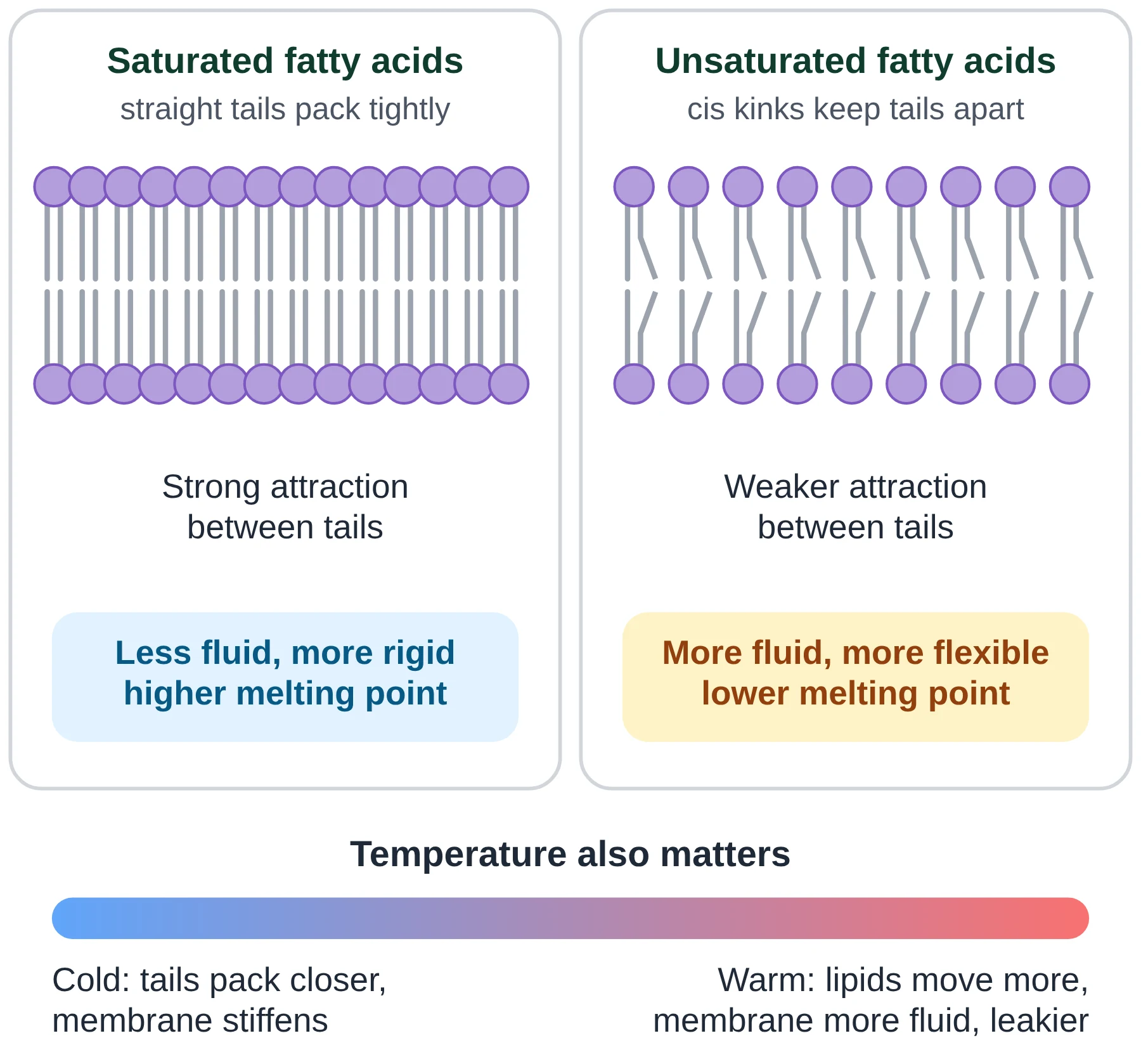
- Saturated fatty acids have no C=C double bonds. Their chains are straight, so they pack closely and attract each other more strongly. Saturated fatty acids have higher melting points and make the membrane less fluid and stronger at higher temperatures.
- Unsaturated fatty acids have one or more C=C double bonds. In membranes these are mostly cis double bonds, which put a kink in the chain. Kinked tails cannot pack closely, so attractions are weaker. Unsaturated fatty acids have lower melting points and keep the membrane fluid and flexible at the temperatures the cell experiences.
- Shorter fatty acid chains also increase fluidity, because shorter chains have less surface to attract one another.
Temperature
As temperature rises, phospholipids gain kinetic energy and move more, so the membrane becomes more fluid and more permeable. If a membrane gets too fluid it becomes leaky and can lose its structure. As temperature falls, tails pack together and the membrane stiffens, like butter in a fridge; transport proteins and enzymes in a rigid membrane work poorly.
Adapting membranes to the habitat
Organisms that cannot control their body temperature adjust the fatty acids in their membranes to suit where they live:
- Fish from polar seas have a higher proportion of unsaturated fatty acids in their membranes than fish from tropical waters, so their membranes stay fluid near 0 °C.
- Bacteria and many plants grown at low temperature increase the proportion of unsaturated (and shorter) fatty acids; when grown warmer they make more saturated fatty acids.
- Microorganisms that live in hot springs have membranes rich in saturated, longer-chain or otherwise specialised lipids, which stay stable at high temperature.
Plant cell membranes, which do not rely on cholesterol, depend especially on this balance of saturated and unsaturated fatty acids.
Fluidity, vesicles and membrane fusion
AHL Fluidity allows the membrane to curve, pinch off and join. In endocytosis a region of plasma membrane folds inwards and pinches off as a vesicle; in exocytosis a vesicle fuses with the plasma membrane and becomes part of it. Vesicles also carry proteins from the rough endoplasmic reticulum to the Golgi apparatus. None of this would be possible if the bilayer were rigid.
Key points
- AHL Saturated tails: straight, close packing, higher melting point, less fluid.
- AHL Unsaturated (cis) tails: kinked, loose packing, lower melting point, more fluid.
- Higher temperature increases fluidity; low temperature stiffens membranes.
- Organisms adjust fatty acid saturation to their habitat temperature.
- Fluidity is needed for vesicle formation and fusion.
Common mistakes
- Saying unsaturated fatty acids ‘have more hydrogen’. They have fewer hydrogen atoms, because of the C=C double bonds.
- Linking fluidity to the heads. Fluidity depends on how the tails pack.
Exam tip
- For ‘explain’ marks, give the full chain: double bond → kink → tails cannot pack closely → weaker attractions → more fluid.
Test yourself (click a question to reveal the answer)
AHL Predict how the membrane composition of a bacterium will change if it is moved from 37 °C to 10 °C, and explain why.
It will make more unsaturated (and shorter) fatty acids. Kinked unsaturated tails pack less closely, which keeps the membrane fluid at the lower temperature.
AHL Why do saturated fatty acids make a membrane less fluid?
Their straight chains pack tightly, so the attractions between neighbouring tails are stronger and phospholipids move less.
7 Cholesterol in the cell membrane
Syllabus link: AHL B2.1.12 Cholesterol and membrane fluidity in animal cells
AHL Cholesterol has a bad name in health news, but in the cell membrane it is essential. Up to about a third or more of the lipid molecules in an animal plasma membrane can be cholesterol.
Position in the membrane
Cholesterol is a steroid, and like phospholipids it is amphipathic, although mostly hydrophobic:
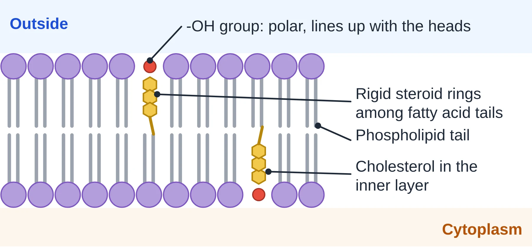
- a single small hydroxyl (–OH) group is polar, so it sits level with the phosphate heads, near the water;
- a flat, rigid group of four steroid rings and a short hydrocarbon tail are non-polar, so they lie among the fatty acid tails in the hydrophobic core.
Cholesterol as a fluidity buffer
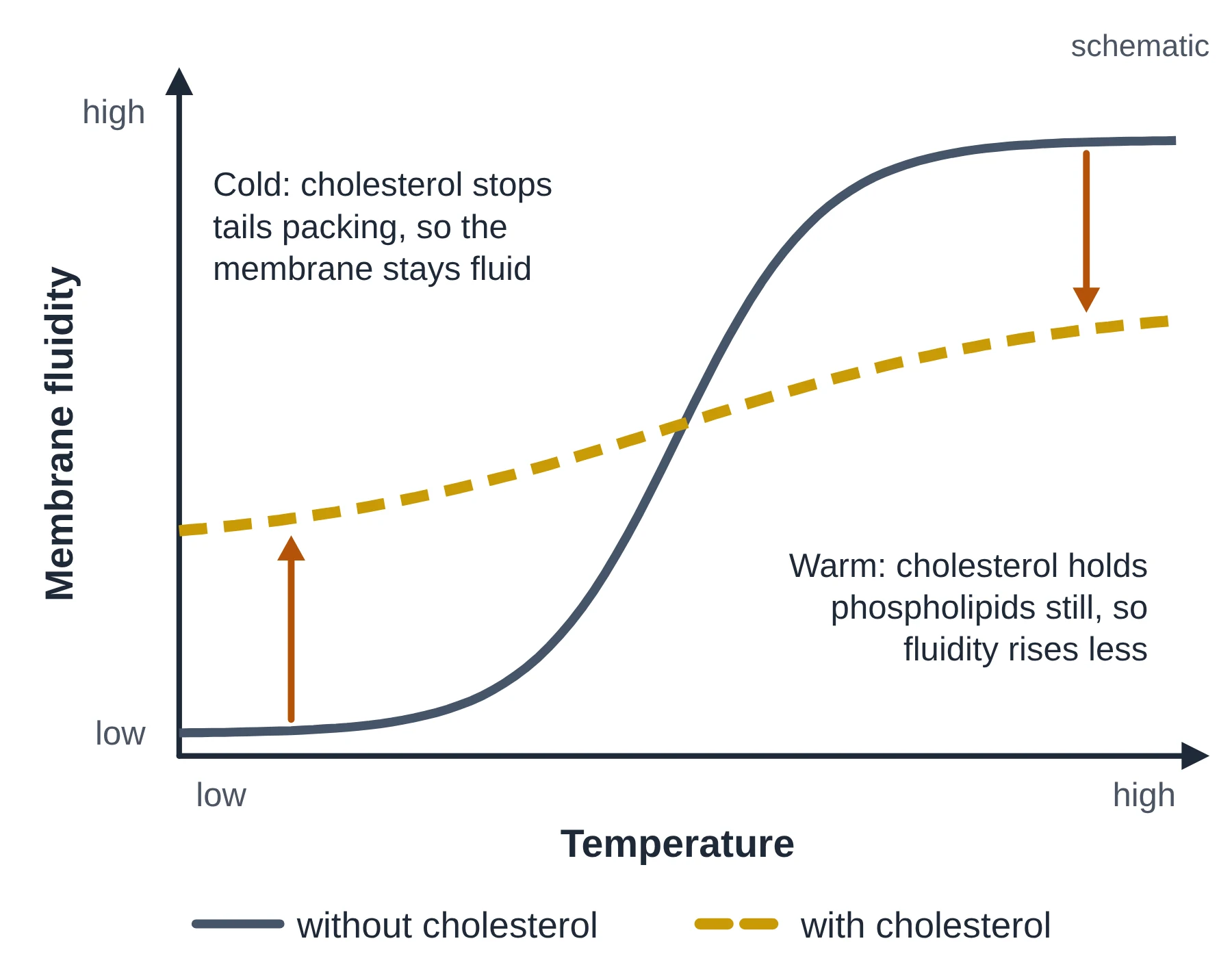
Cholesterol acts as a modulator (adjuster) of fluidity: it keeps the membrane at a useful fluidity across a range of temperatures.
- At higher temperatures the rigid rings hold the nearby parts of the tails still, restricting phospholipid movement. This reduces fluidity and stabilises the membrane, stopping it becoming too fluid and leaky.
- At lower temperatures cholesterol molecules sit between the phospholipids and stop the tails packing closely together and crystallising. This prevents the membrane stiffening, so it stays fluid.
Other effects of cholesterol
- It makes the bilayer less permeable to small water-soluble molecules and ions, by filling gaps between the tails.
- It adds mechanical stability, which matters for animal cells, because they have no cell wall.
Which cells have cholesterol?
Cholesterol is typical of animal cell membranes. Plant membranes contain only small amounts of cholesterol; instead they have related sterols (phytosterols) that do a similar job. Fungi use a different sterol, ergosterol. Most prokaryotes have no sterols at all. This is why the IB syllabus links cholesterol with animal cells.
Key points
- AHL Cholesterol is amphipathic: –OH among the heads, rings and tail among the fatty acid tails.
- It reduces fluidity at higher temperature and prevents stiffening at lower temperature (a fluidity buffer).
- It also lowers permeability to ions and small polar molecules.
- Typical of animal membranes; plants use similar sterols.
Common mistakes
- Writing ‘more cholesterol always makes the membrane less fluid’. That is true when warm, but at low temperature cholesterol keeps the membrane more fluid.
- Saying plant membranes contain no sterols at all. They have phytosterols that play a similar role.
- Placing cholesterol among the heads only. Only its small –OH group is at head level.
Test yourself (click a question to reveal the answer)
AHL Explain why the –OH group of cholesterol is found close to the phosphate heads.
The –OH group is polar (hydrophilic), so it is attracted to water and the polar heads; the rest of the molecule is hydrophobic and lies among the tails.
AHL Describe the effect of cholesterol on membrane fluidity at high and at low temperatures.
At high temperature it restricts phospholipid movement, reducing fluidity. At low temperature it stops tails packing closely, preventing the membrane from becoming rigid.
8 Membrane permeability
Syllabus link: B2.1.2 Lipid bilayers as barriers; B2.1.3 Simple diffusion across membranes; B2.1.8 Selectivity in membrane permeability
The cell membrane must let in what the cell needs (such as oxygen, glucose and ions) and let out waste (such as carbon dioxide and urea). Which substances cross, and how fast, depends on two things: the properties of the bilayer and the proteins in it.
Crossing the bilayer directly
Some particles pass straight between the phospholipids by simple diffusion, down their concentration gradient, with no protein and no ATP. How fast depends on the particle’s size and how well it dissolves in lipid (non-polar particles dissolve best):
- Small non-polar molecules (O2, CO2, N2) and lipid-soluble molecules such as steroid hormones dissolve in the hydrophobic core and cross quickly.
- Small uncharged polar molecules such as water, urea and glycerol can cross, but much more slowly.
- Large polar molecules such as glucose and sucrose cross the bilayer only extremely slowly.
- Ions such as Na+, K+, Cl− and H+ are charged and surrounded by a shell of water molecules. The bilayer is almost completely impermeable to them.
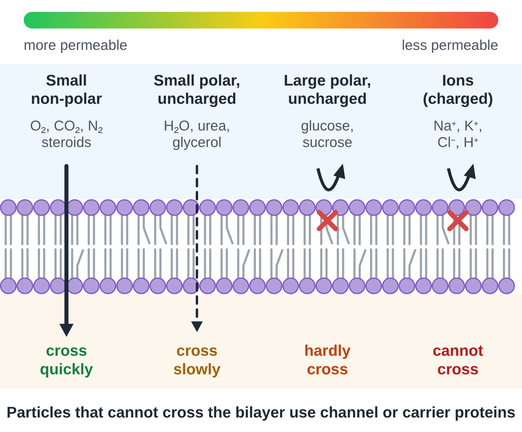
Note that polar and charged are not the same thing. Water is polar but has no overall charge, and it crosses slowly. Ions carry a full charge and are blocked.
Crossing with the help of proteins
Large and charged particles do cross membranes, but not through the lipid; they use transport proteins. These proteins carry ions such as Na+, K+ and Cl−, as well as amino acids, sugars, nucleotides and even water (aquaporins):
- Channel proteins and carrier proteins allow facilitated diffusion, which is passive and goes down the gradient.
- Pump proteins carry out active transport, using ATP to move particles against their gradient.
- Large particles and droplets can be moved in bulk by vesicles (endocytosis and exocytosis).
Selective permeability
Simple diffusion is not selective: anything small and non-polar enough will cross. Selectivity comes from proteins. Each channel, carrier or pump is specific for one substance or a few related ones, so a cell controls exactly what crosses by choosing which transport proteins to make and put into its membrane. That is why different cells, and different membranes in the same cell, have different permeabilities.
Details of diffusion, osmosis, facilitated diffusion, active transport and vesicles are covered in our Membrane Transport chapter.
Key points
- The hydrophobic core lets small non-polar molecules through and blocks ions and large polar molecules.
- Rate of simple diffusion depends on size and lipid solubility.
- Transport proteins (channels, carriers, pumps) let ions and polar molecules cross.
- Simple diffusion is not selective; proteins make membranes selectively permeable.
Common mistakes
- Saying water cannot cross the bilayer. It can, slowly; aquaporins make it much faster.
- Saying ‘polar (charged)’ as if they mean the same. Many polar molecules have no net charge.
Test yourself (click a question to reveal the answer)
Arrange in order of how fast they cross a pure phospholipid bilayer, fastest first: glucose, CO2, K+, water.
CO2 > water > glucose > K+.
Explain why the plasma membrane is described as selectively permeable rather than just partially permeable.
Its permeability is controlled: specific channel, carrier and pump proteins let particular substances cross, so the cell chooses what passes, not only particle size and polarity.
9 Cell membrane components at a glance
Swipe sideways to see the whole table →
| Component | Structure | Where in the membrane | Main role |
|---|---|---|---|
| Phospholipid | Glycerol + 2 fatty acids + phosphate group; amphipathic | Two layers: heads outwards, tails inwards | Forms the bilayer; hydrophobic core is the barrier |
| Integral protein | Amphipathic protein with hydrophobic region | Embedded in the core; often spans the membrane | Channels, carriers, pumps, receptors, CAMs, enzymes |
| Peripheral protein | Protein held by weak attractions | On the inner or outer surface | Enzymes, signalling, linking to the cytoskeleton |
| Glycoprotein | Protein + carbohydrate chain | Integral protein; sugar faces outside | Recognition, adhesion, receptors |
| Glycolipid | Lipid + carbohydrate chain | Outer layer only | Recognition (e.g. ABO antigens), adhesion |
| Cholesterol | Steroid with one –OH group | Between phospholipids, in both layers (animal cells) | AHL Buffers fluidity; reduces permeability |
Memory hook: lipids make the barrier, proteins do the work, sugars give the identity.
10 Quick check
Ten questions: six multiple choice and four short answer. Try them all before opening the answers.
-
Which part of a phospholipid is hydrophilic?
- A. The fatty acid tails
- B. The phosphate head
- C. The C=C double bonds
- D. The whole molecule
-
Which observation was evidence against the Davson–Danielli model?
- A. Membranes look three-layered in electron micrographs
- B. Membranes are barriers to ions
- C. Freeze-fracture faces show particles in the bilayer core
- D. Membranes contain phospholipids
-
Where are the carbohydrate chains of glycoproteins found?
- A. On the cytoplasmic side
- B. In the hydrophobic core
- C. On both sides equally
- D. On the extracellular side
-
Which molecule crosses a pure phospholipid bilayer fastest?
- A. Oxygen
- B. Glucose
- C. Sodium ions
- D. Amino acids
-
AHL A membrane with a high proportion of unsaturated fatty acids will be:
- A. Less fluid, because tails pack closely
- B. More fluid, because kinked tails pack loosely
- C. More fluid, because unsaturated fatty acids have higher melting points
- D. Unaffected, because fluidity depends only on proteins
-
AHL Which statement about cholesterol is correct?
- A. It is found among the phosphate heads only
- B. It increases fluidity at high temperatures
- C. It is amphipathic and lies among the tails with its –OH group near the heads
- D. It is the main lipid in plant membranes
-
Distinguish between integral and peripheral membrane proteins. (2 marks)
-
Draw and label a diagram of the fluid mosaic model of a membrane. (5 marks)
-
Outline the cell fusion experiment and what it showed about membranes. (3 marks)
-
AHL Explain how cholesterol helps keep animal cell membranes working over a range of temperatures. (3 marks)
Show answers
- B. The phosphate head is polar or charged, so it interacts with water. The tails are hydrophobic.
- C. Particles in the core show that proteins pass into the bilayer, not just coat it. The three-layered appearance was at first taken as support for the model.
- D. Carbohydrates of glycoproteins and glycolipids face the outside of the cell.
- A. O2 is small and non-polar, so it dissolves in the hydrophobic core.
- B. Cis double bonds kink the tails, so they pack loosely and have lower melting points.
- C. Its polar –OH group sits at head level and its rigid rings among the tails.
- Integral proteins are embedded in the hydrophobic core of the bilayer, often spanning it [1]; peripheral proteins are attached to one surface only and are not embedded in the core [1].
- One mark each, up to 5: phospholipid bilayer with heads and tails shown; hydrophilic heads and hydrophobic tails labelled; integral protein spanning the bilayer; peripheral protein on a surface; glycoprotein with carbohydrate on the outside; cholesterol among the tails.
- Mouse and human membrane proteins were labelled with green and red fluorescent antibodies [1]. After the cells were fused, the colours started on separate halves but became fully mixed within about 40 minutes at 37 °C [1]. This shows that membrane proteins can move sideways, so the membrane is fluid (mixing was much slower at low temperature) [1].
- At high temperature cholesterol restricts the movement of phospholipids, reducing fluidity so the membrane does not become too fluid or leaky [1]. At low temperature it stops the fatty acid tails packing closely together, so the membrane does not become rigid [1]. Fluidity therefore changes less with temperature: cholesterol acts as a buffer [1].
11 Glossary
© competitorspoint.com · IB Biology notes. Diagrams and text are original to competitorspoint.com.
Ready to practise? Download the Cell Membrane practice questions with answers (free for signed-up members).
⬇ Download practice questions (PDF)