
Looking for a clear, high-resolution labeled diagram of human kidney for your biology studies, presentations, or revision notes? Below, you can download a premium, copyright-free cross-section illustration of the kidney, designed to meet global academic standards like AP Biology, IB Biology, A-Levels, and NCERT Class 11 (Chapter 16).
📥 Download High-Res Kidney Diagram (Free PNG)
Labeled Diagram of Human Kidney Features:
- It is the primary excretory organ
- Retroperitoneal: Human kidney is present in the abdominal cavity, but only the ventral portion of the kidney is covered by peritoneum.
- Human kidney is dorso-lateral in position.
- It is kiney shaped structure positioned between 12th thoracic vertebrae and 3rd lumbar vertebrae.
- The length, width and depth of the kindey are 10 -12 x 5 – 7 x 2 -3 cm
Explaining the Labeled Diagram of Human Kidney Structures
To master your biology exams (such as AP Biology, IB Biology, or NCERT layouts), make sure you can identify and explain these core components shown in the diagram:
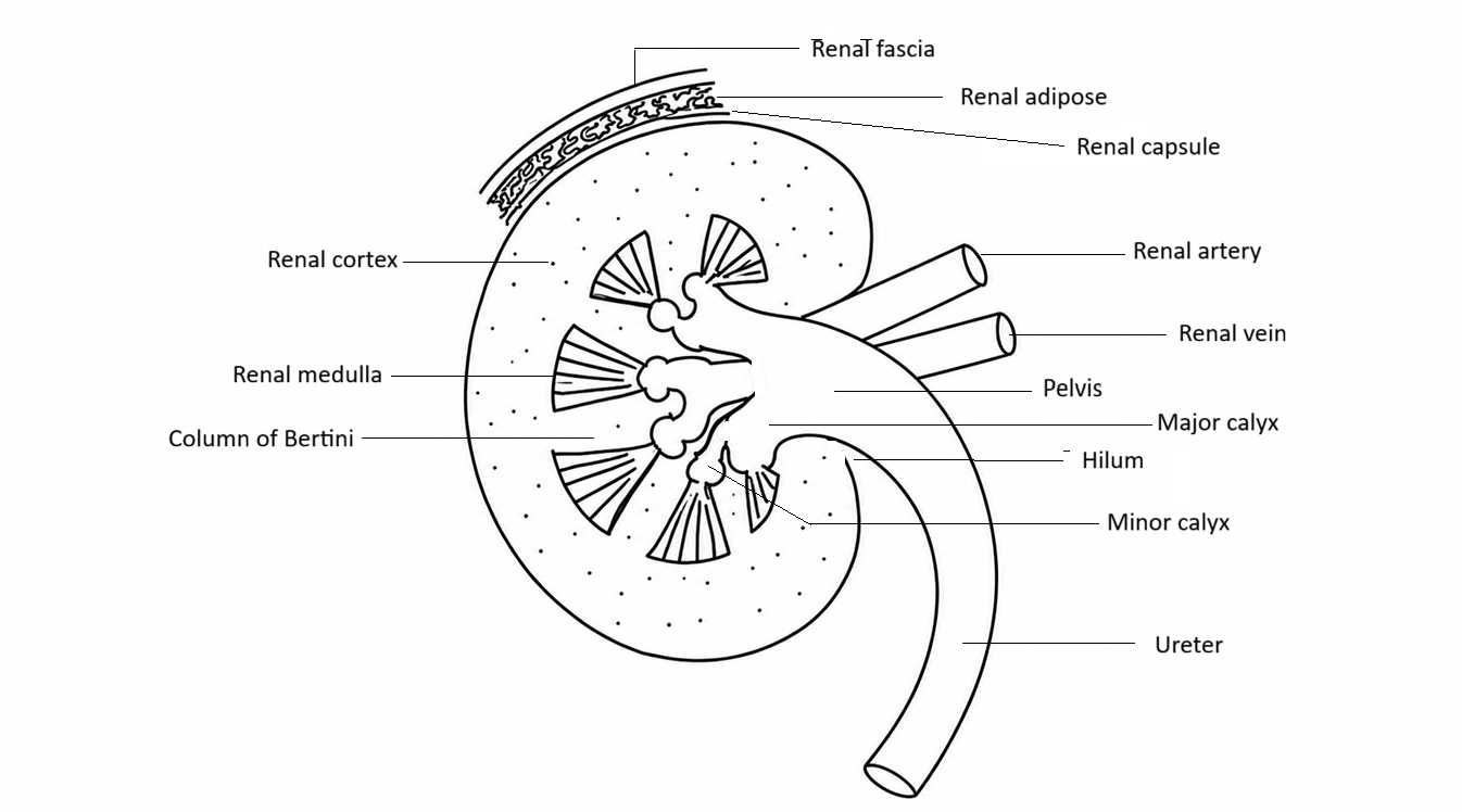
- Protective covering of the kidney: Renal fascia (outermost) + Renal adipose (middle layer) + Renal capsule (innermost layer)
- Renal cortex: It is that portion of the kidney that lies between renal capsule and renal medulla.
- Renal medulla: It is divided into many meduallry pyramids
- Renal column of Bertini: The portion of renal cortex embedded in the medually region is called the renal column of Bertini
- Minor calyx: A small cup-shaped cavity that surrounds the tip (renal papilla) of a renal pyramid. It receives urine produced by the kidney tissue.
- Major calyx: A larger chamber formed by the joining of two or more minor calyces. It collects urine from the minor calyces and drains it into the renal pelvis.
- Pelvis: In the human kidney, the renal pelvis is a funnel-shaped structure that collects urine from the major calyces and channels it into the ureter
Renal pyramid → Minor calyx → Major calyx → Renal pelvis → Ureter → Urinary bladder
8. Renal artery: Oxygenated blood enters into the kidney
9. Rnal vein: deoxygenated blood flows out from the kidney
Step-by-Step Guide: How to Draw a Human Kidney Cross-Section: Labeled Diagram of Human Kidney
- For many high school biology exams, AP practicals, or board revisions, you might be asked to sketch the internal structure of the kidney by hand. Use this simple breakdown to draw it quickly:
- 1. The Bean Outline: Start by drawing a large asymmetric bean shape on your paper. The indented inner curve is called the hilum, where the blood vessels enter.
2. The Cortex Boundary: Lightly sketch a parallel line just inside your main outline. This creates a clear outer border layer representing the renal cortex.
3. The Pyramids: Inside the inner line, draw 6 to 8 neatly spaced triangular or cone-shaped structures pointing inward. These represent the renal medullary pyramids.
4. The Collection Funnel: In the central open space near the hilum, draw an open funnel network that connects the points of all your triangles. This represents the renal pelvis.
5. The Outlet Tubes: From the center funnel, extend a tube downward to represent the ureter. Right next to it, draw two interacting circular vessels representing the renal artery and renal vein.
High-Yield Points for AP, IB, and Entrance Exams
- When studying the urinary system or human osmoregulation, examiners frequently test the functional relationships of these structures:
- Filtration Site: Ultrafiltration occurs exclusively within the renal cortex because that is where the Bowman’s capsules and glomeruli are located. If a question asks where podocytes or fenestrated capillaries are found, the answer is always the cortex.
- Concentration Gradient: The loops of Henle inside the renal pyramids use a countercurrent multiplier mechanism. This builds a massive sodium chloride gradient in the inner medulla, allowing humans to reabsorb water and excrete hypertonic (concentrated) urine to prevent dehydration.
- Collecting Ducts: Multiple nephrons empty their fluid into shared collecting ducts traveling straight down through the pyramids. This fluid dumps into the minor calyces, passes through the renal pelvis, and moves into the urinary bladder via peristaltic waves along the ureters.