
Nervous System IB Biology: 16 Easy Notes (C2.2, C3.1)
In nervous system IB Biology notes, the nervous system is the network of neurons that detects stimuli, carries information as fast electrical impulses and coordinates responses. It has a central part, the brain and spinal cord, and a peripheral part of nerves. The IB guide calls this neural signalling (C2.2).
1 What is the nervous system? (C3.1.3)
Syllabus link: IB Biology (first assessment 2025) C2.2 Neural signalling: C2.2.1 to C2.2.7 (SL), C2.2.8 to C2.2.16 (HL only); C3.1 Integration of body systems: C3.1.3 to C3.1.11 and C3.1.14 to C3.1.16 (SL). Hormones (C3.1.12, C3.1.13), system integration (C3.1.1, C3.1.2) and plant responses (C3.1.17 to C3.1.23, HL) are covered in their own chapters.
IB HL = detail the IB guide expects at Higher Level only
Every organ in your body works with the others. During a sprint, breathing deepens, the heart speeds up and blood is sent to the leg muscles. Two systems link these changes: the nervous system, which sends electrical impulses along neurons, and the endocrine system, which sends hormones in the blood. The IB guide calls the first neural signalling and asks you to compare it with hormonal signalling (C3.1.3).
The working unit is the neuron, a cell specialised to detect, receive and transmit stimuli. Hydra has only a net of neurons, insects have a brain and ganglia, and vertebrates have the most developed nervous system; the human brain holds about 86 billion neurons. Every nervous response follows the same path: stimulus → receptor → sensory neuron → CNS → motor neuron → effector → response. For the other chapters in this unit, see the Human Physiology chapters hub.
Key idea
- Signals are electrical along a neuron and usually chemical between neurons (at a synapse).
- Nervous responses are fast, short-lived and precisely targeted.
2 Divisions of the human nervous system (C3.1.4 to C3.1.7)
The central nervous system (CNS) is the brain and spinal cord, the site of information processing and control. The peripheral nervous system (PNS) is all the nerves linking the CNS to the body: 12 pairs of cranial nerves and 31 pairs of spinal nerves. PNS fibres are of two kinds:
- Afferent (sensory) fibres carry impulses from tissues and organs to the CNS.
- Efferent (motor) fibres carry impulses from the CNS to muscles and glands.
Somatic and autonomic nervous systems
The somatic nervous system relays impulses from the CNS to skeletal muscles and is largely voluntary. The autonomic nervous system carries impulses to the involuntary organs and smooth muscles, the heart and glands. It has two divisions that usually have opposite effects:
- Sympathetic: “fight or flight”. It raises heart rate, dilates the pupils and slows digestion; its nerves leave the thoracic and lumbar spinal cord.
- Parasympathetic: “rest and digest”. It slows the heart and stimulates digestion; its nerves leave the brain (mainly the vagus nerve) and the sacral spinal cord.
The gut wall has its own enteric nervous system, which controls peristalsis (section 10). The division of labour matters for IB: the spinal cord integrates unconscious processes (C3.1.5), sensory neurons bring input to the spinal cord and cerebral hemispheres (C3.1.6), and motor neurons carry output from the cerebral hemispheres to muscles (C3.1.7).
3 Neuron structure and types of nerve cells (C2.2.1, C3.1.8)
The neuron is the structural and functional unit of the nervous system, with three main parts (IB C2.2.1):
- Cell body (cyton): cytoplasm, a nucleus and organelles, including Nissl’s granules (rough ER and ribosomes).
- Dendrites: short, branched fibres, also with Nissl’s granules, that carry impulses towards the cell body.
- Axon: one long fibre that carries impulses away from the cell body. Its branches end in synaptic knobs holding vesicles of neurotransmitter, at a synapse or a neuromuscular junction. It has no Nissl’s granules.
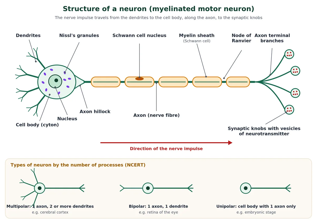
Types of neurons
By function (IB C3.1.5 to C3.1.7): sensory neurons carry impulses to the CNS, interneurons (relay neurons) link neurons inside the CNS, and motor neurons carry impulses to effectors.
Myelinated and unmyelinated nerve fibres
In a myelinated fibre, Schwann cells wrap the axon in many layers of lipid-rich membrane, the myelin sheath; the gaps between neighbouring Schwann cells are the nodes of Ranvier. These fibres occur in spinal and cranial nerves. In an unmyelinated fibre a Schwann cell encloses the axon without forming myelin; these are common in the autonomic and somatic systems. In the CNS, myelin is made by oligodendrocytes. Schwann cells and oligodendrocytes are glial cells, which support neurons. Mature neurons normally do not divide, so CNS damage heals poorly (see how specialised cells such as neurons differentiate).
Nerves (IB C3.1.8)
- A nerve is a bundle of nerve fibres inside a protective sheath; a transverse section shows both myelinated and unmyelinated fibres.
- Most nerves are mixed, with fibres of both sensory and motor neurons. A neuron is one cell; a nerve contains fibres of many neurons.
4 Resting potential and the sodium-potassium pump (C2.2.2)
A neuron that is not conducting an impulse has a polarised membrane: the inside is negative relative to the outside. This potential difference is the resting potential, about −70 mV (values from −60 to −80 mV are quoted).
How the resting potential is generated (IB C2.2.2)
- Pumping: the sodium-potassium pump uses ATP to move 3 Na+ out for every 2 K+ in, by active transport against the concentration gradients. This keeps Na+ high outside and K+ high inside.
- Unequal leakage: the resting membrane is much more permeable to K+ than to Na+, so K+ diffuses out through leak channels, carrying positive charge out.
- Trapped negative charge: large negatively charged proteins in the axoplasm cannot leave.
The pump adds slightly to the negative value, because it moves three positive charges out for every two in, but the main cause is the K+ leak. Each ion crosses through its own channel proteins in the plasma membrane.
Swipe the table sideways to see every column.
| Ion | Inside the axon (approx.) | Outside (approx.) | Resting membrane |
|---|---|---|---|
| Na+ | 15 mmol dm−3 | 145 mmol dm−3 | Nearly impermeable |
| K+ | 140 mmol dm−3 | 5 mmol dm−3 | Fairly permeable |
| Proteins (−) | High | Very low | Impermeable |
5 Action potential: how a nerve impulse is generated (C2.2.3, C2.2.8, C2.2.10)
A nerve impulse is an action potential: a brief reversal of the membrane potential that travels along the nerve fibre (IB C2.2.3). It is electrical because positive ions move across the membrane. When a stimulus is applied, that patch of membrane becomes freely permeable to Na+; Na+ rushes in and the inside becomes positive. The membrane is depolarised for about 1 to 2 ms.
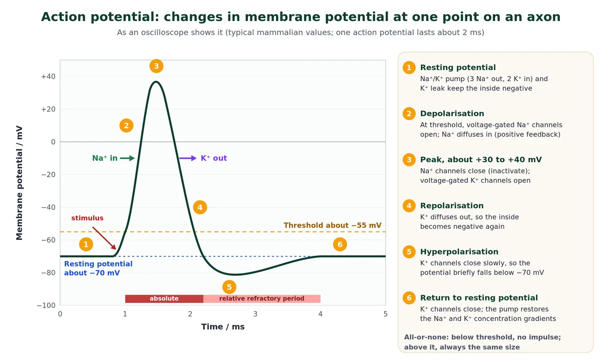
Stages of an action potential IB HL C2.2.8
- Resting potential (about −70 mV): voltage-gated channels are closed.
- Depolarisation: if a stimulus raises the potential to the threshold, about −55 mV, voltage-gated Na+ channels open and Na+ diffuses in. This opens more Na+ channels: positive feedback.
- Peak (about +30 to +40 mV): Na+ channels close and voltage-gated K+ channels open.
- Repolarisation: K+ diffuses out and the inside becomes negative again.
- Hyperpolarisation: K+ channels close slowly, so the potential briefly falls to about −80 mV.
- Recovery: the K+ channels close and the pump restores the ion gradients.
Threshold, all-or-none and the refractory period
A stimulus below threshold gives no action potential; any stimulus that reaches threshold gives a full one of the same size. This is the all-or-none principle. A stronger stimulus produces more impulses per second, not bigger ones. Straight after an impulse, during the absolute refractory period (about 1 ms), the Na+ channels are inactivated and the membrane cannot fire at all; in the relative refractory period only a stronger stimulus works. The refractory period keeps impulses separate and makes them travel in one direction.
IB HL C2.2.10 Reading an oscilloscope trace
- Time (ms) is on the x-axis and membrane potential (mV) on the y-axis. The steep rise is depolarisation, the fall is repolarisation and the dip below the resting line is hyperpolarisation.
- A resting neuron gives a flat line at about −70 mV; each action potential is a spike to the same peak (all-or-none).
- Frequency: count the spikes in a stated time. For example, 12 action potentials in 40 ms is 12 ÷ 0.040 s = 300 impulses per second.
- A stronger stimulus shows as spikes closer together (higher frequency), never as taller spikes. Read the time scale (ms or s) before calculating.
For a clinical account of the channels involved, see the StatPearls review of action potential physiology on NCBI Bookshelf.
6 Conduction of nerve impulses and saltatory conduction (C2.2.4, C2.2.9, C2.2.11)
Propagation by local currents IB HL C2.2.9
Where the axon is depolarised (point A) the inside is positive; just ahead (point B) it is still negative. Na+ that entered at A diffuses along the inside towards B, while on the outside positive charge moves from B towards A. These local currents raise B to threshold, so an action potential is generated at B, and so on along the axon: a wave of depolarisation and repolarisation. The refractory region behind stops it going backwards.
Saltatory conduction IB HL C2.2.11
Myelin is an electrical insulator. In a myelinated fibre the voltage-gated channels and pumps are clustered at the nodes of Ranvier, so action potentials occur only there and the impulse jumps from node to node, about 1 mm apart. This saltatory conduction (Latin saltare, to jump) is much faster and uses less ATP, because far less membrane depolarises.
Speed of nerve impulses (IB C2.2.4)
Swipe the table sideways to see every column.
| Fibre | Diameter | Speed (approx.) | Reason |
|---|---|---|---|
| Small unmyelinated (human pain fibre) | Under 1.5 µm | 0.5 to 2 m s−1 | Narrow; every patch of membrane depolarises |
| Squid giant axon (unmyelinated) | About 0.5 mm | About 25 m s−1 | Wide axon, low resistance to local currents |
| Large myelinated (human motor neuron) | 10 to 20 µm | Up to about 120 m s−1 | Saltatory conduction |
Myelination and a larger axon diameter both increase speed. Squid giant axons, used for fast escape, are still slower than thin myelinated human fibres.
Application of skills: correlation
- Conduction speed is positively correlated with axon diameter and negatively correlated with animal size.
- The correlation coefficient r (−1 to +1) gives the direction and strength; R2 gives the fraction of the variation explained (R2 = 0.81 means 81 %). Correlation does not prove causation.
7 Synapse: how the impulse passes between cells (C2.2.5 to C2.2.7, C2.2.13, C2.2.14)
A synapse is a junction between two neurons, or between a neuron and an effector cell such as a muscle fibre (IB C2.2.5). A synapse with a skeletal muscle fibre is a neuromuscular junction.
At an electrical synapse the pre- and postsynaptic membranes are very close and current flows directly between them; transmission is faster, but such synapses are rare in humans. At a chemical synapse a fluid-filled synaptic cleft, about 20 nm wide, separates the membranes, and a neurotransmitter carries the signal. The IB guide covers only chemical synapses.
Steps in synaptic transmission (IB C2.2.5 to C2.2.7)
- An action potential arrives at the synaptic knob and depolarises its membrane.
- Voltage-gated calcium channels open and Ca2+ diffuses in, acting as a signal inside the neuron.
- Vesicles fuse with the presynaptic membrane and release neurotransmitter by exocytosis.
- The neurotransmitter diffuses across the cleft and binds to receptors on the postsynaptic membrane.
- With acetylcholine, the receptors are ion channels; Na+ enters and the membrane depolarises: an excitatory postsynaptic potential (EPSP). If threshold is reached, an action potential starts.
- Acetylcholinesterase in the cleft hydrolyses acetylcholine into choline and acetate; choline is reabsorbed and recycled. Other transmitters, such as dopamine, are pumped back into the presynaptic neuron (reuptake).
Transmission is one-way, because vesicles are only in the presynaptic knob and receptors only on the postsynaptic membrane. Acetylcholine works at many synapses, including every neuromuscular junction. Excitatory neurotransmitters, such as acetylcholine, depolarise the postsynaptic membrane (an EPSP, SL C2.2.7); inhibitory ones hyperpolarise it (an IPSP, HL C2.2.13). For a fuller textbook account, see OpenStax Biology 2e on how neurons communicate.
IB HL C2.2.13 and C2.2.14 Inhibition and summation
- Inhibitory neurotransmitters such as GABA open Cl− or K+ channels, so the postsynaptic membrane is hyperpolarised: an inhibitory postsynaptic potential (IPSP).
- One EPSP is usually too small to reach threshold. Spatial summation adds inputs from several presynaptic neurons at once; temporal summation adds repeated impulses from one neuron.
- EPSPs and IPSPs are added together with an all-or-none result: an action potential fires only if the net depolarisation reaches threshold.
- Worked example: resting potential −70 mV, threshold −55 mV. Three excitatory neurons each give an EPSP of +6 mV (+18 mV) while one inhibitory neuron gives an IPSP of −5 mV. Net change = +13 mV, so the membrane reaches only −57 mV and no action potential fires. Remove the IPSP and it reaches −52 mV, so it fires.
- Summation lets one postsynaptic neuron integrate many inputs; this is how neurons in the brain “make decisions”.
8 Drugs, pesticides, pain and consciousness (C2.2.12, C2.2.15, C2.2.16)
IB HL C2.2.12 Exogenous chemicals at synapses
- Exogenous chemicals come from outside the body. Some block transmission, some prolong it; the IB guide uses a pesticide that blocks transmission and a drug that blocks reuptake.
- Neonicotinoids (such as imidacloprid) are insecticides that bind to nicotinic acetylcholine receptors in insects. Acetylcholinesterase cannot break them down, so they stay bound, overstimulate the receptors and then block transmission, paralysing and killing the insect. They bind far more strongly to insect receptors than to mammalian ones. Harm to bees led the European Union to ban outdoor use of three neonicotinoids in 2018.
- Cocaine blocks the transporters that reabsorb dopamine into the presynaptic neuron, so dopamine builds up in the cleft and keeps stimulating the postsynaptic neuron, causing euphoria and, with repeated use, addiction.
Perception of pain IB HL C2.2.15
Pain receptors are free nerve endings of sensory neurons in the skin. They have channels for positive ions that open in response to high temperature, acid or chemicals such as capsaicin from chilli peppers. Positive ions enter, threshold is reached, and impulses travel to the brain, where pain is perceived.
Consciousness IB HL C2.2.16
No single neuron is conscious. Consciousness, our awareness of ourselves and our surroundings, emerges from the interactions of billions of neurons in the brain: an emergent property of the whole system that cannot be predicted from one cell. Evidence: consciousness is lost when activity across the cortex is disrupted (general anaesthesia, deep sleep) even though each neuron is still alive. The same idea of emergence runs through system integration (C3.1.1).
9 Central nervous system: the human brain (C3.1.4, C3.1.10, C3.1.11)
The brain is the body’s central information-processing organ, its “command and control system”. It controls voluntary movement, balance, vital organs, temperature, hunger, thirst and circadian rhythms, and processes senses, speech, memory, emotions and thought. The IB guide stresses that it combines information from several inputs and is the organ of learning and memory (C3.1.4).
Forebrain
- Cerebrum: the largest, most developed part. A deep cleft divides it into left and right cerebral hemispheres, joined by the corpus callosum. The folded cerebral cortex is grey matter (neuron cell bodies) with motor, sensory and association areas; association areas handle intersensory associations, memory and communication. Beneath it lies white matter of myelinated tracts.
- Thalamus: wrapped by the cerebrum; a major coordinating centre for sensory and motor signals.
- Hypothalamus: at the base of the thalamus. It controls body temperature, hunger and thirst, acts as the body’s master clock and has neurosecretory cells that release hypothalamic hormones, linking it to the hypothalamus and pituitary gland system.
- Limbic system: inner parts of the hemispheres with the amygdala and hippocampus. With the hypothalamus it regulates sexual behaviour, emotions (excitement, pleasure, rage, fear) and motivation.
Hindbrain
- Pons: fibre tracts that interconnect regions of the brain; also helps regulate breathing.
- Cerebellum: its very convoluted surface makes room for many neurons, about 80 % of all those in the brain. It coordinates skeletal muscle contraction for smooth, accurate movement and maintains balance and posture (IB C3.1.10).
- Medulla oblongata: joins the spinal cord; contains centres for breathing, cardiovascular reflexes and gastric secretions, plus swallowing, coughing and vomiting.
The midbrain, pons and medulla form the brain stem, which connects the brain to the spinal cord.
Sleep, circadian rhythms and melatonin (IB C3.1.11)
The suprachiasmatic nucleus of the hypothalamus receives impulses from light-sensitive cells in the retina. In darkness it signals the pineal gland to secrete melatonin; light suppresses secretion. Melatonin is therefore high at night and low by day, a diurnal pattern that promotes sleepiness and sets the 24-hour sleep-wake cycle. Jet lag happens because the melatonin rhythm is still on home time. This is a neural input controlling a hormone, so it also appears in the pineal gland and melatonin notes.
10 Spinal cord, nerves and integration of body systems (C3.1.5 to C3.1.7, C3.1.14 to C3.1.16)
The spinal cord, about 45 cm long in adults, runs from the medulla through the vertebral canal, protected by the vertebrae, meninges and CSF. Its arrangement is the reverse of the cerebrum: inner, butterfly-shaped grey matter around the central canal, and outer white matter of tracts running to and from the brain. Each spinal nerve joins the cord by a dorsal root (sensory fibres; the dorsal root ganglion holds their cell bodies) and a ventral root (motor fibres).
Input, processing and output (IB C3.1.5 to C3.1.7)
- Input: sensory neurons carry impulses from receptor cells to the spinal cord and cerebral hemispheres.
- Processing: the spinal cord integrates unconscious processes such as reflexes; the cerebral hemispheres handle conscious ones.
- Output: motor neurons carry impulses from the cerebral hemispheres to muscles, which contract.
Unconscious control of heart, breathing and gut (IB C3.1.14 to C3.1.16)
- Heart rate (C3.1.14): baroreceptors in the aorta and carotid arteries monitor blood pressure; chemoreceptors monitor blood pH, O2 and CO2. Low pressure, high CO2 or low pH → the cardiovascular centre in the medulla sends impulses along sympathetic nerves to the SA node → faster, stronger beats. High pressure → more impulses along the vagus (parasympathetic) nerve → slower beats. The change reverses the stimulus: negative feedback. See how the cardiac cycle and heart rate are controlled.
- Ventilation rate (C3.1.15): during exercise, extra CO2 lowers blood pH; chemoreceptors in the medulla and in the carotid and aortic bodies detect it. The respiratory centre in the medulla sends more impulses to the diaphragm and intercostal muscles, so breathing becomes faster and deeper until CO2 and pH return to normal: negative feedback (see regulation of breathing and lung volumes).
- Peristalsis (C3.1.16): swallowing and egestion are under voluntary control by the CNS; peristalsis between them is involuntary, run by the enteric nervous system, a network of neurons in the gut wall. Stretch of the wall by food triggers contraction of circular muscle behind the bolus and relaxation ahead of it. The CNS can modulate the ENS through autonomic nerves (see digestion and absorption in the gut).
11 Reflex action and the reflex arc (C3.1.9)
A reflex action is a rapid, automatic, involuntary response to a stimulus. The pathway of impulses is the reflex arc. Many reflexes are integrated in the spinal cord, so the response starts before the brain is aware of the stimulus. The IB guide uses the pain withdrawal reflex, with skeletal muscle as the effector (C3.1.9).
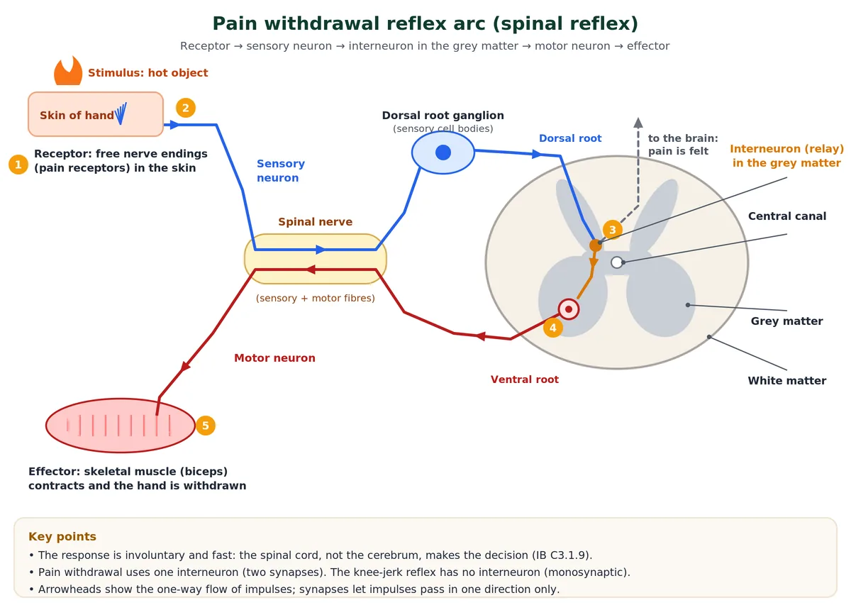
- Receptor: a free nerve ending in the skin of the hand detects a harmful stimulus, such as a hot object.
- Sensory neuron: carries impulses through the dorsal root (cell body in the dorsal root ganglion) into the grey matter.
- Interneuron: one relay neuron in the grey matter passes the impulse to a motor neuron across synapses.
- Motor neuron: carries impulses out through the ventral root to the arm.
- Effector: the biceps contracts and the hand is withdrawn.
Impulses also travel up to the brain, so pain is felt just after the hand moves. Skeletal muscle is the effector: the motor neuron releases acetylcholine at the neuromuscular junction, the muscle fibre depolarises and contracts. The response is involuntary because the spinal cord, not the cerebral hemispheres, makes the connection.
Knee-jerk reflex
A tap below the kneecap stretches the quadriceps muscle. Stretch receptors (muscle spindles) send impulses along a sensory neuron that synapses directly with a motor neuron in the spinal cord, and the quadriceps contracts, kicking the lower leg forward. With no interneuron and one synapse, the knee-jerk is monosynaptic; the pain withdrawal reflex is polysynaptic.
Syllabus note
- The pain reflex arc is SL content for IB (C3.1.9); learn it with skeletal muscle as the effector. The knee-jerk is a useful extra example of a stretch reflex, not a guide requirement.
12 Nervous system vs endocrine system and other comparisons (C3.1.3)
The IB guide asks you to distinguish nervous from hormonal signalling (C3.1.3). The hormone side is covered in the endocrine system and hormones chapter.
Swipe the table sideways to see every column.
| Feature | Nervous system | Endocrine system |
|---|---|---|
| Signal | Electrical impulses; neurotransmitters at synapses | Hormones |
| Route | Along neurons | In the blood |
| Target | Specific muscles and glands | Any cell with the right receptor; often many organs |
| Speed | Milliseconds | Seconds to hours |
| Duration | Short | Longer lasting |
| Example | Pain withdrawal reflex | Blood glucose control, growth, puberty |
Some responses use both: sympathetic nerves act within a fraction of a second, while adrenaline from the adrenal medulla spreads and prolongs the message (C3.1.12). The hypothalamus is the main link between the two systems: it is part of the brain but controls the pituitary gland (C3.1.13).
Sympathetic vs parasympathetic nervous system
Swipe the table sideways to see every column.
| Feature | Sympathetic | Parasympathetic |
|---|---|---|
| Role | “Fight or flight” | “Rest and digest” |
| Origin | Thoracic and lumbar spinal cord | Brain (mainly the vagus nerve) and sacral cord |
| Transmitter at organ | Mostly noradrenaline | Acetylcholine |
| Heart rate | Increases | Decreases |
| Pupil | Dilates | Constricts |
| Bronchioles | Dilate | Constrict |
| Digestion | Inhibited | Stimulated |
13 Common exam traps
Mistakes examiners see most often in nervous system IB Biology answers.
Watch out for these
- “The pump makes the resting potential by itself.” The pump builds the gradients; the negative value is mainly due to K+ leaking out and to trapped negative proteins.
- “Na+ is pumped in during depolarisation.” Na+ diffuses in through voltage-gated channels; no ATP is used at that moment. The pump moves Na+ out.
- “A stronger stimulus gives a bigger action potential.” All-or-none: it gives a higher frequency of identical impulses.
- “The impulse jumps along the myelin.” Action potentials occur at the nodes of Ranvier, where the myelin is absent.
- “Neurotransmitter is actively transported across the cleft.” It diffuses. It is released by exocytosis, which is triggered by Ca2+ entry, not by Na+.
- “Acetylcholinesterase is in the presynaptic knob.” It acts in the synaptic cleft, at the postsynaptic membrane.
- Grey and white matter swap Cerebrum: grey outside, white inside. Spinal cord: grey inside, white outside.
- Knee-jerk has no interneuron Do not draw a relay neuron in a knee-jerk arc; the pain withdrawal arc has one.
14 Key terms
The nervous system IB Biology terms you need to describe neural signalling in an exam answer.
| Term | Meaning |
|---|---|
| Neuron | Nerve cell that carries electrical impulses; cell body, dendrites and axon. |
| Myelin sheath | Lipid-rich insulating layer made by Schwann cells (PNS) or oligodendrocytes (CNS). |
| Resting potential | Potential across the membrane of a resting neuron, about −70 mV, inside negative. |
| Threshold potential | Potential (about −55 mV) at which voltage-gated Na+ channels open. |
| Action potential | Rapid depolarisation and repolarisation of the membrane that forms a nerve impulse. |
| Refractory period | Short time after an action potential when the membrane cannot fire again, or only with a strong stimulus. |
| Saltatory conduction | Conduction from node to node along a myelinated fibre. |
| Synapse | Junction between two neurons or a neuron and an effector. |
| Acetylcholinesterase | Enzyme in the synaptic cleft that hydrolyses acetylcholine to choline and acetate. |
| EPSP / IPSP | Depolarisation / hyperpolarisation of the postsynaptic membrane caused by a neurotransmitter. |
| Summation | Adding together of postsynaptic potentials (spatial or temporal). |
| Melatonin | Pineal hormone secreted in darkness that promotes sleep and sets circadian rhythms. |
| Exogenous chemical | Substance from outside the body that alters synaptic transmission, e.g. neonicotinoids, cocaine. |
| Reflex arc | Pathway of a reflex: receptor, sensory neuron, (interneuron), motor neuron, effector. |
15 Common doubts about the nervous system
Questions students ask us most often about nervous system IB Biology topics.
What are the main parts of the human nervous system?
The central nervous system (brain and spinal cord) and the peripheral nervous system (cranial and spinal nerves). The PNS has a somatic division, which controls skeletal muscles, and an autonomic division, with sympathetic and parasympathetic parts.
What is the difference between a resting potential and an action potential?
The resting potential is the steady potential across the membrane of a resting neuron, about −70 mV, inside negative. An action potential is a brief reversal to about +30 to +40 mV as Na+ enters, followed by repolarisation as K+ leaves.
What is the role of Na+ in the generation of an action potential?
At threshold, voltage-gated Na+ channels open and Na+ diffuses rapidly into the axon, reversing the polarity so the inside becomes positive. This depolarisation is the action potential; local currents of Na+ then depolarise the next part of the axon.
Why is the inside of a resting neuron negative?
The sodium-potassium pump moves 3 Na+ out for 2 K+ in, building ion gradients. The resting membrane is far more permeable to K+, so K+ leaks out with its positive charge, while negatively charged proteins stay inside.
How is a nerve impulse transmitted across a chemical synapse?
The impulse opens calcium channels in the synaptic knob; Ca2+ enters and vesicles release neurotransmitter, such as acetylcholine, by exocytosis. It diffuses across the cleft and binds to receptors that open ion channels. Na+ entry depolarises the postsynaptic membrane and, at threshold, starts a new action potential. Acetylcholinesterase then breaks the acetylcholine down.
Why does conduction in a myelinated fibre happen faster than in an unmyelinated fibre?
Myelin insulates the axon, so action potentials occur only at the nodes of Ranvier and the impulse jumps from node to node (saltatory conduction). In an unmyelinated fibre every part of the membrane depolarises in turn. Speeds reach about 120 m/s in myelinated fibres but only 0.5 to 2 m/s in thin unmyelinated ones.
Which part of the human brain is the most developed, and which acts as the master clock?
The cerebrum is the largest and most developed part. The hypothalamus, through its suprachiasmatic nucleus, acts as the master clock that sets the body’s circadian rhythms.
What is the difference between the cerebrum and the cerebellum?
The cerebrum (forebrain) controls voluntary actions, sensation, thought, memory and speech. The cerebellum (hindbrain) coordinates muscle contraction for smooth movement and maintains balance and posture.
What is a reflex arc?
A reflex arc is the pathway of a reflex action: receptor, sensory neuron, usually an interneuron in the spinal cord grey matter, motor neuron and effector. In the pain withdrawal reflex, a pain receptor in the skin triggers contraction of the biceps before the brain registers pain.
How is the nervous system different from the endocrine system?
The nervous system sends electrical impulses along neurons to specific effectors, giving fast, short-lived, localised responses. The endocrine system sends hormones in the blood, giving slower, longer-lasting and often widespread responses.
What is the difference between an EPSP and an IPSP?
An excitatory postsynaptic potential (EPSP) is a depolarisation of the postsynaptic membrane, usually caused by Na+ entering, which moves it towards threshold. An inhibitory postsynaptic potential (IPSP) is a hyperpolarisation, caused by Cl− entering or K+ leaving, which moves it away from threshold. The neuron fires only if their sum reaches threshold.
How does melatonin control the sleep-wake cycle?
In darkness the suprachiasmatic nucleus of the hypothalamus signals the pineal gland to secrete melatonin; light stops secretion. High melatonin at night promotes sleep and low melatonin by day promotes waking, so the 24-hour circadian rhythm is kept in step with day and night.
16 Practice questions
Tap each nervous system IB Biology question to reveal the answer.
Test yourself on the nervous system
1. Name the two parts of the human nervous system and state what each contains.
The CNS (brain and spinal cord) and the PNS (cranial and spinal nerves).
2. (IB SL) Describe how the sodium-potassium pump contributes to the resting potential. [3 marks]
Uses ATP to move 3 Na+ out and 2 K+ in; creates gradients (Na+ high outside, K+ high inside); K+ leaks out faster than Na+ leaks in, so the inside becomes negative, about −70 mV.
3. (IB HL) Explain the roles of voltage-gated channels in depolarisation and repolarisation. [4 marks]
At threshold voltage-gated Na+ channels open; Na+ diffuses in (depolarisation); at the peak Na+ channels close and voltage-gated K+ channels open; K+ diffuses out (repolarisation); slow closing of K+ channels causes hyperpolarisation.
4. What does the all-or-none principle mean?
Below threshold there is no action potential; at or above threshold there is always a full one of the same size. Stimulus strength is coded by frequency.
5. (IB SL) State two factors that increase the speed of a nerve impulse.
A myelin sheath (saltatory conduction) and a larger axon diameter.
6. (IB SL) Outline the role of calcium ions at a synapse. [2 marks]
Depolarisation opens voltage-gated Ca2+ channels and Ca2+ diffuses in; Ca2+ makes vesicles fuse with the membrane and release neurotransmitter by exocytosis.
7. (IB HL) Explain how neonicotinoid pesticides kill insects. [3 marks]
They bind to nicotinic acetylcholine receptors on the postsynaptic membrane; acetylcholinesterase does not break them down, so they stay bound; receptors are overstimulated and transmission is blocked, causing paralysis and death.
8. (IB SL) Which part of the brain contains centres for respiration, cardiovascular reflexes and gastric secretions?
The medulla oblongata, part of the hindbrain and the brain stem.
9. (IB SL) List, in order, the components of a pain withdrawal reflex arc.
Pain receptor (free nerve ending) → sensory neuron → interneuron in the spinal cord grey matter → motor neuron → effector (skeletal muscle).
10. (IB SL) Distinguish between the roles of the nervous system and the endocrine system in sending messages. [3 marks]
Nervous: electrical impulses along neurons; endocrine: hormones in the blood; nerves reach specific effectors, hormones act on any target cell with receptors; nervous responses are faster and shorter-lived.
11. (IB HL) Three excitatory synapses each produce an EPSP of +6 mV and one inhibitory synapse an IPSP of −5 mV on a neuron at −70 mV with a threshold of −55 mV. Deduce whether an action potential is generated. [2 marks]
Net change = 18 − 5 = +13 mV, so the membrane reaches −57 mV; this is below threshold, so no action potential (all-or-none).
12. (IB SL) Explain how ventilation rate increases during exercise. [3 marks]
More CO2 from respiration lowers blood pH; chemoreceptors in the medulla and carotid/aortic bodies detect it; the medulla sends more impulses to the diaphragm and intercostal muscles, so breathing is faster and deeper (negative feedback).
Ready to practise? Download the Nervous System IB question set (C2.2 and C3.1, SL and HL) with markscheme.
© competitorspoint.com · Biology notes. Diagrams are original to competitorspoint.com.