During 19th century, scientists didn’t know the genetic material. Scientists of that time believed that the genetic material could be protein, RNA, DNA, or polysaccharides. Many experiments were done across the globe in search for the genetic material.
A molecule that can act as a genetic material must fulfill the following characteristics:
- The genetic material must be able to make its exact copy – Replication
- The genetic material should be structurally and chemically stable
- The genetic material must provide the scope for change (mutation) required for evolution
- The genetic material must be able to express itself in the form of Mendelian characteristics
Following experiments were done to prove that DNA was the genetic material:
Griffith Experiment in Search for the Genetic Material
Frederick Griffith was working on pneumococcus bacteria because there was pneumonia epidemic in Spain and he wanted to make vaccine against it. That is the reason why Griffith used pneumoccous as his experimental material in search for the genetic material. Griffith in 1928 did a series of experiments with bacteria Diplococcus pneumonia / Streptococcus pneumonia / Pneumococcus pnumoniae – all are the alternative names of the same bacteria causing pneumonia. It is a gram +ve bacteria.
When we talk about bacteria – they have a cell envelope consisting of tightly bound three layered structure:
- Glycocalyx (outermost)
- Cell wall (middle layer)
- Plasma membrane (innermost)
The outermost covering – GLYCOCALYX is further differentiated into two types:
- Slime layer: loose sheath – forms rough surface — composed of exopolysaccharides, glycoproteins and glycolipids. Slime layer is loosely bound to the cell wall. It mainly aids in adherence, and protects the cell from dehydration and nutrient loss.
- Capsule: thick and tough – forms smooth surface — composed of polysaccharides, which is thicker than the slime layer. Capsule is tightly bound to the cell wall. Acts as a virulence factor that helps to evade phagocytosis.
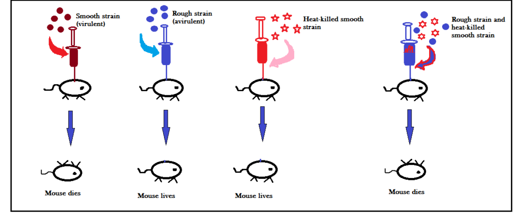
The bacterium chosen by Griffith (Streptococcus pneumoniae) had two strains R and S. R stands for rough and S stands for smooth. The rough strain was avirulent (did not cause pneumonia) and the smooth strain was virulent (caused pneumonia).

When smooth strain was injected in the mouse – the mouse died. On the other hand, when rough strain was injected in the mouse – the mouse survived. In the third experiment, when heat-killed smooth strain was injected in the mouse – the mouse survived. However, when rough strain and heat-killed smooth strain was injected in the mouse – the mouse couldn’t survive.
And when Griffith examined the dead mice body – he found that the body contained living R-type and living S-type bacteria. It means, the chemical present in heat-killed S-type bacteria transformed living R-type into S-type by forming capsule. Griffith called this chemical as TRANSFORMING PRINCIPLE. However, he couldn’t analyze the chemical to know whether the chemical was a carbohydrate, protein, or nucleic acid responsible for transformation.
The three scientists Avery, McLeod and McCarty worked together (1933-44) to determine the biochemical nature of transforming principle in Griffith’s experiment.
Avery, McLeod and McCarty Experiment in Search for the Genetic Material

In the first experiment, scientists took S-strain extract and mixed it with live R strain and enzyme that could destroy lipids present within the cell. The strain when given to the mouse, the mouse couldn’t survive. Transformation was seen because the live R type changed to S type; however, lipid was not responsible for this transformation.
In the second experiment, the S-strain was mixed with live R strain and enzyme that destroys polysaccaharides. When injected to the mouse – the mouse died. Transformation was seen because the live R type changed to S type; however, polysaccharide was not responsible for this transformation.
In the third experiment, the S-strain extract was mixed with live R strain and the enzyme responsible for destroying proteins. When injected to the mouse – the mouse died. Transformation was seen because the live R type changed to S type; however, protein was not responsible for this transformation.
In the fourth experiment, the S strain extract was mixed with R strain and the enzyme responsible for destroying proteins. When injected to the mouse – the mouse died. Transformation was seen because the live R type changed to S type; however, lipid was not responsible for this transformation.
In the fifth experiment, the S strain extract was mixed with R strain and the enzyme responsible for destroying DNA. When injected to the mouse – the mouse survived. Transformation was not seen because the live R type couldn’t change to S type. It means, DNA was responsible for transformation.
The experiments conducted by Avery, Mcleod and McCarty were definitive, but many scientists were still reluctant to accept DNA as the genetic material. Additional evidence to prove DNA as a genetic material was provided in 1952 by Hershey and Chase.
Hershey and Chase Experiment in Search for the Genetic Material
The scientists used T2 bacteriophage as their experimental material. T2 is a virus that infects bacteria. The principle behind the experiment was that the infecting T2 phage must inject into the bacterium the specific information that is responsible for the reproduction of new viral particles. If they could find out what material the phage injects into the host – they could determine the genetic material of the phage.
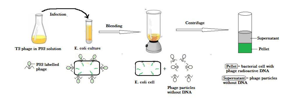
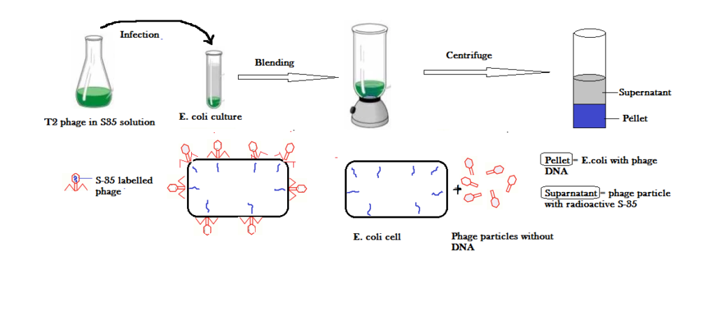
The T2 virus body consists of linear double stranded DNA and protein coat. It means, the virus has only two components i.e., DNA and protein. Scientists decided to label DNA and protein using radioisotopes of phosphorus and sulfur. They used P32 to label DNA as DNA consists of phosphorus and S35 radioisotope to label protein as protein consists of sulfur containing amino acids. Following experimental steps were performed:


- Radioactive labeling: In this step, T2 phages were kept in a separate flask containing P32 and S35 radioisotope. After some time, the DNA is labeled with P32 and the T2 protein coat is labeled with S35 radioisotope.
- Infection: The P32 labeled phage is put into the E. coli culture media for infection to take place. Likewise, the S35 labeled phageis also put into the E. coli culture media for infection to take place. They are then given sufficient time for infection.
- Blending / Agitation: After infection, the infected E. coli with phage particles are agitated in a kitchen blender so as to separate the empty phage particles from the bacterial cells.
- Centrifugation: After blending, the bacterial cells are completely separated from the phage ghosts and then measured the radioactivity in the two fractions. When P32-labeled phages were used to infect E. coli, most of the radioactivity was seen inside the bacterial cell indicating that the phage DNA entered into the cell. The radioactivity is seen in the pellet because bacterial cells are heavier than phage particles. And phage particles float as supernatant. On the other hand, when S35-labeled phages were used to infect E. coli – most of the radioactive material ended up in the phage ghosts indicating that the phage protein never entered the bacterial cell. In the centrifuge, the radioactivity is seen in the supernatant and not the pellet because the supernatant consists of radioactive phage particles.
Conclusion: This proves that DNA is the hereditary material. The phage proteins are just the structural packaging material that is not allowed to enter into the host after delivering viral DNA into the E. coli cell.

