
Biology › Molecular basis of inheritance › Structure of DNA
Structure of DNA (Deoxyribonucleic Acid)
Learn the structure of DNA from nucleotides and nitrogenous bases to the Watson-Crick double helix, Chargaff’s rules, base pairing, DNA vs RNA and how DNA is packed into chromosomes, with NCERT values for NEET, IB A1.2 links, diagrams, FAQs and a quick-check quiz.
1 Introduction: what is the structure of DNA?
Syllabus link: NCERT XII Molecular Basis of Inheritance (6.1 The DNA); IB A1.2 Nucleic acids
NCERT = value or statement as given in NCERT Class XII (use in NEET/CBSE answers) IB = IB Biology A1.2 link
What is the basic structure of DNA (deoxyribonucleic acid), and why does it matter so much? DNA is the genetic material of almost all living organisms. The structure of DNA explains how genetic information is stored, copied and passed on. This chapter explains the structure of DNA step by step, from a single nucleotide to a whole chromosome, with NCERT values for NEET and CBSE and links to IB Biology A1.2.
The structure of DNA was first approached through the extraction of nuclein by Friedrich Miescher. Later, it was identified as nucleic acid, a polymer of nucleotides. These nucleotides are composed of phosphate, sugar and nitrogenous bases. Watson and Crick’s model of DNA in 1953 demonstrated its double-helix form, a critical structure for heredity and genetic expression. DNA’s intricate design allows the encoding and transfer of genetic information essential for life.
In 1869, Miescher extracted a substance from the nuclei of pus cells (leucocytes). He named that substance NUCLEIN. At that time nobody knew it was the hereditary material; that was shown much later by Griffith (1928), Avery, MacLeod and McCarty (1944) and Hershey and Chase (1952). Later on, Richard Altmann studied the chemical nature of nuclein and confirmed that the substance is acidic in nature, and that is why he called nuclein nucleic acid (1889).
Key points on the structure of DNA
- DNA is a polymer (polynucleotide) of deoxyribonucleotides.
- Each nucleotide = phosphate + deoxyribose sugar + nitrogenous base.
- Two antiparallel strands form a right-handed double helix (Watson and Crick, 1953).
- A pairs with T and G pairs with C (complementary base pairing).
- In eukaryotes the DNA is packed with histones into nucleosomes and chromosomes.
2 Nucleotides: the building blocks of DNA
Syllabus link: NCERT XII 6.1.1 Structure of polynucleotide chain; IB A1.2.2-A1.2.3
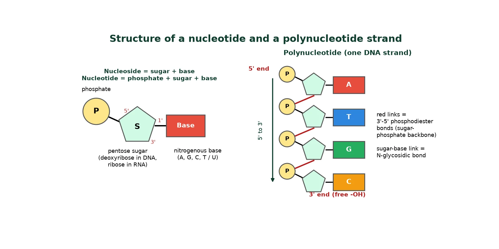
Nucleic acids are polymers of nucleotides. When we look at the structure of a nucleotide, it is made from three parts: a phosphate group, a pentose (5-carbon) sugar and a nitrogenous base.
Formula to remember
Nucleotide = Phosphate + Pentose sugar + Nitrogenous base
Nucleoside = Pentose sugar + Nitrogenous base

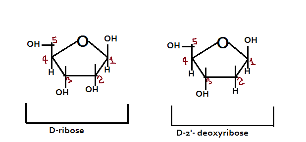
It is important to mention that there are two types of pentose sugar: D-ribose (found in RNA) and D-2′-deoxyribose (found in DNA). Deoxyribose has a hydrogen (-H) instead of a hydroxyl (-OH) group on carbon 2′, which is why it is called ‘deoxy’. The carbons of the sugar are numbered 1′ to 5′ to tell them apart from the atoms of the base.
IB IB A1.2 drawing tip
In IB exams, draw a nucleotide with a circle for phosphate, a pentagon for the sugar and a rectangle for the base. The phosphate joins carbon 5′ and the base joins carbon 1′. In a strand the phosphate of one nucleotide joins carbon 3′ of the next.
3 Nitrogenous bases in the structure of DNA
Syllabus link: NCERT XII 6.1.1; IB A1.2.4
There are two groups of nitrogenous bases: purines and pyrimidines.
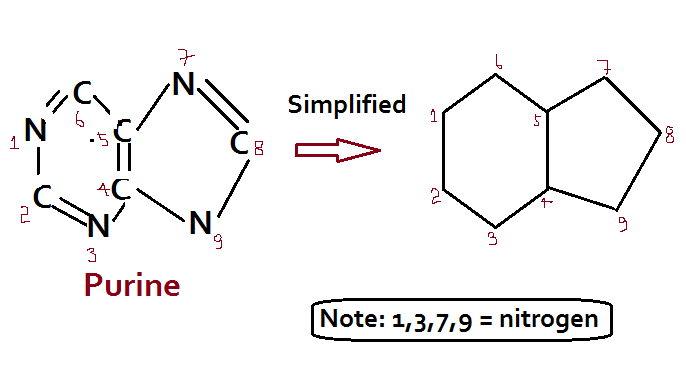
- Purines (adenine, A; guanine, G) have a double ring (a six-membered ring fused to a five-membered ring).
- Pyrimidines (cytosine, C; uracil, U; thymine, T) have a single six-membered ring.
- Mnemonic: CUT the Py (pyrimidines are C, U and T). ‘Pure As Gold’ for purines A and G.
- DNA contains A, G, C and T. RNA contains A, G, C and U (uracil replaces thymine; thymine is 5-methyluracil).
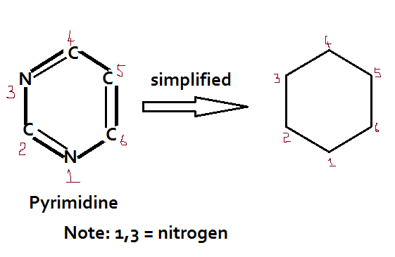
Attachment of bases in nucleotides
The nitrogenous bases are attached to the 1′ carbon of the sugar at the N-9 position of purines and the N-1 position of pyrimidines. The bond formed between the sugar and the nitrogenous base is the N-glycosidic bond in the beta configuration. When a phosphate is then linked to the 5′-OH of the nucleoside by a phosphoester bond, a nucleotide is formed.
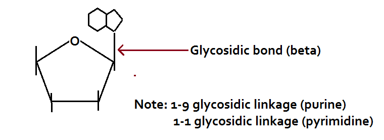
Nomenclature of nitrogenous bases
Each base gives its name to a nucleoside and a nucleotide. With ribose: adenosine, guanosine, cytidine and uridine. With deoxyribose: deoxyadenosine, deoxyguanosine, deoxycytidine and deoxythymidine. Adding phosphate gives, for example, deoxyadenosine monophosphate (dAMP).

4 Phosphodiester bonds: the backbone in the structure of DNA
Syllabus link: NCERT XII 6.1.1; IB A1.2.5 and A1.2.15 (AHL directionality)
When one nucleotide attaches to another, a bond is formed between the 3′-OH group of the sugar of the first nucleotide and the 5′-phosphate group of the next nucleotide. This 3′-5′ phosphodiester bond forms by a condensation reaction, releasing one molecule of water. Repeating this builds a polynucleotide with a sugar-phosphate backbone and bases sticking out to the side.
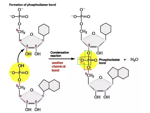
A polynucleotide therefore has polarity: one end has a free phosphate on carbon 5′ (the 5′ end) and the other has a free -OH on carbon 3′ (the 3′ end). The sequence is always written 5′ to 3′.
Worked question
Q. How many water molecules are released when 100 nucleotides polymerise into a single linear strand? (a) 50 (b) 49 (c) 99 (d) 100
Answer: (c) 99. Use (n – 1), where n is the number of nucleotides, because each new phosphodiester bond releases one water molecule. (For a double-stranded linear DNA of n base pairs, it is 2(n – 1); a circular molecule has one extra bond per strand.)
5 Discovering the structure of DNA: Franklin, Wilkins and Chargaff
Syllabus link: NCERT XII 6.1.2 Packaging and 6.1 (Chargaff, Wilkins, Franklin); IB A1.2.14 (AHL)
The three-dimensional structure of DNA was proposed by James Watson and Francis Crick in 1953. However, the basis of this structure was given by the work of other scientists, especially Rosalind Franklin, Maurice Wilkins and Erwin Chargaff.
Rosalind Franklin and Maurice Wilkins: X-ray diffraction
Rosalind Franklin worked at King’s College London in the same unit as Maurice Wilkins (as a colleague, not his assistant). She used X-ray diffraction to study wet fibres of DNA and made marked advances in the technique. Her famous ‘Photo 51’ (taken with Raymond Gosling) showed an X-shaped pattern that suggested several features of the structure of DNA:
- The molecule is a helix with a constant width of 2 nm (20 Å).
- The bases are stacked 0.34 nm (3.4 Å) apart.
- The pattern repeats every 3.4 nm (34 Å), so there are 10 base pairs in each turn.
- The phosphate groups lie on the outside of the molecule.
The length of one complete helical turn is called the pitch (3.4 nm in B-DNA).

Erwin Chargaff: base composition
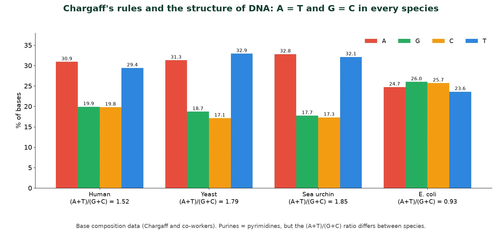
Chargaff pioneered many of the biochemical techniques for the isolation, purification and measurement of nucleic acids from living cells. It was known that DNA contained four nitrogenous bases: A, T, G and C. Chargaff analysed the base composition of DNA isolated from many different species. His results showed that:
Chargaff’s rules (double-stranded DNA)
% adenine = % thymine; % guanine = % cytosine
[A] + [G] = [T] + [C], so [A + G] / [T + C] = 1 (total purines = total pyrimidines)
The ratio [A + T] / [G + C] varies between species but is constant within a species.
For example, for humans [A + T] / [G + C] is about 1.52 (greater than 1, AT-rich), while for E. coli it is about 0.93 (close to 1). It is important to note that Chargaff’s rule holds only for double-stranded DNA; single-stranded DNA (as in phage φX174) and RNA do not follow it.
IB Nature of science (AHL)
Chargaff’s data falsified the earlier tetranucleotide hypothesis, which said DNA was a simple repeat of the four bases in equal amounts and so could not carry information. Watson and Crick then built physical models that fitted both Chargaff’s ratios and Franklin’s X-ray data, a good example of model building in science.
6 The Watson-Crick double helix: salient features
Syllabus link: NCERT XII 6.1.1; IB A1.2.6-A1.2.7
In 1953, Watson and Crick gave the three-dimensional double-helix model of DNA. The hallmark of this structure of DNA is that A always pairs with T and G with C, joined by hydrogen bonds. This finding fitted Chargaff’s data exactly. Watson, Crick and Wilkins received the Nobel Prize in Physiology or Medicine in 1962 for the discovery of the molecular structure of DNA. Franklin had died in 1958, and the prize is not given after death.

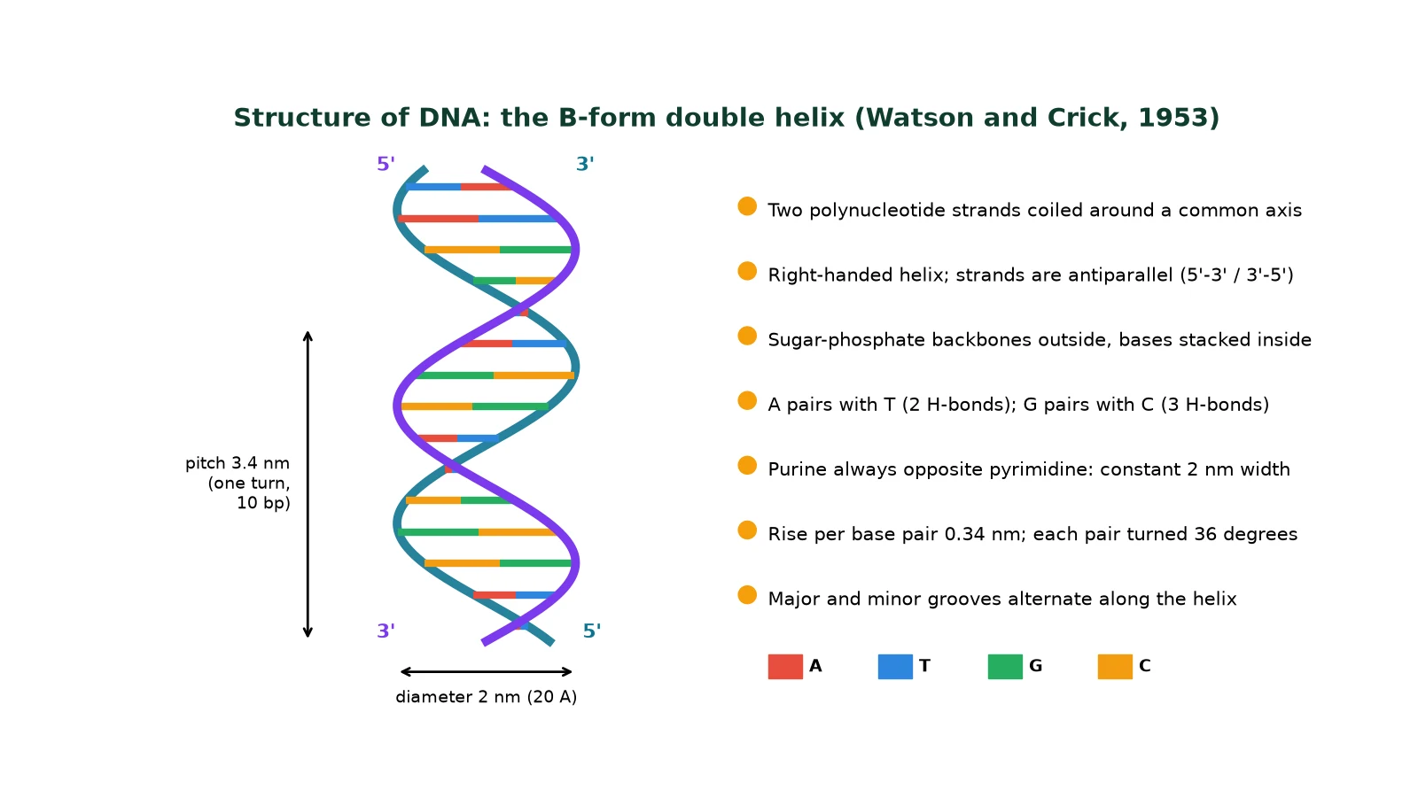
Salient features of the double-helix structure of DNA
- DNA is made of two polynucleotide chains. The sugar-phosphate forms the backbone, and the bases project inside.
- The two strands have antiparallel polarity: if one chain runs 5′ → 3′, the other runs 3′ → 5′.
- The bases of the two strands are paired by hydrogen bonds: adenine pairs with thymine by two hydrogen bonds, and guanine pairs with cytosine by three hydrogen bonds.
- A purine always pairs with a pyrimidine, so the distance between the two strands stays almost the same (about 2 nm) along the whole length.
- The two chains are coiled in a right-handed fashion. The pitch of the helix is 3.4 nm (34 Å), with about 10 bp per turn, so the distance between consecutive base pairs is about 0.34 nm.
- The plane of one base pair stacks over the next. This base stacking, with hydrogen bonds, stabilises the structure of DNA.
- The twisted strands create alternating major and minor grooves, where proteins can read the base sequence.
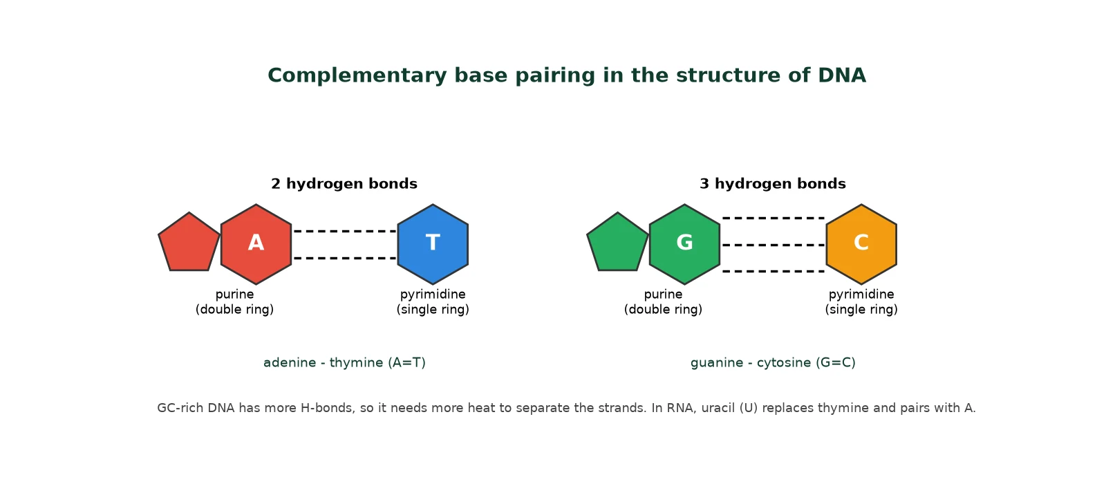
Common mistakes about the structure of DNA
- The H-bonds are between bases; the backbone bonds are covalent phosphodiester bonds.
- Antiparallel does not mean the strands are identical; they are complementary.
- NCERT uses 10 bp per turn; modern measurements give about 10.5 bp per turn in cells. Use 10 in NEET.
IB Why the structure of DNA fits its function
Complementary base pairing lets each strand act as a template, so DNA can be copied accurately (semi-conservative replication) and repaired. The sequence of bases can vary without limit, so a strand of n bases has 4n possible sequences, giving enormous storage capacity. The genetic code is shared by nearly all organisms, which is evidence for universal common ancestry.
7 Forms of DNA: A, B, C and Z
Syllabus link: Extension for NEET and competitive exams
DNA can adopt different forms depending on hydration, salt concentration and base sequence. The Watson-Crick structure of DNA is B-DNA, the main form in cells.
Swipe the table sideways to see all columns.
| DNA form | Helical diameter | Base pairs per turn | Helix rotation | Vertical rise per bp |
|---|---|---|---|---|
| A-DNA | about 23 Å | 11 | Right-handed | about 2.6 Å |
| B-DNA | 20 Å | 10 (10.5 in solution) | Right-handed | 3.4 Å |
| C-DNA | about 19 Å | 9.33 | Right-handed | 3.3 Å |
| Z-DNA | about 18 Å | 12 | Left-handed (zig-zag backbone) | about 3.7 Å |
Z-DNA, found in alternating GC sequences, is the only left-handed form.
8 DNA vs RNA: comparing the structure of DNA and RNA
Syllabus link: NCERT XII 6.1, 6.2; IB A1.2.8
The structure of DNA and RNA is similar: both are nucleic acids built from nucleotides linked by phosphodiester bonds, but they differ in structure and function.
Swipe the table sideways to see all columns.
| Feature | DNA | RNA |
|---|---|---|
| Sugar | 2′-deoxyribose | Ribose (has 2′-OH) |
| Bases | A, G, C, T | A, G, C, U |
| Strands | Usually double-stranded helix | Usually single-stranded (can fold on itself) |
| Base pairing | A-T and G-C | A-U and G-C (within folded regions) |
| Stability | Chemically stable; good for storage | 2′-OH makes it reactive and easily hydrolysed |
| Location (eukaryote) | Nucleus, mitochondria, chloroplasts | Made in the nucleus, works mostly in the cytoplasm |
| Main role | Stores genetic information | mRNA, tRNA, rRNA: protein synthesis; some act as enzymes (ribozymes) |
NCERT NCERT point
RNA was the first genetic material (the RNA world hypothesis). DNA evolved from RNA because the 2′-OH makes RNA reactive and less stable, and thymine (5-methyluracil) in place of uracil gives DNA extra stability. RNA can also act as a catalyst, but it mutates faster.
9 Packaging the structure of DNA in the cell
Syllabus link: NCERT XII 6.1.2 Packaging of DNA helix; IB A1.2.12 (AHL)
We know that the distance between two consecutive base pairs is 0.34 nm (0.34 x 10⁻⁹ m). With this information we can calculate the length of the DNA double helix in a cell: multiply the total number of base pairs by the distance between consecutive base pairs.
Worked calculation (NCERT)
The total number of base pairs in a diploid human cell is 6.6 x 10⁹ bp.
Length = 6.6 x 109 x 0.34 x 10-9 m = about 2.2 metres.
For E. coli (4.6 x 10⁶ bp): length = 4.6 x 106 x 0.34 x 10-9 m = about 1.36 mm.
NCERT gives the dimension of a typical nucleus as about 10-6 m (in reality most nuclei are a few micrometres, about 5-10 x 10-6 m). It seems impossible to fit DNA 2.2 m long into it, yet DNA is very well accommodated in the nucleus. How is this possible? The answer lies in DNA packaging.
Packaging in prokaryotes
Prokaryotes such as E. coli have no defined nucleus. Their DNA is not scattered throughout the cell: the negatively charged DNA is held with some positively charged proteins in a region called the nucleoid, organised in large loops.
Histone and non-histone proteins
In eukaryotes, DNA is packaged in a very complex manner. Two types of protein are required: histone proteins and non-histone chromosomal (NHC) proteins.
- Histone proteins are of low molecular weight and basic in nature (positively charged). They are rich in the basic amino acids lysine and arginine, whose positive side chains bind the negative phosphate groups of DNA.
- Non-histone chromosomal proteins are of higher molecular weight and mostly acidic in nature (negatively charged), being rich in acidic amino acids such as aspartic acid and glutamic acid.
- There are five kinds of histone: H1, H2A, H2B, H3 and H4. H1 is very lysine-rich, H2A and H2B are slightly lysine-rich, and H3 and H4 are arginine-rich.
- Non-histone proteins include structural or scaffold proteins (help the higher-order coiling of DNA), enzymes such as DNA polymerase (DNAP) and RNA polymerase (RNAP), and regulatory proteins such as the high mobility group (HMG) proteins, involved in gene expression.
Q. How do regulatory proteins help in gene expression?
DNA is negatively charged and histones are positively charged, so DNA wound tightly around histones is hard for RNA polymerase to reach, and the genes there are not expressed. Regulatory proteins and enzymes chemically modify histones (for example by acetylation, which neutralises their positive charge). This loosens the grip of histones on DNA, opens the chromatin and lets the genes be transcribed. Removing the modification closes the chromatin again. (IB links this to epigenetics.)
10 Mechanism of DNA packaging: nucleosome to chromosome
Syllabus link: NCERT XII 6.1.2; IB A1.2.12 (AHL)
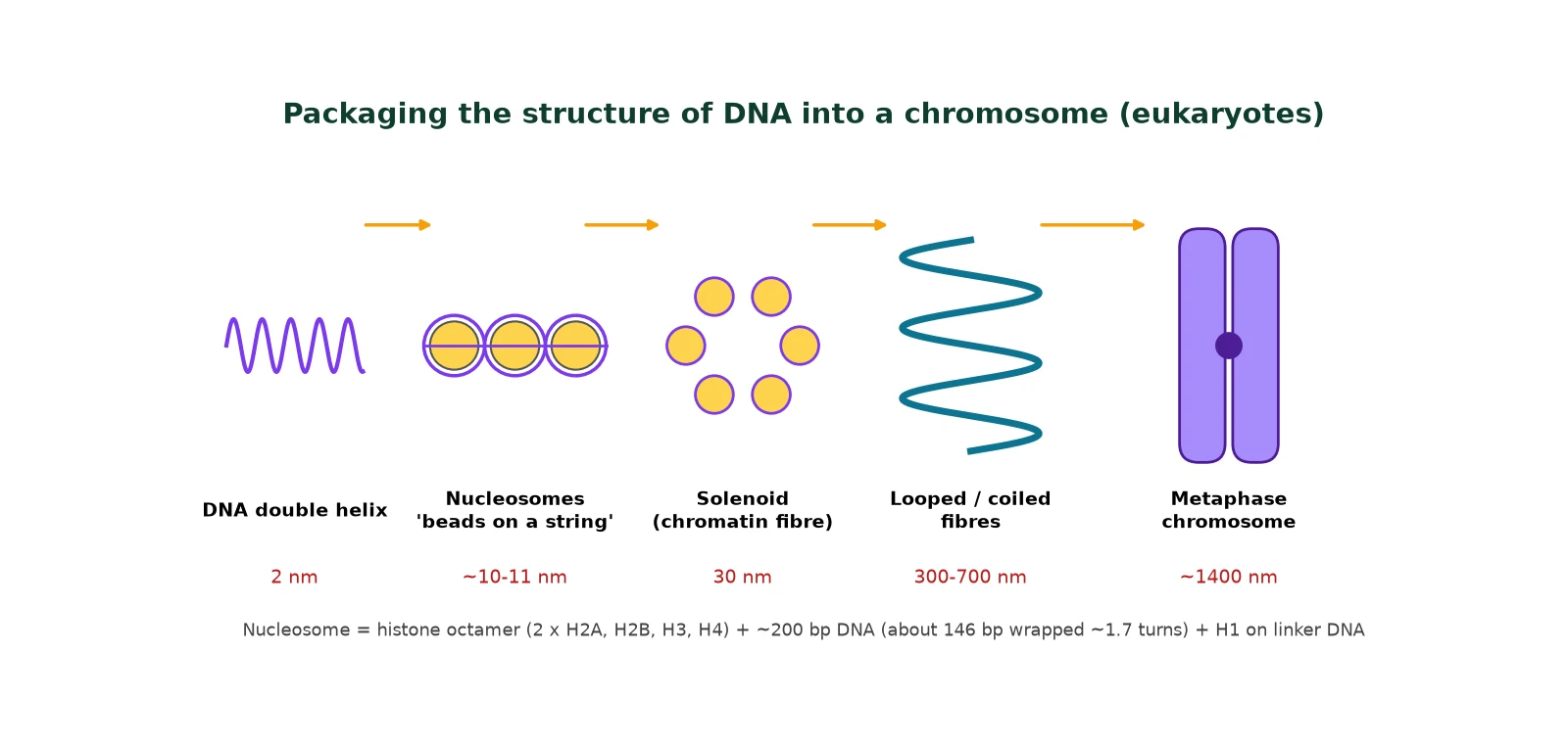
Of the five histones, four (H2A, H2B, H3 and H4) are present in two sets each to form a histone octamer, also called the nu body or core particle. The DNA molecule is wrapped around the octamer about 1¾ turns (about 146-147 bp). The H1 histone binds the linker DNA where it enters and leaves, sealing (clipping) the DNA on the core. Together these form a nucleosome (DNA + histone octamer + H1).
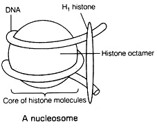
- A typical nucleosome contains about 200 bp of DNA (core plus linker).
- The nucleosome core is about 10-11 nm across and about 6 nm high.
- Repeating nucleosomes give the ‘beads-on-string’ structure seen in the electron microscope, which forms chromatin.
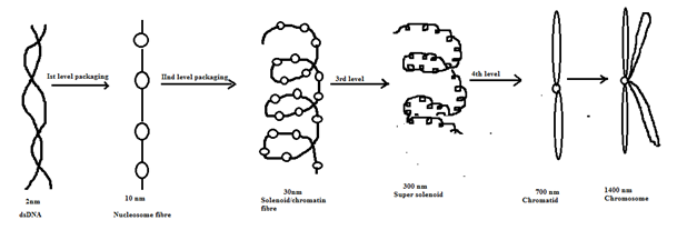
Levels of coiling
- 1st level: the 2 nm DNA helix wraps histone octamers to form the 10 nm nucleosome fibre (beads on a string).
- 2nd level: nucleosomes coil into the 30 nm solenoid chromatin fibre, helped by H1.
- 3rd level: the solenoid forms loops on a scaffold of non-histone proteins, giving a looped fibre of about 300 nm.
- 4th level: further coiling gives a chromatid of about 700 nm diameter.
- After replication, two sister chromatids form a metaphase chromosome of about 1400 nm diameter.
Euchromatin and heterochromatin
Swipe the table sideways to see all columns.
| Feature | Euchromatin | Heterochromatin |
|---|---|---|
| Packing | Loosely packed | Densely packed |
| Staining | Stains light | Stains dark |
| Transcription | Active | Largely inactive |
Original question
Q. Which of the following is true for the two types of chromatin? (a) Heterochromatin is darkly stained while euchromatin is lightly stained (b) Heterochromatin is tightly packed while euchromatin is loosely packed (c) Heterochromatin is transcriptionally inactive while euchromatin is transcriptionally active (d) All of the above
Answer: (d).
11 FAQs on the structure of DNA
What is the basic structure of DNA?
DNA is a double helix of two antiparallel polynucleotide strands. Each strand has a sugar-phosphate backbone, with bases pointing inward and paired A-T and G-C by hydrogen bonds.
Who discovered the double-helix structure of DNA?
James Watson and Francis Crick proposed it in 1953, using X-ray diffraction data from Rosalind Franklin and Maurice Wilkins and the base ratios of Erwin Chargaff.
What are the dimensions in the structure of DNA (B-DNA)?
Diameter 2 nm, 0.34 nm between base pairs, 10 bp per turn and a pitch of 3.4 nm (NCERT values).
Why does G-C pairing make DNA more stable?
G-C pairs have three hydrogen bonds, while A-T pairs have two. GC-rich DNA therefore needs more energy (a higher melting temperature) to separate.
What does antiparallel mean?
The two strands run in opposite directions: one 5′ to 3′ and the other 3′ to 5′.
Does Chargaff’s rule apply to RNA?
No. Chargaff’s rule applies to double-stranded DNA. RNA and single-stranded DNA do not show A = T (or A = U) and G = C.
What is a nucleosome made of?
A histone octamer (two each of H2A, H2B, H3 and H4), about 200 bp of DNA wound around it and linker DNA, and histone H1.
How long is the DNA in a human cell?
About 2.2 m: 6.6 x 109 bp x 0.34 x 10-9 m per bp.
12 Quick check on the structure of DNA
Which base is a purine?
- (a) Cytosine
- (b) Guanine
- (c) Thymine
- (d) Uracil
Show answer
Answer: (b) Purines are adenine and guanine (double ring).
If a DNA sample has 20% adenine, what percentage is cytosine?
- (a) 20%
- (b) 40%
- (c) 60%
- (d) 30%
Show answer
Answer: (d) A = T = 20%, so G + C = 60% and C = 30%.
The bond between the sugar and the base is a(n):
- (a) N-glycosidic bond
- (b) phosphodiester bond
- (c) hydrogen bond
- (d) peptide bond
Show answer
Answer: (a) The base attaches to C1′ by an N-glycosidic bond.
Which histone is NOT part of the nucleosome octamer?
- (a) H2A
- (b) H3
- (c) H1
- (d) H4
Show answer
Answer: (c) H1 binds linker DNA outside the octamer.
13 Conclusion
In conclusion, the structure of DNA plays a fundamental role in the transmission of genetic information, as first outlined by Watson and Crick’s model in 1953. The double-helix structure of DNA ensures the precise encoding and replication of genetic material, which is vital for life. Understanding the structure of DNA continues to be essential in fields such as genetics, medicine and biotechnology, providing insights into heredity and disease mechanisms. Read more on DNA on Wikipedia.
© competitorspoint.com · Biology notes. New diagrams are original to competitorspoint.com.
Ready to practise? Download the Structure of DNA IB and NEET question set with markscheme (50 marks, free for signed-up members).
⬇ Download practice questions (PDF)