
IB Biology › Theme A › A2.3 Viruses
Viruses: IB Biology A2.3
Viruses are non-cellular infectious particles made of a nucleic acid genome inside a protein coat, and they can reproduce only inside a living host cell. In the IB Biology course (first assessment 2025), A2.3 is additional higher level (AHL) material, so it is examined only at HL.
Contents
- Why viruses are a special case
- A2.3.1 Structural features common to viruses
- A2.3.2 Diversity of structure in viruses
- Three guide examples: lambda, coronavirus and HIV
- How viruses depend on host cells
- A2.3.3 The lytic cycle of bacteriophage lambda
- A2.3.4 The lysogenic cycle of bacteriophage lambda
- Lytic versus lysogenic: comparing the two cycles
- A2.3.5 Evidence for several origins of viruses
- A2.3.6 Rapid evolution in viruses
- Evolution of influenza viruses
- Evolution of HIV
- Consequences for treating viral diseases
- Data-based practice: phage growth and viral evolution
- Exam traps and examiner tips
- Key terms flip cards
- Self-check: multiple-choice questions
- Structured exam questions with model answers
- References and further reading
- Common doubts about viruses
Why viruses are a special case in biology HL
Almost everything else in the IB course is about cells. Cells take in nutrients, release energy by respiration, make their own proteins and divide. Viruses do none of these things on their own. A virus particle outside a host is chemically inert: it has no metabolism, takes in nothing and gives out nothing. Only when it enters a suitable host cell does its genome take over part of the cell’s machinery and direct the production of new virus particles. That is why viruses are usually described as non-cellular (acellular) and why most biologists do not class them as living organisms, even though they carry genes, mutate and evolve by natural selection.
The two guiding questions for this topic are how can viruses exist with so few genes? and in what ways do viruses vary? The first is answered by obligate parasitism: a virus can manage with a tiny genome because the host supplies energy, raw materials, ribosomes and most enzymes. Influenza viruses carry roughly ten to fourteen protein-coding genes on eight RNA segments, whereas even the smallest free-living bacteria need several hundred genes. The second question leads to one of the most interesting ideas in the topic: viruses are so varied in genome type, structure and life cycle that they probably did not all evolve from one common ancestor.
How this chapter is organised
Each H2 section matches one understanding from the IB guide (A2.3.1 to A2.3.6), followed by data-based practice, exam traps, flip cards, self-marking questions and structured questions with model answers. Open any “Reveal” box to check your thinking before reading on.
A2.3.1 Structural features common to viruses HL
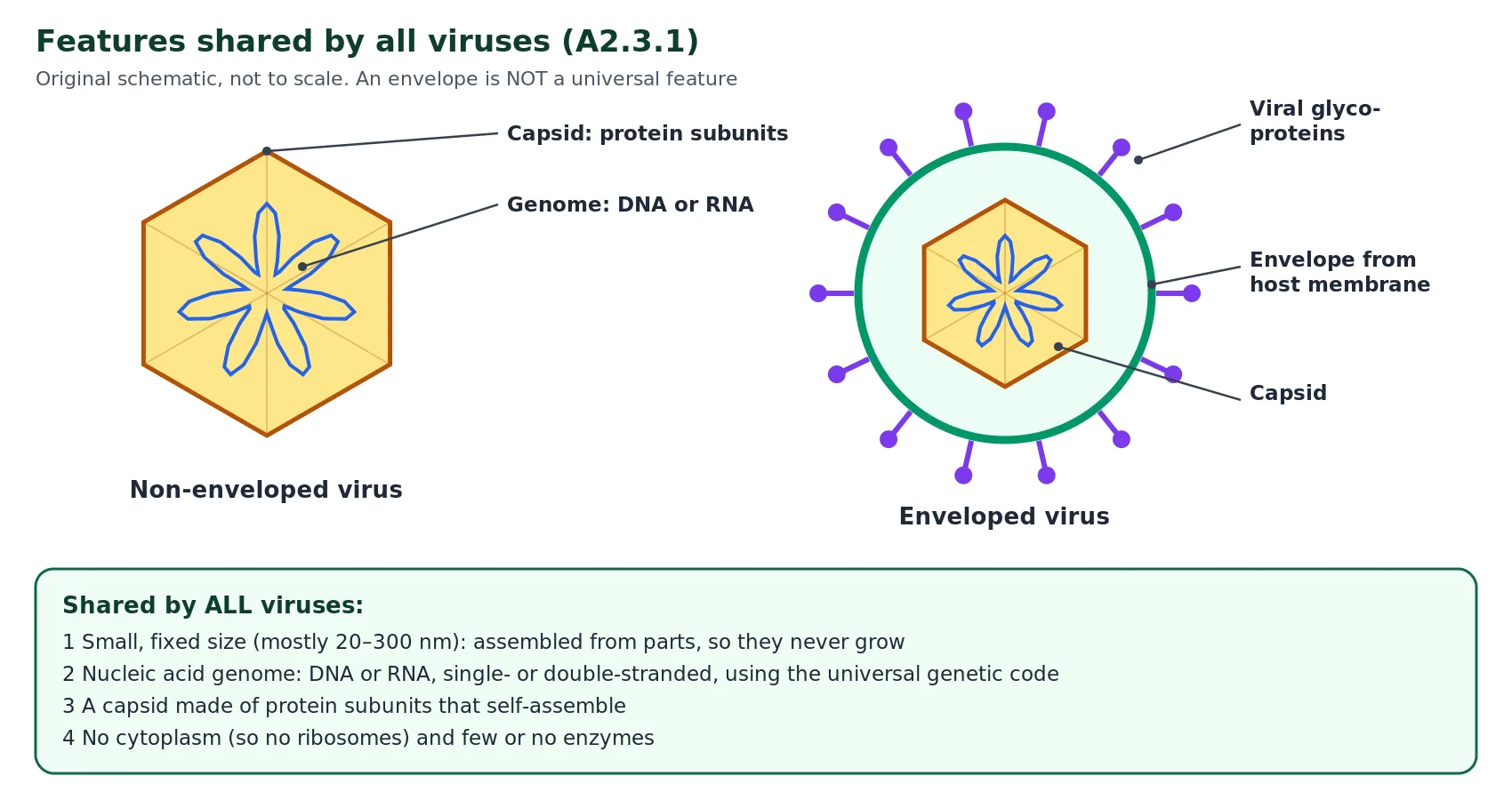
Because viruses are so diverse, the list of features shared by every virus is short. The IB guide names five, and they are worth learning as a set because examiners often ask you to outline them. First, viruses are small and have a fixed size. Most are between about 20 and 300 nanometres across, which is smaller than almost all bacteria and far smaller than a plant or animal cell. A virus does not grow after it is made; it is assembled inside the host from a set number of pre-made parts, rather like a model being clicked together, so the particle is full size as soon as assembly is complete.
Second, every virus has nucleic acid as its genetic material. This may be DNA or RNA, but a particle contains one type, not both. Viral genes use the same genetic code as cells, which is essential because viral proteins are made by the host cell’s ribosomes and tRNA. Third, the nucleic acid is enclosed in a capsid made of protein. The capsid is built from many copies of one or a few kinds of protein subunit (capsomeres). These subunits self-assemble into regular, symmetrical shapes such as icosahedra or helical rods, which is why viruses look so geometric under the electron microscope compared with the irregular outlines of cells.
Fourth, viruses have no cytoplasm. There is no cytosol, no ribosomes and no organelles inside a virus particle. Finally, viruses contain few or no enzymes. The small number of viral enzymes that do exist are linked to infection and replication of the viral genome, for example reverse transcriptase and integrase in HIV, or the enzymes that a phage uses to break open its host. Note what is not on the list: an envelope, a tail, spikes and DNA are all found in some viruses but not in others.
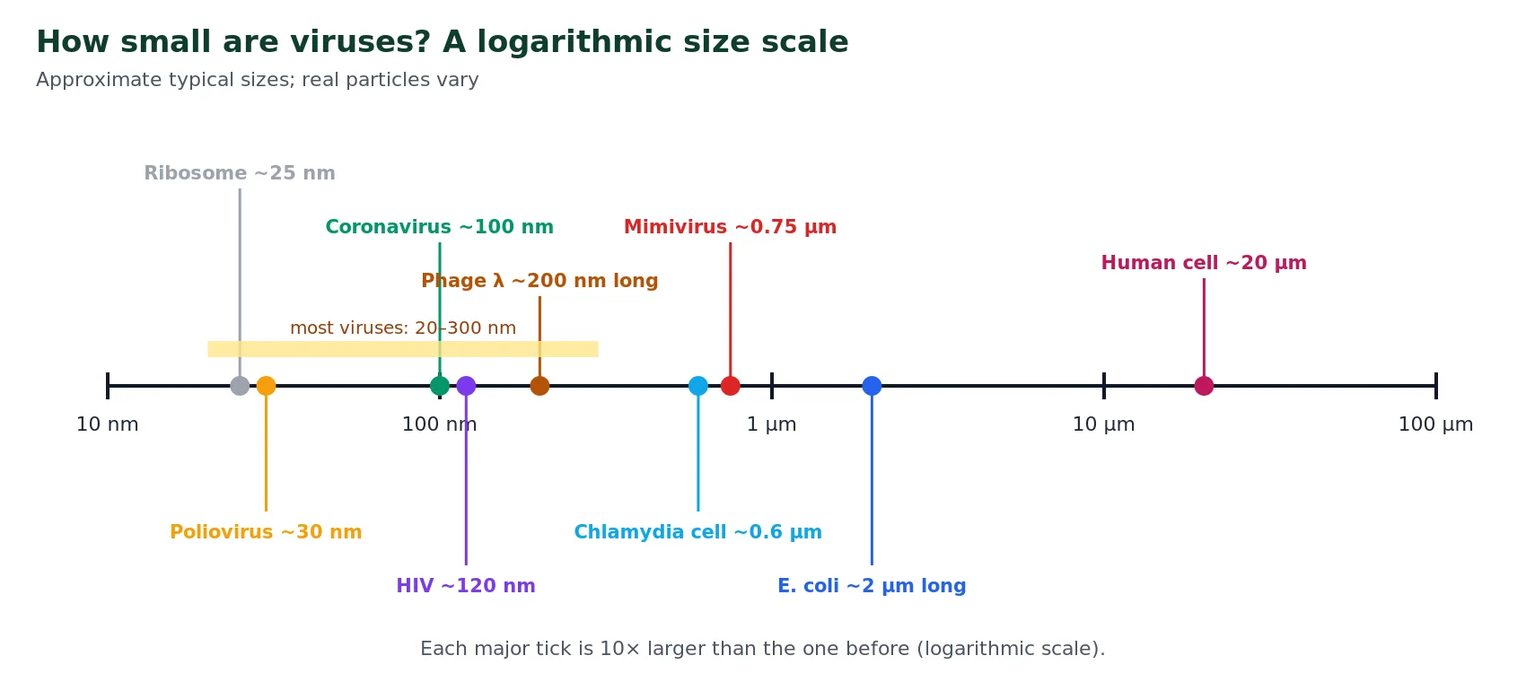
The size scale also shows why viruses could not be seen until the electron microscope was invented. A light microscope cannot resolve objects much smaller than about 200 nm, so most virus particles are below its limit of resolution. (For the cell side of this comparison, see Introduction to Cells (IB A2.2).) Size alone is not a reliable way to separate viruses from cells, however: giant viruses such as mimivirus, which infects amoebae, are larger than some small bacteria such as Chlamydia. The real distinction is structural and functional: a bacterium has cytoplasm, ribosomes and its own metabolism, whereas mimivirus has none of these and still depends on a host cell.
Reveal: Why must a virus be smaller than its host cell?
A virus has to get its genome inside the host cell, and many viruses enter whole or inject their genome through a small region of the cell surface. New particles are also assembled inside the host, so many of them must fit within one cell before release. A small particle with no cytoplasm is also cheap to make: the host can produce hundreds of copies from its resources.
A2.3.2 Diversity of structure in viruses HL
Beyond the shared features, viruses are astonishingly varied, and this diversity is what the IB expects you to appreciate. The most fundamental difference is in the genetic material. A viral genome may be DNA or RNA, and either type can be single-stranded or double-stranded. Genomes may be linear or circular, may be one molecule or split into several segments, and range from a few thousand bases to more than a million base pairs in giant viruses. No single gene is found in every virus, which is unlike cells, where hundreds of genes (for ribosomal RNA, ribosomal proteins and DNA polymerase, for example) are shared across all domains.
Single-stranded RNA viruses show how much variety can exist even within one group. In a positive-sense RNA virus, such as a coronavirus, the genome has the same base sequence as messenger RNA and can be translated directly by host ribosomes on arrival. In a negative-sense RNA virus, such as influenza, the genome is complementary to mRNA, so the virus must carry its own RNA polymerase to transcribe mRNA before any viral protein can be made. Retroviruses such as HIV take a third route: they use reverse transcriptase to copy their RNA into double-stranded DNA, which is inserted into a host chromosome and then transcribed like a host gene.
The second major difference is whether a virus is enveloped or non-enveloped. In enveloped viruses, the capsid is surrounded by a membrane. Its phospholipid bilayer, with the same fluid mosaic structure described in Cell Membrane (IB B2.1), comes from the host cell, usually taken from the plasma membrane as new particles bud out, although some viruses acquire their envelope from internal membranes. The proteins embedded in the envelope, which are mostly glycoproteins, are coded for by viral genes. These glycoproteins bind to specific receptors on the next host cell, which helps to explain why each virus infects only certain cell types. Enveloped viruses are particularly common among those that infect animal cells, whereas most viruses of bacteria and plants are non-enveloped, with the protein capsid as their outer surface.
Viruses also differ in capsid shape (icosahedral, helical, or complex with a head and tail), in the type of host (bacteria, archaea, protists, fungi, plants or animals) and in how they leave the host cell: some burst the cell open (lysis), while many enveloped viruses bud out through the plasma membrane. A virus that infects bacteria is called a bacteriophage, or phage for short.
The IB guide names three examples to illustrate diversity. Between them they cover a DNA virus and two RNA viruses, a non-enveloped virus and two enveloped ones, and hosts from bacteria to human immune cells. Use the tabs below to compare them, then study each labelled diagram.
Bacteriophage lambda (λ) infects the bacterium Escherichia coli. Its genome is one linear molecule of double-stranded DNA (48 502 base pairs) with short single-stranded “sticky” ends. It is non-enveloped: an icosahedral protein head is joined to a long, flexible tail that ends in a tail fibre. Lambda can follow either the lytic or the lysogenic cycle.
Coronaviruses, including SARS-CoV-2, which causes COVID-19, infect animal cells. The genome is one molecule of single-stranded, positive-sense RNA of about 30 000 bases, among the largest RNA genomes known. The RNA is coated with nucleocapsid protein inside an envelope studded with spike glycoproteins that give the crown-like outline. SARS-CoV-2 is a zoonosis: it most probably passed to humans from another mammal.
HIV (human immunodeficiency virus) infects helper T cells and other cells carrying the CD4 receptor. Each particle carries two copies of single-stranded, positive-sense RNA (about 9 700 bases) inside a cone-shaped capsid, with the enzymes reverse transcriptase and integrase. It is enveloped; the envelope glycoprotein gp120 binds CD4. As a retrovirus, it copies its RNA into DNA that is integrated into a host chromosome.
| Feature | Phage lambda | Coronavirus | HIV |
|---|---|---|---|
| Nucleic acid | DNA | RNA | RNA (copied to DNA in host) |
| Strands | Double | Single (+) | Single (+), two copies |
| Envelope | No | Yes | Yes |
| Capsid | Icosahedral head + tail | Helical nucleocapsid | Cone-shaped |
| Host | E. coli | Mammal (and bird) cells | Human CD4 cells |
| Key enzyme carried | None needed at entry | RNA polymerase coded in genome | Reverse transcriptase, integrase |
| Leaves host by | Lysis | Exocytosis/budding | Budding |
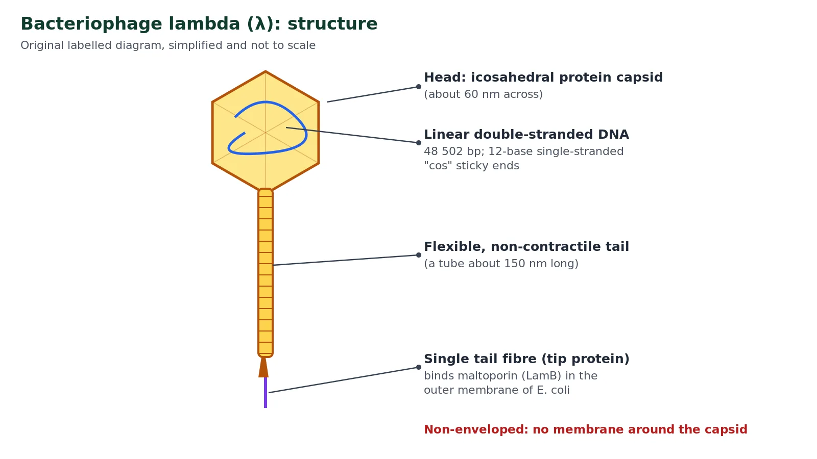
Label practice: phage lambda
Without looking at Figure 3, choose the correct label for each numbered part, then press Check labels. (1) The protein structure at the top that holds the genome; (2) the molecule inside it; (3) the long hollow tube; (4) the structure at the very tip that recognises the host.
Show the answers
1 icosahedral protein head (capsid); 2 double-stranded DNA; 3 flexible, non-contractile tail tube; 4 tail fibre that binds maltoporin (LamB). Lambda has no envelope and no RNA.
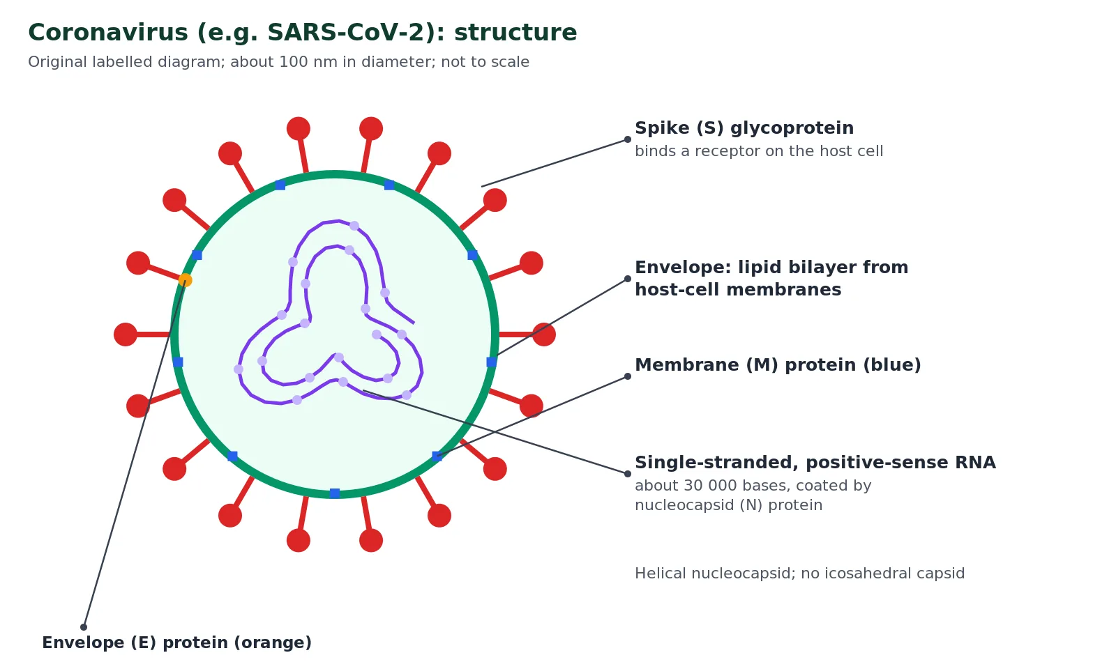
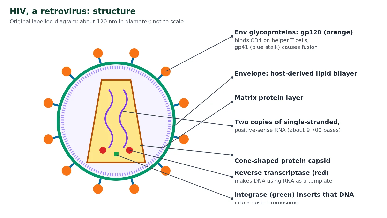
How viruses depend on host cells HL
Before looking at the life cycles, it helps to be clear about what the host provides, because the IB guide asks you to appreciate that viruses rely on a host cell for energy supply, nutrition, protein synthesis and other life functions. A virus particle carries no ATP and cannot make any; the energy for copying the viral genome and for building proteins comes from the host cell’s respiration. The nucleotides used to copy viral DNA or RNA and the amino acids used to make capsid proteins come from the host’s own pools or from the breakdown of host molecules. Viral proteins are translated on host ribosomes using host tRNA, aminoacyl-tRNA synthetases and translation factors.
This dependence explains the answer to the first guiding question. A virus needs genes only for the things that the host cannot or will not do for it: building the capsid, getting into and out of the cell, and, in some viruses, copying an unusual genome (an RNA genome or an RNA-to-DNA step) or controlling which host genes are switched on. Everything else is borrowed. Viruses with larger genomes, such as the poxviruses and mimivirus, carry genes for more of these functions themselves and are correspondingly less dependent on their hosts, but even they cannot make ribosomes or generate ATP.
Exam link
If a question asks why viruses are called obligate intracellular parasites, give the reason as well as the label: they can only reproduce inside a host cell because they lack the ribosomes, enzymes and energy-releasing metabolism needed to make proteins and copy their genome.
A2.3.3 The lytic cycle of bacteriophage lambda HL
In a lytic cycle the virus reproduces inside the host cell and then bursts it open (lysis), killing it and releasing many new virus particles. The IB guide asks you to use bacteriophage lambda as the example, so learn the stages specifically for lambda and E. coli. Use the stepper to work through the sequence one stage at a time; if scripts are switched off, all the stages are shown as a list.
Stepper: lytic cycle of lambda
-
1. Attachment (adsorption)
Proteins at the tip of lambda’s tail bind to maltoporin (LamB), a protein channel in the outer membrane of E. coli that normally lets maltose and other sugars into the cell. This binding is specific: bacteria lacking this protein are not infected.
-
2. Entry of the genome
Lambda’s linear DNA passes through the hollow tail and across the bacterial membranes into the cytoplasm. The empty protein head and tail stay outside the cell; only the genome enters.
-
3. Circularisation
The two single-stranded “sticky” (cos) ends of the DNA are complementary, so they base-pair and the gap is sealed by host DNA ligase. The genome becomes a circle. At this point lambda “decides” between the lytic and lysogenic routes.
-
4. Replication of viral DNA
Using host nucleotides and enzymes together with a few viral proteins, the lambda DNA is copied many times. Later in the cycle a rolling-circle method produces long chains of repeated genomes that are cut into single genome lengths during packaging.
-
5. Transcription and translation of viral genes
Viral genes are transcribed by host RNA polymerase and the mRNA is translated on host ribosomes. Early proteins help with DNA replication and control; later, large amounts of head and tail proteins and lysis proteins are made. Host ATP and amino acids are consumed throughout.
-
6. Self-assembly
Head proteins assemble into empty capsids, a single genome length of DNA is pumped into each head, and separately assembled tails are attached. No energy-releasing reactions of the virus are involved: the host provides the ATP.
-
7. Lysis and release
Viral holin proteins make holes in the inner membrane, allowing a viral endolysin to digest the peptidoglycan cell wall; other proteins disrupt the outer membrane. The cell bursts and releases about 100 new phages, which can infect neighbouring bacteria.
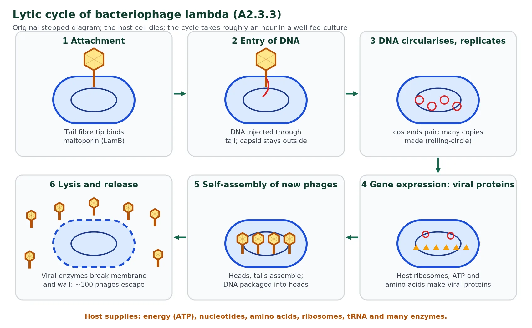
A phage following the lytic cycle is described as virulent because it kills its host. This has a cost for the virus: every round destroys the cells it depends on, so the phage can persist only if it keeps finding new, uninfected hosts. If a phage destroyed an entire host population it would have nowhere left to reproduce. Many viruses of plants and animals also reproduce in a lytic way, spreading from cell to cell within the host organism, but in a multicellular host the immune system usually brings the infection under control.
A2.3.4 The lysogenic cycle of bacteriophage lambda HL
Lambda is a temperate phage, meaning it can also follow a quieter route, the lysogenic cycle, again using lambda as the IB example. Attachment, entry and circularisation happen exactly as in the lytic cycle. If conditions favour lysogeny, a viral repressor protein (CI) is made that switches off the genes needed for the lytic cycle. The viral enzyme integrase then inserts the circular lambda DNA into the bacterial chromosome at one specific site, by recombination between a short sequence in the phage DNA and a matching sequence in the host DNA. The integrated viral DNA is now called a prophage, and the bacterium carrying it is a lysogen.
While it is a prophage, lambda makes no new virus particles and does not harm its host. Every time the bacterium replicates its chromosome before binary fission, the prophage is copied too, so both daughter cells inherit it. In this way the viral genome can be passed down through many generations of bacteria, spreading vertically with its host rather than by infection. The repressor protein also makes the lysogen immune to infection by other lambda phages, because it switches off the incoming genomes in the same way.
The lysogenic state is not permanent. If the host cell is stressed, particularly if its DNA is damaged (for example by ultraviolet radiation), the lambda repressor is destroyed. The lytic genes are then expressed, the prophage is cut out of the chromosome, and the virus enters the lytic cycle, eventually lysing the cell. This is called induction. In effect, the virus “abandons ship” when its host looks unlikely to survive.
Stepper: lysogenic cycle of lambda
-
1. Attachment and entry
As in the lytic cycle: tail proteins bind maltoporin and the DNA enters the cell.
-
2. Circularisation and repression
The cos ends join to form a circle. The viral repressor protein is produced and blocks transcription of the lytic genes.
-
3. Integration
Viral integrase inserts the lambda DNA at one specific site in the E. coli chromosome. It is now a prophage.
-
4. Replication with the host
The prophage is replicated with the bacterial chromosome and inherited by both daughter cells at every division. The host is not harmed.
-
5. Induction
DNA damage or other stress inactivates the repressor. The prophage is excised and the lytic cycle begins, ending in lysis.
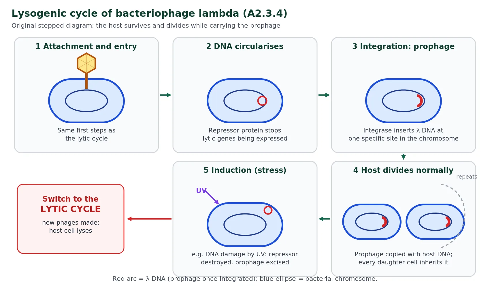
Lysogeny can even benefit the host. Prophages sometimes carry genes picked up from a previous host or genes of their own that give the bacterium a new characteristic. Movement of genes between bacteria by phages increases genetic diversity in bacterial populations and can speed up their evolution.
Reveal: Why might lysogeny be favoured when there are few bacteria nearby?
If hosts are scarce, new phages released by lysis may not find a cell to infect and will be wasted. By integrating as a prophage, the viral genome is safely replicated each time the host divides, so it spreads with the bacterial population until conditions improve. In lambda, high numbers of phage per cell and poor nutrient conditions for the host tend to favour lysogeny.
Lytic versus lysogenic: comparing the two cycles HL
A comparison question will expect differences and at least one similarity. Both cycles start with the same attachment, entry and circularisation steps, and both depend on host enzymes. The table summarises the key contrasts, and the sorting activity lets you test yourself.
| Feature | Lytic cycle | Lysogenic cycle |
|---|---|---|
| Fate of host cell | Killed by lysis | Survives and divides |
| Viral DNA | Stays separate; copied many times | Integrated into host chromosome as a prophage |
| New virus particles | About 100 released per cell | None while lysogenic |
| Key viral protein | Lysis proteins (holin, endolysin) | Repressor and integrase |
| How the viral genome spreads | Infecting new cells (horizontal) | Inherited by daughter cells (vertical) |
| Phage described as | Virulent | Temperate |
| Similarity | Same attachment to maltoporin, DNA entry and circularisation; both use host enzymes, nucleotides and energy | |
Sorting activity: lytic, lysogenic or both?
Choose a category for each statement, then press Check.
Show the answers
Lysogenic: inserted into chromosome; copied whenever host divides; integrase. Lytic: host bursts; about 100 phages released; head and tail proteins made in quantity. Both: binding maltoporin; DNA becomes circular.
A2.3.5 Evidence for several origins of viruses from other organisms HL
All cellular life shares hundreds of genes and the same basic biochemistry, which is strong evidence that all cells descend from one last universal common ancestor (LUCA). Viruses are different. There is no gene present in all viruses, and they vary in almost every structural feature: DNA or RNA, single- or double-stranded, enveloped or not, icosahedral, helical or tailed. The IB guide makes the point directly: the diversity of viruses suggests several possible origins, rather than one ancestral virus.
If viruses did evolve several times, how can they share any features at all? The guide’s answer is convergent evolution. All viruses share an extreme form of obligate parasitism: they must get a genome into a host cell, have it copied by the host, and package the copies for transfer to the next cell. That shared way of life creates the same selection pressures, so unrelated lineages could independently arrive at the same solutions, such as a protective protein capsid, small size and the loss of cytoplasm and metabolism. These are similarities of function, not of ancestry, just as the wings of birds and insects are similar because both fly.
A further piece of evidence is that viruses use the same genetic code as living organisms. This fits best with the idea that viruses arose from cells, or at least evolved alongside them, because a virus whose genes used a different code could not have its proteins made by host ribosomes. Three main hypotheses try to explain where viruses came from, and they are not mutually exclusive; different groups of viruses may have arisen in different ways.
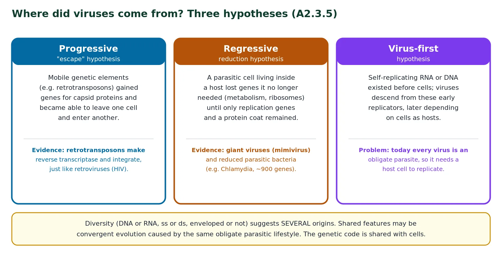
The progressive or “escape” hypothesis proposes that viruses were built up step by step from pieces of host genetic material that gained the ability to move between cells. The best evidence comes from retrotransposons, sequences that make up a large part of eukaryotic genomes. A retrotransposon is transcribed to RNA, its own reverse transcriptase makes a DNA copy, and the copy is inserted at a new place in the chromosomes. This is strikingly similar to how retroviruses such as HIV copy and integrate their genomes. A retrotransposon that acquired genes for capsid and envelope proteins would, in effect, be a retrovirus.
The regressive or reduction hypothesis proposes that viruses descend from cells that became parasites inside other cells and then lost more and more of their own functions until only the genes needed for replication and packaging remained. Two lines of evidence support it. First, some bacteria are already well along this path: Chlamydia can reproduce only inside host cells and has a genome of about one million base pairs with fewer than 900 protein-coding genes, far fewer than most free-living bacteria. Second, giant viruses such as mimivirus have genomes of about 1.2 million base pairs and code for some proteins involved in translation, which looks like the remains of a once more independent organism.
The virus-first hypothesis suggests that self-replicating RNA or DNA molecules existed before the first cells and that viruses descend from them. Its main weakness is that every virus known today needs a host cell to reproduce, so it is hard to see how viruses could have existed before cells. When evaluating the hypotheses, notice that the shared genetic code and obligate parasitism favour origins from cells, while the enormous diversity favours several independent origins rather than one.
Reveal: How would you use evidence to evaluate the regressive hypothesis?
For: obligate intracellular bacteria such as Chlamydia show that reduction of a cell towards dependence on a host really happens; giant viruses carry genes for functions most viruses lack. Against or limitations: no known intermediate form links a cell to a typical small virus; many small RNA viruses have no genes that look like the remains of a cell; the hypothesis does not explain RNA viruses well. A balanced conclusion is that reduction may explain some large DNA viruses but probably not all viruses.
A2.3.6 Rapid evolution in viruses HL
Viruses are not alive in the usual sense, but they have genes, they vary and they are subject to natural selection, so they evolve. Some viruses evolve so fast that heritable changes can be detected within one infected person over a few months. The guide asks for reasons for very rapid rates of evolution, and three are usually given.
First, generation time is very short. Evolution happens between generations, so the more generations per year, the faster it can proceed. A lambda lytic cycle takes under an hour, and HIV completes a replication cycle in about a day, compared with roughly 25 years for a human generation. Second, viral populations are enormous; an untreated HIV infection produces billions of new particles a day, so even rare mutations appear frequently. Third, mutation rates are high, especially in RNA viruses. RNA polymerases and reverse transcriptases generally lack the proofreading activity of DNA polymerase, so copying errors are not corrected.
Variation alone does not produce evolution; it must be acted on by strong selection pressures. Host immune systems recognise viral surface proteins as antigens, and antibodies bind to them. A variant whose surface protein has changed shape may no longer be recognised, so it survives and multiplies while the original form is destroyed. Antiviral drugs exert similar pressure, favouring any variant whose drug-target protein has changed. With short generations, huge numbers and high mutation rates supplying the variation, and immunity and drugs supplying intense selection, viral evolution is unusually rapid.
Evolution of influenza viruses HL
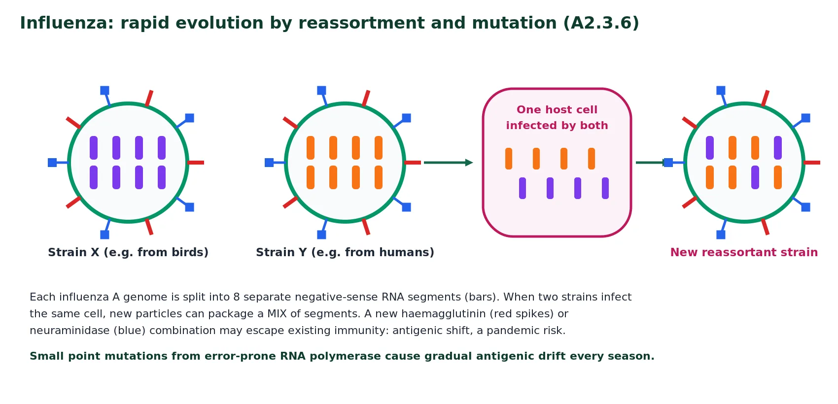
Influenza is caused by an enveloped virus whose genome is negative-sense, single-stranded RNA, split into eight separate segments in influenza A. Its RNA polymerase does not proofread, so each round of replication introduces mutations. Two glycoproteins on the envelope act as the main antigens: haemagglutinin (H), which binds to host cells, and neuraminidase (N), which helps new particles to be released. Strains are named after the types they carry, for example H1N1 or H3N2.
Influenza evolves in two ways. Small, continual changes caused by point mutations in the H and N genes gradually alter the shape of these antigens; this is called antigenic drift. Over a season or two, antibodies made against last year’s strain recognise the drifted virus less well, which is why people can catch influenza repeatedly and why influenza vaccines are reviewed and updated every year. Occasionally, a much larger change happens. If one host cell is infected by two different influenza strains, for example a bird strain and a human strain, the new particles may package a mixture of segments from both. This reassortment can produce a virus with a completely new combination of H and N, a change called antigenic shift. Because few people have immunity to it, a shifted virus can spread worldwide; the 2009 H1N1 pandemic virus contained gene segments originating from pigs, birds and humans.
Evolution of HIV HL
HIV has one of the highest mutation rates known. Its reverse transcriptase, which copies the RNA genome into DNA, has no proofreading function, so copying errors are frequent and are not corrected. A host enzyme, cytidine deaminase, which converts cytosine to uracil in viral nucleic acid as a defence, adds further mutations. When two strains infect one cell, recombination between the two RNA copies packaged in each particle adds still more variety. Combined with a short replication cycle and enormous numbers of particles, these mutations mean that one infected person soon carries a diverse population of genetically different variants, sometimes called a quasispecies.
This diversity has serious consequences. Variants with changes in the envelope glycoproteins escape the antibodies made against earlier forms, so the immune system is always one step behind and the infection becomes long-lasting rather than cleared. Mutations in the env gene can also allow the virus to bind different host-cell proteins and so infect different cell types. Most importantly for treatment, variants carrying mutations that alter the target enzymes of antiretroviral drugs, such as reverse transcriptase or protease, are selected whenever a single drug is used.
Consequences for treating diseases caused by rapidly evolving viruses HL
The guide asks you to consider what rapid evolution means for treatment, and this is a favourite source of extended-response questions. The first point is that antibiotics are useless against viruses. Antibiotics target bacterial structures and processes, such as cell-wall synthesis or 70S ribosomes, which viruses do not have. Using antibiotics for viral infections gives no benefit and adds to selection for antibiotic-resistant bacteria.
Second, vaccines may need frequent updating. A vaccine trains the immune system to recognise particular antigens. When those antigens drift, as in influenza, protection falls, so the composition of influenza vaccines for the northern and southern hemispheres is reviewed every year using global surveillance data, and each seasonal vaccine contains several strains. For HIV, the variability of the envelope glycoproteins within and between patients is one of the main reasons that no effective vaccine has yet been produced.
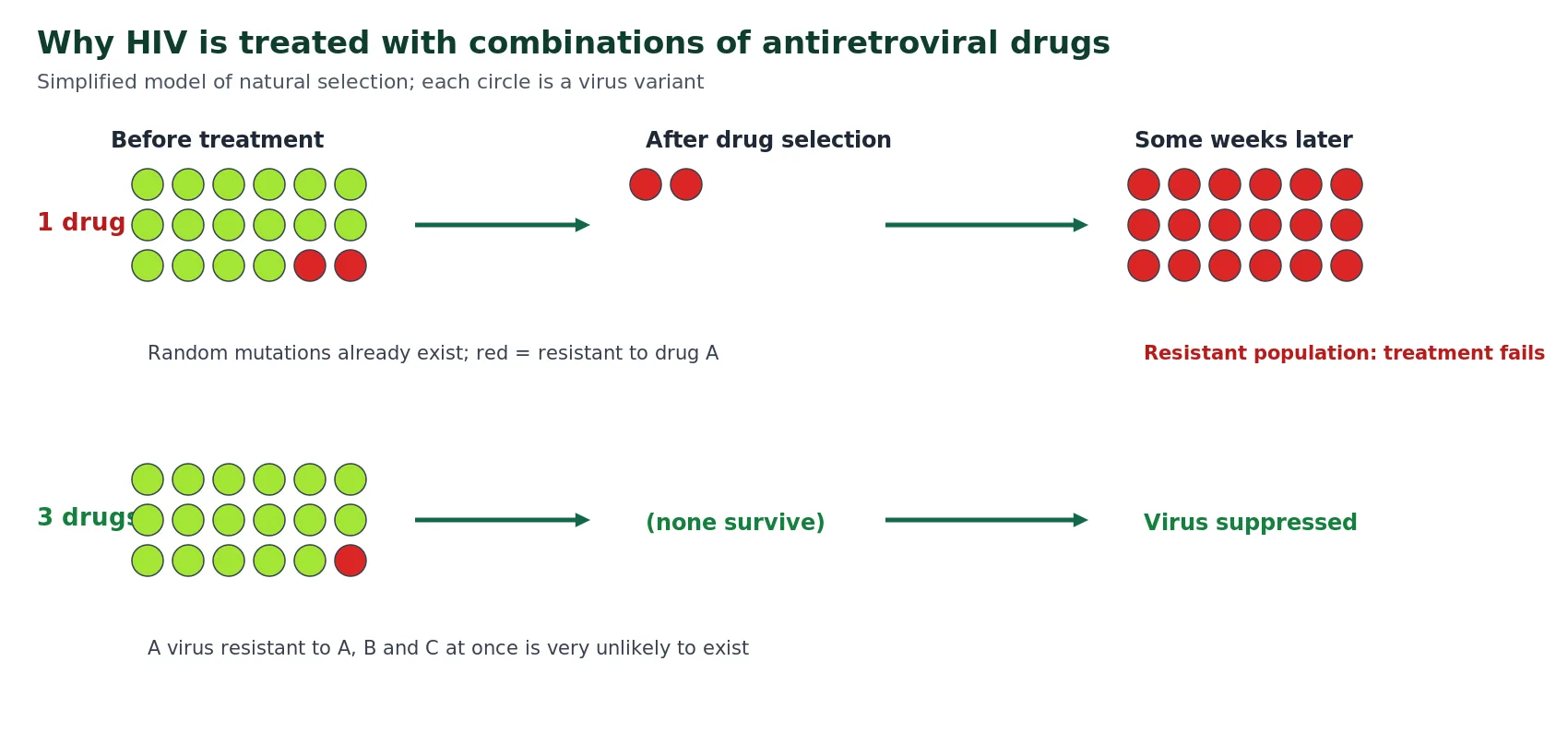
Third, antiviral drugs lose effectiveness as resistant variants are selected. HIV shows this most clearly. Early treatment with a single reverse-transcriptase inhibitor often stopped working within months, because variants with a resistance mutation already existed in the patient and were selected by the drug. Modern antiretroviral therapy (ART) uses a combination of usually three drugs acting on different targets. A variant would need resistance mutations against all three at once to survive, which is extremely unlikely, so viral replication is suppressed. Treatment must be taken consistently and for life, because missed doses allow replication to resume and give resistant variants the chance to emerge. Even then, integrated DNA in long-lived resting cells cannot be removed by current drugs, which is why HIV can be controlled but not cured.
Link to the nature of science
Influenza vaccine strain selection is a real-world example of scientists making predictions with uncertainty: strains must be chosen months before the season begins, based on surveillance data from many countries, and the match is sometimes poor. Discuss how international cooperation and data sharing reduce this uncertainty.
Paper 1B and Paper 2 data questions on viruses often use growth curves, tables of mutation counts or patient data. Work through these scaffolded examples. All data here are invented for practice, but they are realistic in scale.
Data question 1: one-step growth of a phage
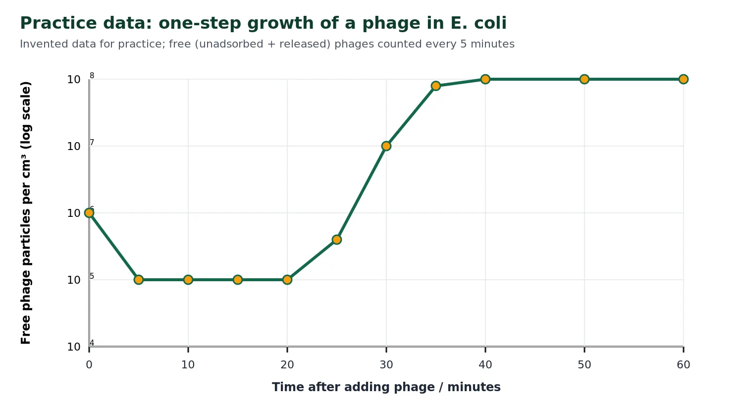
(a) Explain the fall in free phage between 0 and 5 minutes. [2]
Hint, then model answer
Hint: where have the phages gone? Are they destroyed?
Model answer: phages attach to receptors (maltoporin) on the bacteria and inject their DNA ✔, so they are no longer counted as free particles; about 90% (9.0 × 10⁵) have adsorbed ✔.
(b) Estimate the latent period (time from infection to the first release of new phages). [1]
Model answer
About 20 minutes (accept 20–25 min) ✔: free phage starts to rise only after 20 minutes. During this period DNA is replicated and new phages are assembled inside the cells.
(c) Calculate the burst size, the mean number of phages released per infected cell. Show your working. [2]
Scaffold: step-by-step working
Step 1: infected cells = phages adsorbed = 1.0 × 10⁶ − 1.0 × 105 = 9.0 × 105 cm−3.
Step 2: phages released = plateau − unadsorbed phages = 1.0 × 10⁸ − 1.0 × 10⁵ ≈ 1.0 × 108 cm−3.
Step 3: burst size = 1.0 × 10⁸ ÷ 9.0 × 10⁵ ≈ 110 phages per cell ✔✔ (accept 110–111; one mark for correct method with an arithmetic slip).
(d) Explain why the number of free phages stops rising after about 40 minutes. [2]
Model answer
All the infected cells have lysed by about 40 minutes, so no more phages are being released ✔; because the culture was diluted, released phages do not start a second round of infection, so the number does not rise again ✔.
Data question 2: comparing rates of evolution
| Gene | Length / bases | Base substitutions fixed in 10 years |
|---|---|---|
| Influenza haemagglutinin (H3) | 1 700 | 85 |
| A human gene coding for a structural protein | 1 600 | 0 |
Practice data, invented for this question but similar in scale to published estimates.
(a) Calculate the rate of change in the haemagglutinin gene in substitutions per base per year. [2]
Model answer
85 ÷ (1 700 × 10) ✔ = 5.0 × 10⁻³ substitutions per base per year ✔.
(b) Suggest two reasons for the difference between the two genes. [2]
Model answer
Any two: influenza RNA polymerase does not proofread, so the mutation rate is higher ✔; many more viral generations occur in 10 years than human generations (none or less than one) ✔; immune selection favours new haemagglutinin variants, so mutations in this antigen are fixed ✔; enormous viral population sizes ✔.
(c) Explain the consequence for influenza vaccination. [2]
Model answer
Changes in haemagglutinin alter its shape so antibodies/memory cells from earlier vaccines or infections no longer bind well (antigenic drift) ✔; vaccines must therefore be reformulated each year using strains predicted to circulate ✔.
Data question 3: HIV and single-drug treatment
| Weeks of single-drug treatment | 0 | 2 | 4 | 8 | 12 |
|---|---|---|---|---|---|
| Viral load / % of starting value | 100 | 3 | 8 | 60 | 95 |
| Drug-resistant variants / % of virus | 0.1 | 5 | 40 | 95 | 99 |
Practice data for one hypothetical patient.
(a) Describe the changes in viral load. [2]
Model answer
Viral load falls sharply to 3% within 2 weeks ✔, then rises again, returning almost to the starting value (95%) by week 12 ✔.
(b) Explain these changes using the data on resistant variants. [3]
Model answer
Resistant variants were already present before treatment (0.1%), produced by random mutation during error-prone reverse transcription ✔. The drug killed/prevented replication of sensitive variants, so viral load fell ✔. Resistant variants survived and replicated, increasing to 99% of the population: natural selection, so viral load rose again ✔.
(c) Suggest why combination therapy with three drugs prevents this pattern. [2]
Model answer
A variant would need mutations giving resistance to all three drugs simultaneously ✔, which is extremely improbable, so no variant can replicate and the population is suppressed ✔.
Exam traps and examiner tips HL
✗ Calling a virus a cell or an organism
✓ Write “virus particle” or “virion”, and describe viruses as non-cellular. Never say a virus “grows” or “respires”.
✗ Listing an envelope as a universal feature
✓ Only some viruses are enveloped. Phage lambda is not. The universal list is: small fixed size, nucleic acid, protein capsid, no cytoplasm, few or no enzymes.
✗ Saying the envelope is made by the virus
✓ The phospholipids come from the host cell membrane; only the embedded glycoproteins are coded by viral genes.
✗ Writing that lambda injects its whole body
✓ Only the DNA enters; the empty head and tail remain outside the cell.
✗ Describing HIV as following the lysogenic cycle
✓ Use phage lambda for both cycles, as the guide specifies. HIV integrates as a provirus but releases new particles by budding without bursting the cell, so it does not fit either phage cycle neatly.
✗ Saying antibiotics can treat viral infections
✓ Antibiotics target bacterial structures absent from viruses; antiviral drugs target viral enzymes or proteins.
✗ Confusing drift and shift
✓ Drift is gradual point mutation; shift is a sudden new H or N combination, usually from reassortment, and causes pandemics.
✗ Saying the drug causes resistance mutations
✓ Mutations are random and already present; the drug selects resistant variants.
Key terms flip cards HL
Tap or press Enter on a card to see the definition. Try to say the definition aloud before you flip.
CapsidFlip
The protein coat around a viral genome, built from many repeating protein subunits that self-assemble.
EnvelopeFlip
A membrane around the capsid of some viruses; phospholipids from the host cell with viral glycoproteins embedded.
BacteriophageFlip
A virus that infects bacteria, such as phage lambda in E. coli.
Lytic cycleFlip
Viral reproduction ending with the host cell bursting open and releasing new virus particles.
Lysogenic cycleFlip
A cycle in which viral DNA integrates into the host chromosome and is replicated with it, without producing new particles.
ProphageFlip
Phage DNA integrated into the bacterial chromosome.
InductionFlip
Switching of a prophage into the lytic cycle, often triggered by host DNA damage.
RetrovirusFlip
An RNA virus that uses reverse transcriptase to make a DNA copy of its genome, which is integrated into a host chromosome.
Reverse transcriptaseFlip
An enzyme that synthesises DNA using RNA as a template; it does not proofread.
Convergent evolutionFlip
Independent evolution of similar features in unrelated lineages because of similar selection pressures.
Antigenic driftFlip
Gradual change in viral surface antigens caused by accumulated point mutations.
Antigenic shiftFlip
A sudden major change in influenza surface antigens, usually by reassortment of RNA segments.
Self-check: multiple-choice questions HL
Choose one answer for each question, then mark them all at once. Each question also has its own answer and explanation box, which works even if interactive scripts are disabled.
Structured exam questions with model answers HL
Attempt each question on paper before revealing the model answer. Ticks (✔) show where a mark would be awarded; you need one valid point per mark.
1. Outline the structural features that are common to all viruses. [4]
Show model answer (markscheme style)
Any four: small size (mostly 20–300 nm) ✔; fixed size because particles are assembled and do not grow ✔; nucleic acid as genetic material, either DNA or RNA ✔; genome enclosed in a capsid made of protein subunits ✔; no cytoplasm/no ribosomes ✔; few or no enzymes ✔.
Do not credit envelope, tail or DNA alone.
2. Compare and contrast the lytic and lysogenic cycles of bacteriophage lambda. [4]
Show model answer (markscheme style)
Similarities (max 2): both begin with attachment of tail proteins to maltoporin ✔; DNA injected and circularised in both ✔; both use host enzymes/ribosomes/energy ✔.
Differences (max 3): lytic kills host by lysis whereas lysogenic host survives ✔; lytic produces about 100 new phages per cell, lysogenic produces none while dormant ✔; lysogenic DNA integrated into host chromosome by integrase as a prophage, lytic DNA stays separate and is replicated many times ✔; prophage inherited by daughter cells at division ✔.
3. Explain why viruses can only reproduce inside a host cell. [3]
Show model answer (markscheme style)
Viruses have no ribosomes, so they cannot synthesise proteins and must use host ribosomes and tRNA ✔; they have no metabolism/respiration so cannot produce ATP; energy comes from the host ✔; they lack raw materials and most enzymes, so nucleotides, amino acids and polymerases are provided by the host ✔.
4. Discuss the evidence for several origins of viruses. [5]
Show model answer (markscheme style)
Viruses are very diverse: DNA or RNA, single- or double-stranded, enveloped or not ✔; no gene is shared by all viruses, unlike cells ✔; so they are unlikely to share one common ancestor ✔. Shared features (capsid, small size, no cytoplasm) can be explained by convergent evolution due to obligate parasitism ✔. Retrotransposons resemble retroviruses, supporting origins from mobile genetic elements ✔; reduced obligate bacteria and giant viruses support regressive origins ✔. Shared genetic code suggests viruses arose from/with cells ✔. Conclusion: different groups may have arisen by different routes ✔.
5. Explain the reasons for rapid evolution of influenza viruses and the consequences for controlling influenza. [6]
Show model answer (markscheme style)
RNA polymerase lacks proofreading so mutation rate is high ✔; short generation time/many generations per year ✔; large populations ✔; mutations in haemagglutinin/neuraminidase genes change antigens (antigenic drift) ✔; segmented genome allows reassortment when two strains infect one cell ✔; transmission between birds, pigs and humans brings new segments (antigenic shift) ✔; immune selection favours new antigen variants ✔. Consequences: people can be reinfected ✔; vaccines must be updated annually and contain several strains ✔; risk of pandemics from novel strains ✔.
6. Explain why HIV infection can be controlled but not cured with current treatments. [5]
Show model answer (markscheme style)
Reverse transcriptase does not proofread, so the mutation rate is very high ✔; host cytidine deaminase adds further mutations ✔; many variants arise in one patient, including some resistant to any single drug ✔; single drugs select resistant variants so treatment fails ✔; combination therapy requires simultaneous resistance to several drugs, so replication is suppressed ✔; variable envelope proteins evade antibodies and hinder vaccine development ✔; integrated proviral DNA persists in resting cells, which drugs cannot remove ✔.
References and further reading HL
These authoritative sources support specific facts in this chapter. The explanations, diagrams and questions here are original.
- International Baccalaureate: DP Biology course page and subject brief: course structure, themes and levels for first assessment 2025.
- NCBI Nucleotide: Enterobacteria phage lambda, complete genome (NC_001416.1): 48 502 bp linear double-stranded DNA genome.
- NCBI Bookshelf: Bacteriophage lambda terminase and the mechanism of viral DNA packaging: 12-base cohesive (cos) ends and circularisation of the genome.
- Chatterjee & Rothenberg (2012), Interaction of bacteriophage λ with its E. coli receptor, LamB. Viruses (PMC): tail fibre protein binding to maltoporin (LamB).
- CDC: How flu viruses can change: “drift” and “shift”: antigenic drift, antigenic shift and annual vaccine review.
- Stephens et al. (1998), Genome sequence of an obligate intracellular pathogen of humans: Chlamydia trachomatis. Science (PubMed): reduced genome of an obligate intracellular bacterium.
Common doubts about viruses HL
Are viruses living or non-living?
Most biologists describe viruses as non-living or non-cellular because they have no metabolism, no cytoplasm and cannot reproduce on their own. They do have genes, mutate and evolve by natural selection, so they sit on the boundary of life.
Is the virus topic in IB Biology SL or HL?
A2.3 Viruses is additional higher level (AHL) content in the IB Biology guide for first assessment 2025, so it is examined only at higher level.
Do all viruses have an envelope?
No. Enveloped viruses such as HIV, influenza and coronaviruses have a membrane taken from the host cell, but many viruses, including bacteriophage lambda and most plant viruses, are non-enveloped.
Can a virus contain both DNA and RNA?
A virus particle carries either DNA or RNA as its genome, not both. Retroviruses such as HIV have an RNA genome that is copied into DNA only after it enters a host cell.
Why is phage lambda used for both the lytic and lysogenic cycles?
Lambda is a temperate phage that can follow either route after infecting E. coli, so it shows both cycles and the switch between them, and it is the example named in the IB guide.
What is the difference between a prophage and a provirus?
A prophage is phage DNA integrated into a bacterial chromosome. A provirus is the DNA copy of a retrovirus such as HIV integrated into a eukaryotic host chromosome.
Why can people catch influenza more than once?
Influenza antigens change through antigenic drift and occasionally antigenic shift, so antibodies from earlier infections or vaccines no longer recognise the new strain well.
Why do antibiotics not work against viruses?
Antibiotics target bacterial structures such as cell walls and 70S ribosomes. Viruses do not have these, so antibiotics cannot affect them; antiviral drugs target viral proteins instead.