
IB Biology › Theme A › A1.2 Nucleic acids
Nucleic Acids IB Biology Notes (A1.2)
Nucleic acids (DNA and RNA) are long, unbranched polymers of nucleotides that store, copy and express hereditary information in every living cell. These nucleic acids IB Biology notes cover topic A1.2, which is studied by all IB Biology students; sections marked HL (A1.2.11–A1.2.15) are additional higher level content.
Contents
- Why nucleic acids matter
- A1.2.1 DNA as the genetic material
- A1.2.2 Components of a nucleotide
- A1.2.3 The sugar–phosphate backbone
- A1.2.4 Bases: the basis of a code
- Purine and pyrimidine structure
- A1.2.5 RNA as a polymer
- A1.2.6 DNA as a double helix
- Bonds in nucleic acids: summary
- A1.2.7 Differences between DNA and RNA
- A1.2.8 Complementary base pairing
- A1.2.9 Limitless capacity for information
- A1.2.10 A universal genetic code
- A1.2.11 Directionality (HL)
- A1.2.12 Purine–pyrimidine pairing (HL)
- A1.2.13 Nucleosomes (HL)
- A1.2.14 Hershey–Chase experiment (HL)
- A1.2.15 Chargaff’s data (HL)
- Nature of science and TOK links
- Common mistakes and examiner tips
- Key terms flip cards
- Self-check: multiple-choice questions
- Exam-style and data-based questions
- References and further reading
- Nucleic acids IB Biology: FAQs
Why nucleic acids matter SL & HL
Every cell has to solve the same problem: it must keep a permanent record of how to build itself, copy that record accurately before it divides, and read parts of it on demand to make the proteins it needs at a particular moment. Nucleic acids are the molecules that do this. Deoxyribonucleic acid (DNA) is the long-term store of hereditary information, and ribonucleic acid (RNA) carries copies of that information to where it is used, helps translate it into protein and, in some cases, acts as a catalyst. The name comes from their discovery in cell nuclei in the nineteenth century, although we now know they are found throughout cells, in mitochondria and chloroplasts, and in viruses.
The IB guide frames the topic around two guiding questions: how does the structure of nucleic acids allow hereditary information to be stored? and how does the structure of DNA facilitate accurate replication? Keep both in mind as you read. Almost every structural detail in this chapter, from the covalent backbone to the hydrogen bonds between bases, makes sense once you ask what it contributes to storing or copying information. The first ten understandings (A1.2.1–A1.2.10) are for SL and HL students; the last five (A1.2.11–A1.2.15) add molecular detail and historical evidence for HL.
A1.2.1 DNA as the genetic material of all living organisms SL & HL
Genetic material is information that can be copied and passed on, both from a cell to its daughter cells and from parents to offspring. Because it is inherited, it is also called hereditary information. In every known living organism, from bacteria and archaea to fungi, plants and animals, that information is stored as the sequence of bases in DNA. This shared feature is one of the strongest pieces of evidence for the unity of life.
Some viruses seem to break the rule. Coronaviruses, influenza viruses and HIV, for example, use RNA rather than DNA as their genetic material. This does not falsify the statement that all living organisms use DNA, because viruses are not usually classified as living: they have no metabolism and cannot reproduce except by taking over a host cell. (HL students study viruses in detail in Viruses (IB A2.3).) The same point is worth remembering in exams: if a question asks for the genetic material of an organism, the answer is always DNA.
Nature of science: making a claim that can be tested
“All living organisms use DNA as their genetic material” is a universal claim. No number of confirming examples can prove it, but a single living organism with an RNA-only genome would falsify it. RNA viruses are not counter-examples because they fall outside the category of living things, which shows how carefully the terms in a scientific claim must be defined.
A1.2.2 Components of a nucleotide SL & HL
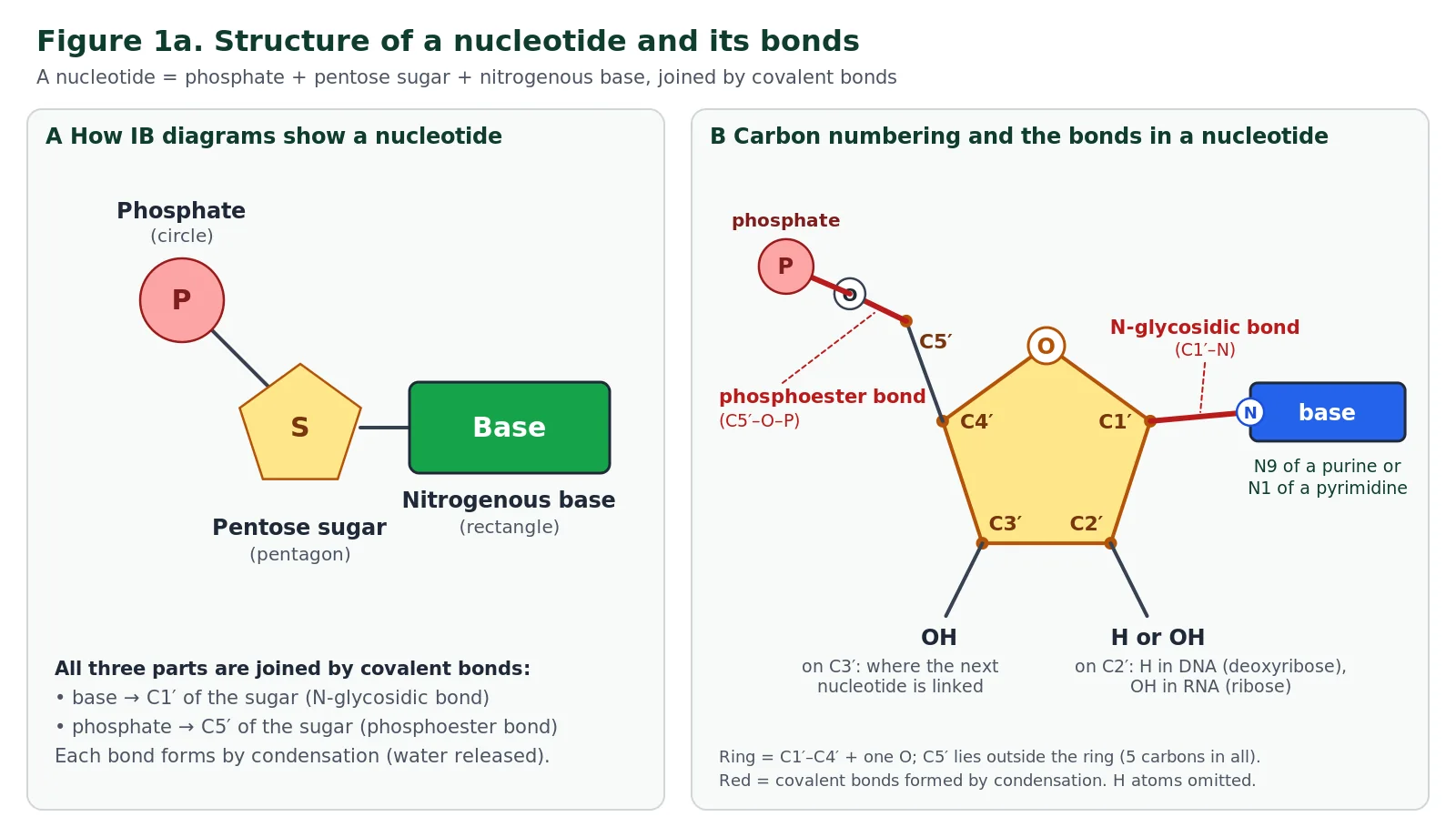
Nucleic acids are polymers, and their monomers are nucleotides. Every nucleotide is built from three parts joined by covalent bonds. The first is a pentose sugar, a sugar with five carbon atoms: deoxyribose in DNA and ribose in RNA. The second is a phosphate group. It is the acidic part of the molecule and carries a negative charge at the pH found in cells, which is why the whole polymer is called a nucleic acid and why DNA is strongly negatively charged. The third is a nitrogenous base, a molecule containing nitrogen arranged in either one ring or two rings of atoms.
To describe how the parts are joined, biologists number the carbon atoms of the pentose with primes (1′, 2′, 3′ and so on) to distinguish them from atoms in the base. The base is attached to carbon 1′ and the phosphate to carbon 5′, which sits outside the ring. Carbon 3′ carries a hydroxyl (–OH) group, and that is where the next nucleotide will be linked. In IB diagrams you are not expected to draw the atoms. Instead, the guide asks you to use simple shapes: a circle for the phosphate, a pentagon for the pentose sugar and a rectangle for the base, placed so that their relative positions are correct.
Exam tip: drawing a nucleotide
Attach the phosphate to the corner of the pentagon on the C5′ side and the base to the opposite side (C1′). Join all three with lines to show covalent bonds, and label each part. A floating circle, pentagon and rectangle that do not touch will not score full marks.
Numbering the pentose: ribose and deoxyribose
A pentose is a sugar with five carbon atoms. In both ribose and deoxyribose, four of those carbons (C1′, C2′, C3′ and C4′) form a five-membered ring together with one oxygen atom, and the fifth carbon (C5′) sits outside the ring as a side branch attached to C4′. Numbering starts at the ring carbon that carries the base (C1′), next to the ring oxygen, and continues round the ring away from the oxygen, so C1′ and C4′ sit on either side of the ring oxygen.
The prime mark (′, read as “prime”) is used for every sugar carbon because the atoms of the base have their own numbers (1, 2, 3 and so on); without primes, “carbon 1” would be ambiguous.
Each numbered carbon has a job. C1′ carries the base. C2′ carries a hydroxyl group (–OH) in ribose but only a hydrogen atom in deoxyribose, which is the single difference between the two sugars and the origin of the name deoxyribose, “ribose with one oxygen removed”. C3′ carries a hydroxyl group in both sugars; this –OH is where the next nucleotide is attached when a strand grows. C4′ is part of the ring and holds C5′. C5′ carries the phosphate group. Remembering “base on 1′, difference on 2′, link on 3′, phosphate on 5′” is enough to explain almost every structural question on nucleotides.
How the three parts are bonded: N-glycosidic and phosphoester bonds
The three parts of a nucleotide are joined by covalent bonds, and each one is made by a condensation reaction in which a molecule of water is removed. The base is attached to the sugar by an N-glycosidic bond (more precisely a β-N-glycosidic bond).
It links carbon 1′ of the pentose to a nitrogen atom in the ring of the base: nitrogen 9 (N9) in a purine (adenine or guanine) or nitrogen 1 (N1) in a pyrimidine (cytosine, thymine or uracil). The name tells you what is joined: “glycosidic” refers to the sugar, and the “N” shows that the sugar is bonded to a nitrogen atom rather than to an oxygen, as it would be in a disaccharide such as maltose.
The phosphate group is attached to the other side of the sugar, on carbon 5′, by a phosphoester bond (also called a phosphate ester bond). Here an oxygen atom bridges the C5′ carbon and the phosphorus atom (C5′–O–P). Because both of these bonds are covalent, a nucleotide is a single, stable molecule, and the base and phosphate do not drift away from the sugar in water. When nucleotides are joined into a chain, this same phosphate forms a second ester bond with the next sugar, turning the phosphoester into a phosphodiester link (see A1.2.3).
Reveal: Why is the bond between sugar and base called a condensation product?
To form the N-glycosidic bond, the –OH on carbon 1′ of the sugar and the H on the ring nitrogen of the base (N9 or N1) are removed together as one water molecule, and the carbon and nitrogen bond directly. Breaking the bond again would need a hydrolysis reaction, which adds water back.
Nucleoside or nucleotide?
A sugar bonded to a base, but with no phosphate, is called a nucleoside; adding one or more phosphate groups to C5′ turns it into a nucleotide. So adenosine (adenine + ribose) is a nucleoside, while adenosine monophosphate (AMP) is a nucleotide. A useful memory aid is that the nucleotide is the one with the phosphate too. The free nucleotides that cells use to build DNA and RNA actually carry three phosphates (they are nucleoside triphosphates such as ATP and dATP), and two of the three are released as the nucleotide is added to a chain. ATP, the cell’s energy currency, is therefore itself a nucleotide.
A1.2.3 Sugar–phosphate bonding and the sugar–phosphate backbone SL & HL
Nucleotides are linked into a chain by covalent bonds between the phosphate group of one nucleotide and the pentose sugar of the next. Because every link is made in the same way, the result is a long, unbranched chain in which sugar and phosphate groups alternate. Running along this chain is an unbroken sequence of covalently bonded carbon, oxygen and phosphorus atoms. This is the sugar–phosphate backbone, and it is found in both DNA and RNA.
The backbone matters because it is strong. Covalent bonds are not broken by the ordinary jostling of molecules at body temperature, so the order of bases attached to the backbone is held in place, sometimes for the whole lifetime of an organism and, in the right conditions, for many thousands of years after death. The bases project sideways from the backbone, and because the backbone itself is identical along its length, it places no restrictions on which base follows which. That is exactly what you want in an information store: a strong, uniform support that can carry any message.
The phosphodiester bond between nucleotides
The bond that links one nucleotide to the next is called a phosphodiester bond. It forms between the hydroxyl group on carbon 3′ of the sugar at the end of the chain and the phosphate group on carbon 5′ of the incoming nucleotide. This is a condensation reaction: an –OH from one partner and an H from the other leave as a molecule of water, and the remaining oxygen bridges the 3′ carbon and the phosphorus.
The phosphate is now joined by ester bonds to two sugars (one through C5′ of its own nucleotide and one through C3′ of the previous nucleotide), which is why the link is called a phosphodiester bond. In full, the repeating link reads C3′–O–P–O–C5′.
Because every phosphodiester bond joins a 3′ carbon to a 5′ carbon, all the nucleotides in a strand face the same way. One end of the strand has a phosphate on C5′ that is not linked to anything (the 5′ end), and the other has a free –OH on C3′ (the 3′ end). New nucleotides can only be added to the 3′ end, so strands are always built in the 5′→3′ direction; HL students study the consequences in A1.2.11. Breaking phosphodiester bonds requires hydrolysis, which is catalysed in cells by nuclease enzymes.
Covalent bonds versus hydrogen bonds
Keep the two kinds of bond in DNA separate. Within each strand, everything is held by strong covalent bonds: N-glycosidic bonds join bases to sugars, and phosphodiester bonds join one nucleotide to the next to form the sugar–phosphate backbone. Between the two strands, complementary bases are held together only by much weaker hydrogen bonds. This is the key to replication: the strands can be separated by breaking hydrogen bonds, using an enzyme or moderate heat, while every strand and its base sequence stays intact. Hydrogen bonds never join nucleotides along a strand.
A1.2.4 Bases in each nucleic acid that form the basis of a code SL & HL
Each nucleotide carries one of four bases, so there are four types of nucleotide in DNA and four in RNA. Three of the bases are shared and one is different. DNA contains adenine (A), cytosine (C), guanine (G) and thymine (T); RNA contains adenine, cytosine, guanine and uracil (U) in place of thymine. You need to know these names, and you should always use the full name at least once before using the letter.
| Base | Letter | Ring structure | Found in DNA? | Found in RNA? |
|---|---|---|---|---|
| Adenine | A | Two rings (purine) | Yes | Yes |
| Guanine | G | Two rings (purine) | Yes | Yes |
| Cytosine | C | One ring (pyrimidine) | Yes | Yes |
| Thymine | T | One ring (pyrimidine) | Yes | No |
| Uracil | U | One ring (pyrimidine) | No | Yes |
Information is stored in the sequence of bases along the strand, in much the same way that meaning is stored in the order of letters in a sentence. A four-letter alphabet sounds limited, but as you will see in A1.2.9 it can encode an effectively unlimited number of different messages. The ring structures behind the terms purine and pyrimidine are explained next; at HL they also explain why the double helix has a constant width (A1.2.12).
Purine and pyrimidine structure
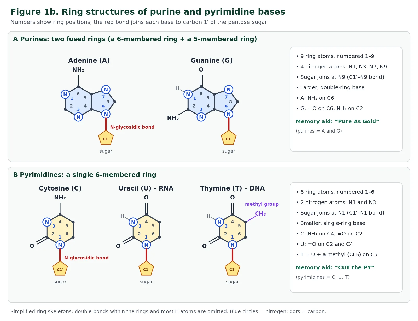
All five bases are flat molecules built from rings of carbon and nitrogen atoms, which is why they are called nitrogenous bases. They fall into two chemical families according to how many rings they have. The purines, adenine (A) and guanine (G), have a double-ring structure: a six-membered ring fused to a five-membered ring, sharing one side. That gives nine ring atoms, numbered 1 to 9, of which four are nitrogen (N1, N3, N7 and N9) and the rest are carbon. The pyrimidines, cytosine (C), thymine (T) and uracil (U), have a single six-membered ring with six atoms numbered 1 to 6, two of which are nitrogen (N1 and N3).
The bases are attached to the sugar through one of their ring nitrogens. In a purine the bond is to N9, on the five-membered ring; in a pyrimidine it is to N1. Each base also carries its own side groups, and these are what make the four bases chemically distinct. Adenine has an amino group (–NH2) on C6, while guanine has a double-bonded oxygen (=O) on C6 and an amino group on C2.
Cytosine has an amino group on C4 and =O on C2. Uracil has =O on both C2 and C4, and thymine is identical to uracil except for an extra methyl group (–CH3) on C5; for this reason thymine is sometimes called 5-methyluracil. The different arrangements of –NH2, =O and N–H groups on the edges of the bases determine which hydrogen bonds each base can form, and so which partner it pairs with.
| Feature | Purines | Pyrimidines |
|---|---|---|
| Bases | Adenine (A), guanine (G) | Cytosine (C), thymine (T), uracil (U) |
| Rings | Two fused rings (6-membered + 5-membered) | One 6-membered ring |
| Ring atoms | 9 (4 of them nitrogen: N1, N3, N7, N9) | 6 (2 of them nitrogen: N1, N3) |
| Atom bonded to C1′ of the sugar | N9 | N1 |
| Relative size | Larger | Smaller |
| Pairs with | A pyrimidine (A–T or A–U; G–C) | A purine (T–A, U–A; C–G) |
Memory aids
“Pure As Gold”: the purines are A and G. “CUT the PY”: C, U and T are the pyrimidines. A third check: the word pyrimidine is the longer word but names the smaller, one-ring bases. For pairing, try “Apple Tree” for A–T and “Green Car” for G–C.
Why does this matter? Every base pair in DNA contains one purine and one pyrimidine: A pairs with T and G pairs with C. A two-ring base plus a one-ring base always spans about the same distance, so the two sugar–phosphate backbones stay a constant distance apart and the helix keeps a uniform width of about 2 nm along its whole length, whatever the sequence. Two purines together would be too wide to fit, and two pyrimidines too narrow to reach across. At SL you need to know which bases are purines and pyrimidines; at HL you also need to explain this link to helix stability (A1.2.12).
Skill: comparing ways of representing a molecule
The bases can be shown as structural formulas (every atom and bond drawn), ball-and-stick models (which show bond angles in three dimensions) or space-filling models (which show the true shape and size of the molecule but hide the bonds). Figure 1b uses a simplified skeleton, which makes ring size and numbering easy to see but leaves out the double bonds and most hydrogens. When answering a question, consider which representation shows the feature you need: ring number and attachment points are clearest in a skeleton, while how two bases fit together is clearest in a space-filling model.
Reveal: Each free base has an N–H group in the same position (N9–H in purines, N1–H in pyrimidines). What happens to it when a nucleotide is assembled?
That hydrogen is lost when the base is bonded to the sugar. The H from the ring nitrogen and the –OH from carbon 1′ of the pentose are removed as water in a condensation reaction, and the nitrogen forms an N-glycosidic bond directly with C1′. Because every base has this nitrogen in a comparable position, all of them can be attached to the sugar in the same way.
A1.2.5 RNA as a polymer formed by condensation of nucleotide monomers SL & HL
RNA is a single, unbranched polymer of nucleotides. There is no fixed limit to its length: some RNA molecules are fewer than a hundred nucleotides long, while others contain many thousands. Whatever the length, every link in the chain is made by the same type of reaction, a condensation reaction. Two molecules are joined into one and a molecule of water is released.
A hydroxyl group from the 5′ phosphate of the incoming nucleotide and the hydroxyl on carbon 3′ of the sugar at the end of the chain take part: one –OH is removed completely, it combines with the hydrogen from the other –OH to form water, and the oxygen that remains forms the new covalent bond between sugar and phosphate. The bond formed is the phosphodiester bond described in A1.2.3.
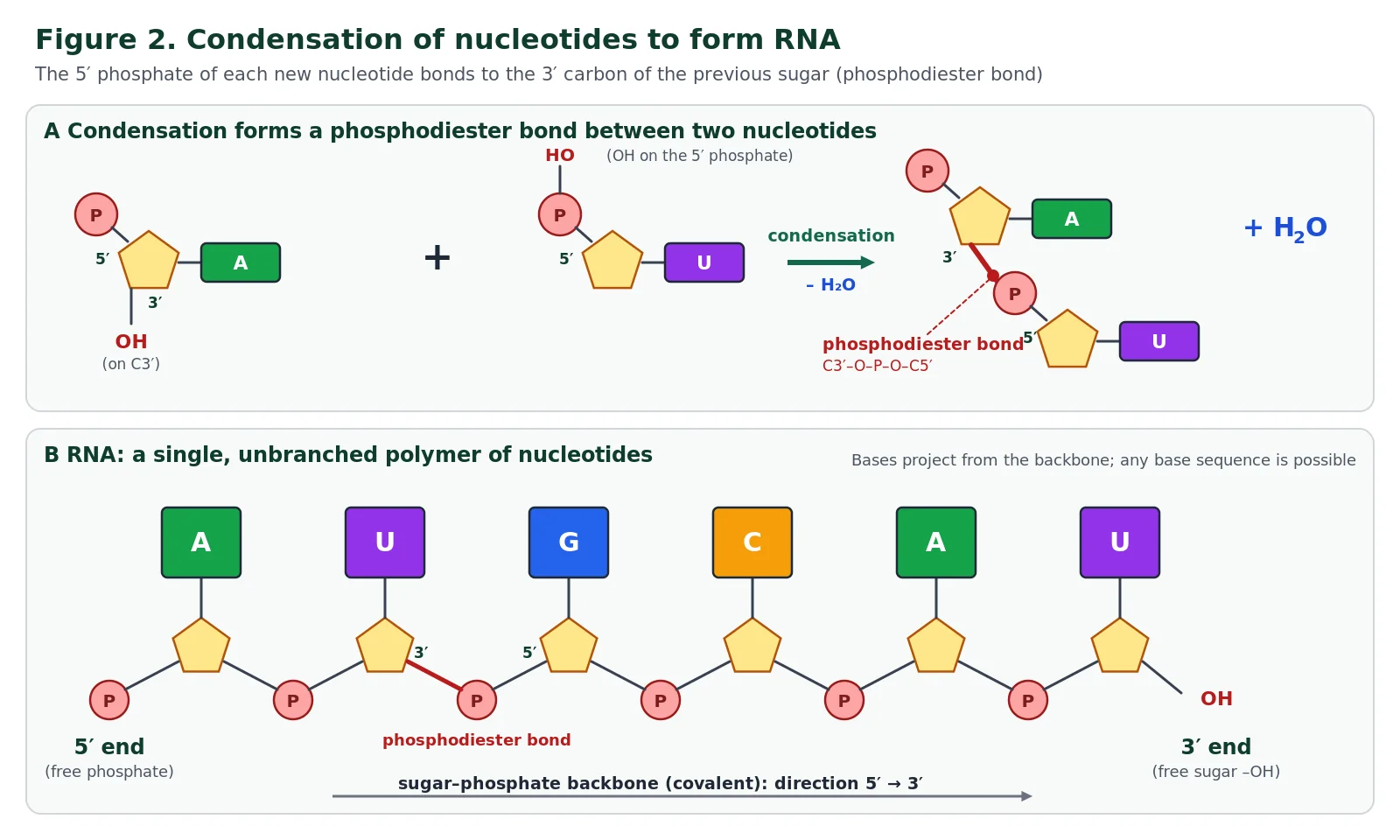
In cells, nucleotides actually arrive as nucleoside triphosphates, and the energy for linking them comes from releasing two of the three phosphates. You do not need this detail for A1.2, but it explains why the reaction can happen in water without running backwards. What you do need is to be able to draw and recognise diagrams of a single nucleotide and a short RNA polymer, using the circle–pentagon–rectangle convention and showing each phosphate linked to the sugars on either side.
Reveal: Why is RNA described as a polymer but not as a protein?
A polymer is any large molecule made of many similar monomers joined by covalent bonds. RNA fits because it is a chain of nucleotide monomers. Proteins are also polymers, but of amino acids joined by peptide bonds. The reaction type (condensation) is the same; the monomers and bonds are different.
A1.2.6 DNA as a double helix of two antiparallel strands SL & HL
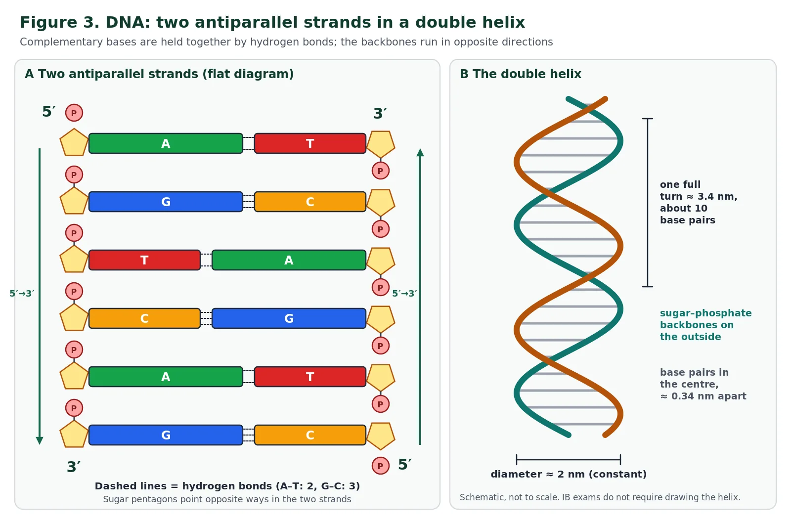
A DNA molecule is made of two strands of nucleotides rather than one. In each strand the sugar is deoxyribose and the bases are adenine, cytosine, guanine and thymine. The two strands are held together by hydrogen bonds between their bases. Adenine on one strand forms hydrogen bonds only with thymine on the other, and guanine only with cytosine. These pairs, A–T and G–C, are called complementary base pairs, and the rule means that the base sequence of one strand fixes the sequence of the other.
The two strands lie alongside each other but run in opposite directions: they are antiparallel. If you follow one strand from its phosphate end to its sugar end, the partner strand runs from sugar end to phosphate end over the same stretch. The bases can only line up correctly for hydrogen bonding when the strands are arranged this way. The paired strands then twist around each other to form a double helix about 2 nm wide. Each complete turn of the helix is roughly 3.4 nm long and contains about ten base pairs, with the sugar–phosphate backbones on the outside and the base pairs stacked inside like the treads of a spiral staircase.
Exam tip: what to draw
The guide says you should draw the two strands antiparallel and show A paired with T and G paired with C, but you do not need to draw the helical shape. The easiest way to show antiparallel strands is to draw the sugar pentagons of one strand the opposite way up to those of the other. The guide also says you need not memorise the number of hydrogen bonds in each pair or the relative sizes of the bases, although both help you understand A1.2.12.
Nature of science: models and collaboration
Watson and Crick’s 1953 model was built by fitting together evidence from several laboratories: X-ray diffraction images from Rosalind Franklin and Maurice Wilkins indicated a helix of constant diameter with the phosphate groups on the outside, and Chargaff’s base ratios suggested pairing. Building physical models allowed them to test which arrangements fitted all the data at once. Franklin died in 1958, before the 1962 Nobel Prize, which cannot be awarded posthumously; her contribution is now widely recognised.
A few more structural details complete the picture, although only the antiparallel strands and the base pairing are needed for drawing. The DNA helix is right-handed: it twists like a normal screw thread.
Because the two backbones are not evenly spaced around the helix, its surface has two spiral grooves of different widths, a wider major groove and a narrower minor groove. The edges of the base pairs are exposed in these grooves, which is where many proteins that switch genes on and off “read” the sequence without separating the strands; this is beyond A1.2 but becomes relevant in gene expression (D2.2). Stability also comes from the stacking of the flat base pairs on top of one another in the centre of the helix, away from water, in addition to the hydrogen bonds between them.
Bonds in nucleic acids: a summary
Questions on nucleic acids often ask you to name a bond and say what it joins. The table brings together every bond you have met so far. Notice that the first three are covalent bonds made by condensation and broken only by hydrolysis, while hydrogen bonds are much weaker attractions that can be broken and remade during replication and transcription.
| Bond | Between | Type | Formed by |
|---|---|---|---|
| N-glycosidic bond | C1′ of the pentose and N9 of a purine or N1 of a pyrimidine | Covalent (within a nucleotide) | Condensation (H2O released) |
| Phosphoester bond | C5′ of the pentose and the phosphate group (C5′–O–P) | Covalent (within a nucleotide) | Condensation |
| Phosphodiester bond | 3′ carbon of one nucleotide’s sugar and the 5′ phosphate of the next (C3′–O–P–O–C5′) | Covalent (joins nucleotides into the sugar–phosphate backbone) | Condensation between the 3′–OH and the 5′ phosphate |
| Hydrogen bonds | Complementary bases on opposite strands: A–T (two) and G–C (three); A–U in RNA pairing | Weak, non-covalent attraction (holds two strands together) | Forms spontaneously when complementary bases line up; broken by helicase or heat |
A1.2.7 Differences between DNA and RNA SL & HL
DNA and RNA are built on the same plan, so it is the differences that examiners ask about. The guide names three. First, the number of strands: DNA usually has two antiparallel strands, whereas RNA usually has only one, although an RNA strand can fold back on itself and form base pairs between its own sections, as in transfer RNA. Second, the bases: DNA contains thymine, while RNA contains uracil instead. Third, the pentose sugar: DNA contains deoxyribose and RNA contains ribose. Deoxyribose has one oxygen atom fewer than ribose, because carbon 2′ carries a hydrogen atom instead of a hydroxyl group. The names follow from the sugars: deoxyribonucleic acid and ribonucleic acid.
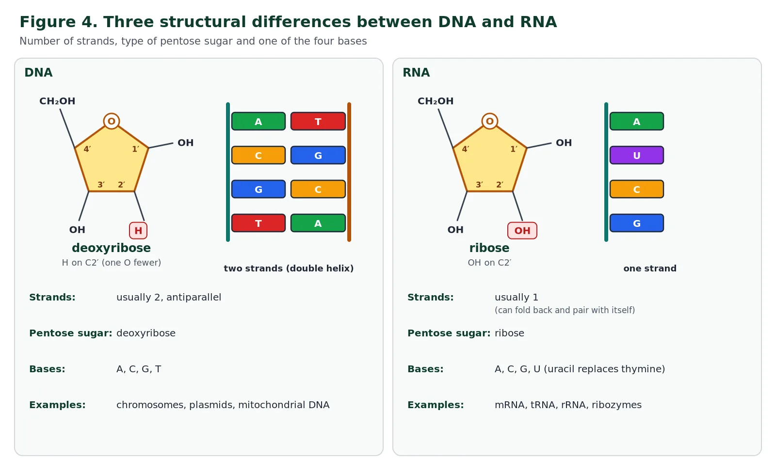
You should also be familiar with some examples of each. DNA occurs as the chromosomes in the nucleus of eukaryotic cells, as the circular chromosome and plasmids of prokaryotes, and inside mitochondria and chloroplasts. RNA occurs as messenger RNA (mRNA), which carries a copy of a gene to the ribosome; transfer RNA (tRNA), which brings amino acids to the ribosome; ribosomal RNA (rRNA), which forms much of the ribosome and catalyses peptide bond formation; and a variety of regulatory RNAs. RNA that acts as a catalyst is called a ribozyme.
Sorting activity: DNA, RNA or both?
Choose a category for each statement, then press Check.
1. Contains deoxyribose
2. Contains uracil
3. Has a sugar–phosphate backbone
4. Usually double-stranded
5. Contains adenine, cytosine and guanine
6. Includes mRNA, tRNA and rRNA
7. Nucleotides linked by condensation reactions
8. Contains thymine
Show the answers
1 DNA; 2 RNA; 3 both; 4 DNA; 5 both; 6 RNA; 7 both; 8 DNA.
A1.2.8 Role of complementary base pairing in replication and expression SL & HL
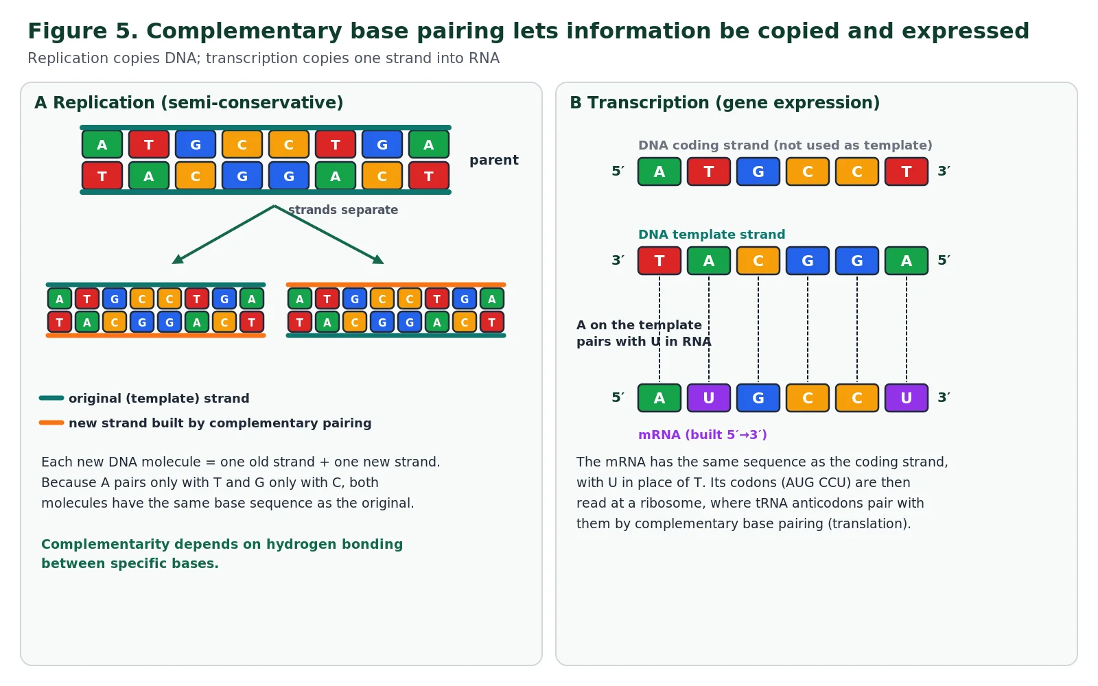
Complementary base pairing depends on hydrogen bonding. The atoms on the edge of each base that can form hydrogen bonds are arranged so that adenine fits thymine (or uracil) and guanine fits cytosine, and other combinations do not bond well. Individually, hydrogen bonds are weak, so the two strands of DNA can be separated without breaking the covalent backbone; collectively, thousands of them hold a long molecule firmly together. This combination of specificity and easy separation is what allows information to be both copied and read.
In replication, which happens during interphase of the cell cycle, the enzyme helicase unwinds the double helix and separates the two strands by breaking the hydrogen bonds between base pairs, and each strand then acts as a template for building a new partner strand. Free nucleotides are added one at a time, and each must carry the base that is complementary to the next base on the template.
Because A can only pair with T and G only with C, the new strand on each template ends up with the same base sequence as the strand that was originally paired with that template. One DNA molecule therefore becomes two identical molecules, each containing one original strand and one new strand. This is semi-conservative replication, and the full mechanism is covered in D1.1.
In gene expression, complementary pairing is used twice. During transcription, one strand of a gene acts as a template for building an RNA copy; the same pairing rules apply, except that adenine on the template pairs with uracil in the RNA. During translation, each three-base codon on mRNA is recognised by the complementary anticodon of a tRNA molecule, which brings the matching amino acid. Both processes are covered in D1.2.
Try it: write the complementary sequences
A DNA template strand reads 3′-T A C G A T T G C-5′.
(a) The complementary DNA strand is 5′-A T G C T A A C G-3′.
(b) An mRNA transcribed from this template is 5′-A U G C U A A C G-3′: the same as the DNA strand in (a), with U in place of T.
Always write the 5′ and 3′ labels at HL; the new strand is antiparallel to its template.
A1.2.9 Diversity of possible DNA base sequences and limitless capacity for storing information SL & HL
Any base can follow any other along a DNA strand, because the sugar–phosphate link is the same whatever the bases are. At each position there are therefore four possibilities. With two positions there are 4 × 4 = 16 possible sequences, with three positions 43 = 64, and in general a sequence of n bases can be arranged in 4n different ways. The number climbs extraordinarily quickly: ten bases already give more than a million possibilities, and a modest gene of a thousand bases has more possible sequences than there are atoms in the observable universe.
| Length of sequence (bases) | Number of possible sequences (4n) |
|---|---|
| 1 | 4 |
| 2 | 16 |
| 3 | 64 |
| 10 | 1 048 576 (over a million) |
| 20 | about 1.1 × 10¹² |
| 1000 | about 10602 |
Two further features make DNA a remarkable storage medium. First, DNA molecules can be almost any length, from a few thousand base pairs in a small plasmid to hundreds of millions in a single human chromosome. Second, DNA is extremely compact. The double helix is only about 2 nm wide, so the roughly 2 metres of DNA in one human body cell (about 6 billion base pairs in a diploid nucleus) fits inside a nucleus only a few micrometres across. Information is stored with great economy in terms of both space and material, which is why researchers are now experimenting with DNA as an archive for digital data.
Real numbers show the range. The human haploid genome is about 3.1 billion base pairs; the smallest human chromosome, the Y chromosome, is about 57 million base pairs, or under 2% of the genome. Some symbiotic bacteria manage with genomes under 200 000 base pairs, some viruses have genomes of only a few thousand bases, and bacterial plasmids can be shorter still, sometimes under 2000 base pairs. Within any of these molecules, the information is carried by the order of bases in genes, sections of DNA whose base sequence codes for a polypeptide or a functional RNA.
Quick calculation
How many different sequences are possible for a stretch of DNA six bases long? 46 = 4096. A six-base sequence on one strand fixes the six complementary bases on the other, so the number of possible double-stranded sequences is still 4096.
A1.2.10 Conservation of the genetic code across all life forms SL & HL
The base sequence of a gene is decoded during protein synthesis. Bases are read in groups of three called codons. Because each of the three positions can hold any of four bases, there are 4 × 4 × 4 = 64 codons. Sixty-one of them specify amino acids, so most amino acids are coded for by more than one codon; one of these, AUG, also acts as the usual start signal. The remaining three are stop codons that end translation. The details of the code are studied in D1.2.
What matters here is that, with only minor exceptions, all living organisms and all viruses use the same genetic code. A codon that means a particular amino acid in a bacterium means the same amino acid in a mushroom, an oak tree and a human. There is no chemical reason why it must be so: many different codes could work equally well, and the number of possible ways of assigning 64 codons to 20 amino acids and stop is astronomically large. The simplest explanation is that the code was established in a common ancestor of all life and has been inherited, almost unchanged, ever since.
Changing the meaning of a codon would alter many proteins at once and would almost always be harmful, so the code has been strongly conserved. The few known variants, found for example in mitochondria and some single-celled organisms, usually involve one codon changing its meaning, most often one of the three stop codons coming to code for an amino acid instead (in human mitochondria, for example, UGA codes for tryptophan rather than “stop”). These are best explained as small changes made during billions of years of evolution, and what is remarkable is how few there are.
When the near-universality of the code became clear in the 1960s, it had a strong effect on the scientists involved. Marshall Nirenberg, who shared the 1968 Nobel Prize for deciphering the code, later described feeling a new sense of kinship with every living thing, since all of them read the same genetic instructions. His reaction is a reminder that scientific findings can change attitudes as well as knowledge.
The universality of the code is therefore treated as evidence for universal common ancestry. It also has practical consequences: a human gene inserted into a bacterium is translated into the same polypeptide, which is how insulin and many other proteins are produced commercially. The guide does not require you to memorise specific exceptions to the code.
Theory of knowledge: language in science
The genetic code is often described as a “language”. Is this a helpful metaphor or a misleading one? Human languages are invented, change rapidly and differ between groups, whereas the genetic code has hardly changed in billions of years. Consider what the metaphor helps us understand, and where it could lead to wrong conclusions.
A1.2.11 Directionality of RNA and DNA HL
Because every nucleotide in a strand is linked in the same way, with the phosphate of one joined to the sugar of the next, all the nucleotides point in the same direction and the strand as a whole has directionality. Its two ends are chemically different. At the 5′ end, the terminal nucleotide has a phosphate group attached to carbon 5′ that is not linked to another sugar.
At the 3′ end, the terminal sugar has a free hydroxyl group on carbon 3′ that is available to bond to another nucleotide. The links within the backbone are therefore described as 5′→3′ linkages. In double-stranded DNA, the two strands are antiparallel, so each end of the molecule has the 5′ end of one strand beside the 3′ end of the other.
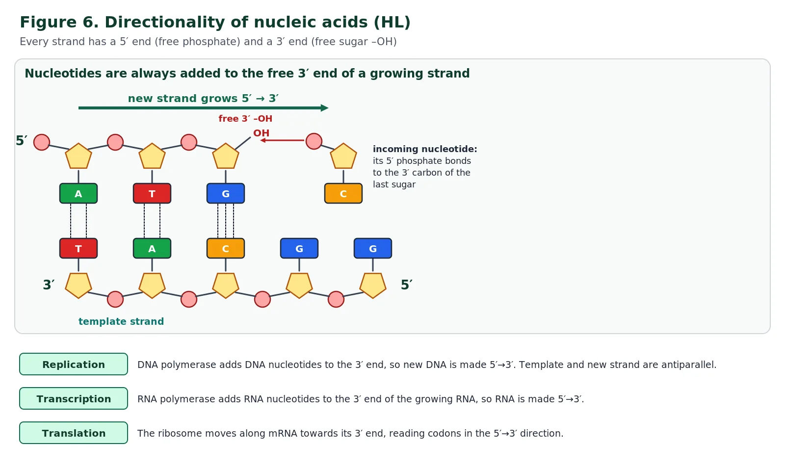
Directionality matters because the enzymes and ribozymes that handle nucleic acids can only bind them the right way round. In replication, DNA polymerase adds each new nucleotide to the 3′ end of the growing strand, linking the 5′ phosphate of the incoming nucleotide to the 3′ carbon of the last deoxyribose, so DNA is always made 5′→3′. Since the two templates are antiparallel, assembly on one template runs in the same direction as the replication fork and on the other runs away from it; this is why replication works differently on the two strands, as explained in D1.1.
In transcription, RNA polymerase also adds nucleotides to the 3′ end, so RNA is built 5′→3′, and only one DNA strand is used as the template. In translation, the ribosome moves along mRNA towards its 3′ end, reading codons 5′→3′.
Exam tip: “5′ to 3′” describes the new strand
Write that nucleotides are added to the 3′ end of the growing strand, so synthesis proceeds 5′→3′. The template is read 3′→5′. Answers that say “the enzyme moves 5′ to 3′ along the template” lose the mark.
A1.2.12 Purine-to-pyrimidine bonding as a component of DNA helix stability HL
The four DNA bases fall into two chemical families. Adenine and guanine are purines, with molecules made of two fused rings of atoms. Cytosine and thymine are pyrimidines, with only one ring (uracil in RNA is also a pyrimidine). A handy memory aid is that the pyrimidines, cytosine and thymine, both contain the letter y. Every complementary pair in DNA, A–T and G–C, contains one purine and one pyrimidine.
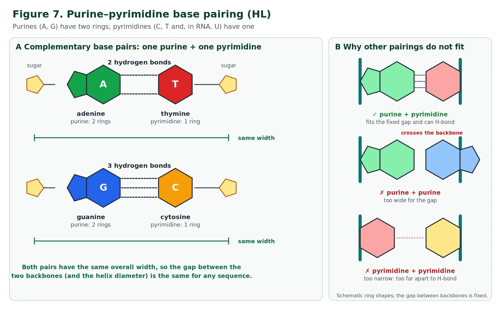
This matters because the two pairs are then the same length. Whichever pair occupies a particular position, the distance between the two sugar–phosphate backbones is the same, so the double helix has a uniform diameter and the same three-dimensional shape regardless of its base sequence. A purine–purine pair would bulge the backbones apart, and a pyrimidine–pyrimidine pair would leave a gap too wide for hydrogen bonds to form. Because the shape does not depend on the sequence, any sequence of bases can be stored in a stable molecule, which links back to the limitless coding capacity in A1.2.9. The flat bases stacked inside the helix also attract each other weakly, adding further stability.
You will often see that A–T pairs are joined by two hydrogen bonds and G–C pairs by three. This is correct and helps explain why DNA with a high proportion of G–C pairs needs more heat to separate its strands, but the IB guide states that you are not required to memorise the numbers of hydrogen bonds.
A1.2.13 Structure of a nucleosome HL
The DNA in a single human nucleus would stretch about 2 metres if laid end to end, yet it has to fit inside a nucleus a few micrometres across and still be accessible for transcription and replication. Eukaryotes solve this by winding their DNA around proteins called histones. Under the electron microscope, partly unpacked chromatin looks like a string of beads, and each bead is a nucleosome.
The structure the guide expects you to know is this. At the centre of a nucleosome is a core of eight histone proteins: two copies each of four different histones (H2A, H2B, H3 and H4), which together form a disc-shaped octamer. The DNA double helix is wrapped around this core about twice (in detail, about 1.7 turns, involving roughly 147 base pairs).
An additional histone, H1, binds to the DNA where it enters and leaves the core, holding the DNA in place on the nucleosome. Between neighbouring nucleosomes is a short, variable length of linker DNA. H1 also helps nucleosomes pack together into more condensed fibres when chromosomes condense before cell division (see Cell Division (IB D2.1)).
Packaging happens in stages. The “beads on a string” of nucleosomes and linker DNA form a fibre about 10 nm wide; with the help of H1 this can coil into a thicker fibre often described as about 30 nm wide, and before mitosis or meiosis these fibres are looped and coiled further (supercoiled) into the compact chromosomes visible with a light microscope.
Histones therefore have three linked roles: packaging DNA into a small volume, helping supercoil chromosomes for cell division, and regulating gene expression. The last role depends on the histone tails, chains of amino acids that project from the nucleosome core; chemical tags added to these tails can loosen or tighten the packaging and so make genes more or less accessible for transcription. Prokaryotes have no nucleosomes: their DNA is not associated with histones and is described as “naked”.
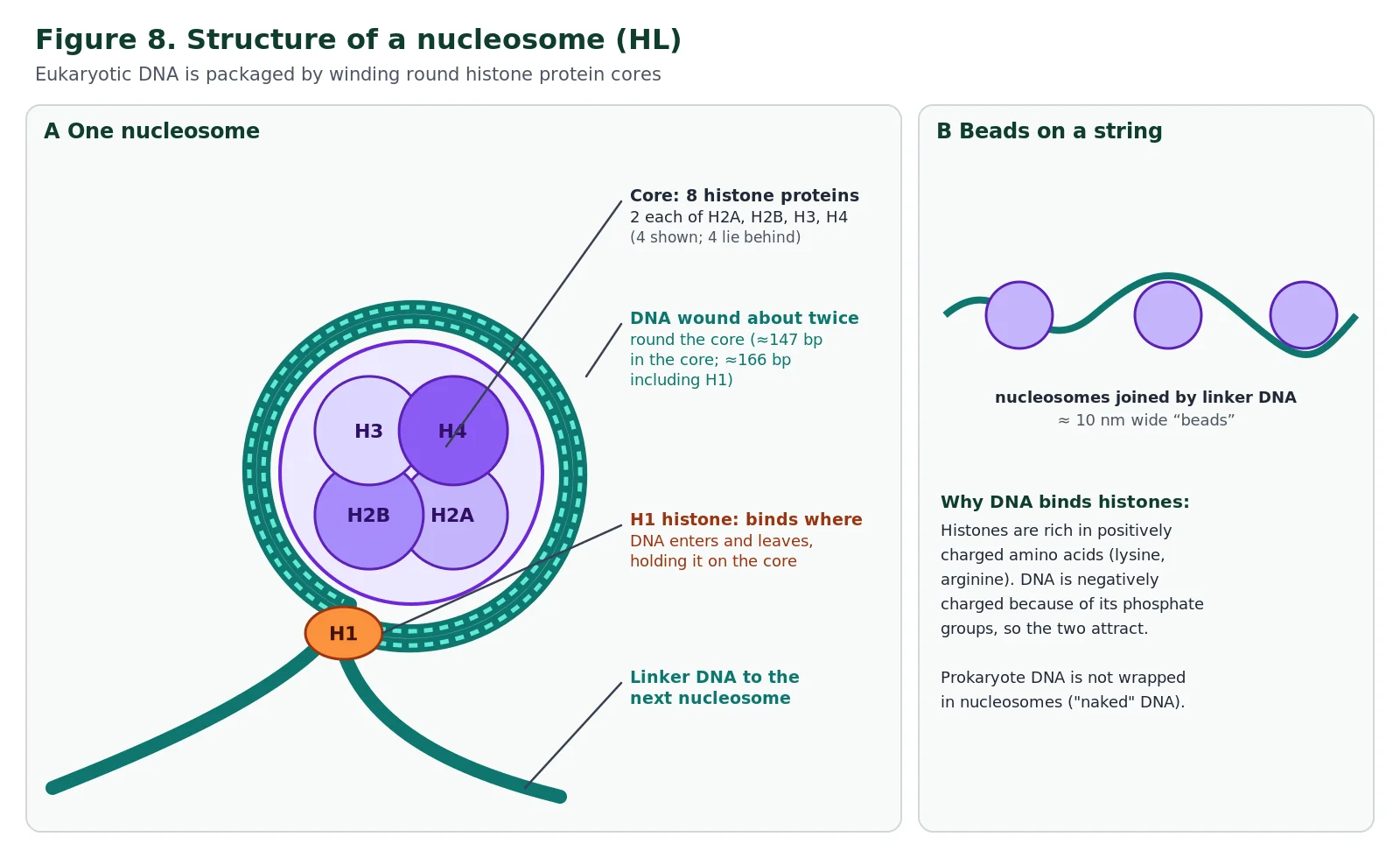
Why does DNA bind so readily to histones? Histones are rich in the amino acids lysine and arginine, whose side chains are positively charged, while DNA is negatively charged along its whole length because of its phosphate groups. The attraction between opposite charges holds DNA on the histone core without any need for particular base sequences. Prokaryotes lack nucleosomes; their DNA is often described as “naked” because it is not wrapped around histones, as you may recall from Introduction to Cells (IB A2.2). Chemical tags added to the tails of histones can loosen or tighten packing and so help regulate which genes are transcribed.
Skill: using molecular visualisation software
The guide requires you to explore a nucleosome in 3D. Open a nucleosome structure in the free RCSB Protein Data Bank viewer (for example entry 1KX5). Rotate the model and try to: (1) count the eight histone chains and find the pairs of identical histones; (2) trace the DNA as it wraps around the core; (3) find the histone tails that project out from the core; (4) colour the structure by charge and notice where positively charged regions of the proteins meet the negatively charged DNA backbone.
A1.2.14 Evidence from the Hershey–Chase experiment for DNA as the genetic material HL
By the 1940s it was clear that genes were carried on chromosomes and that chromosomes contained both protein and DNA, but which of these was the genetic material was still debated. Many biologists favoured protein. Proteins are built from twenty different amino acids and were known to carry out many specific functions, whereas DNA, with only four kinds of nucleotide, was widely thought to be too simple and repetitive to carry complex information (see A1.2.15).
Before Hershey and Chase: Levene, Griffith and Avery
Part of the reason for doubting DNA was the work of Phoebus Levene, a chemist who in the early twentieth century identified deoxyribose as the sugar in DNA, showed that nucleic acids have a sugar–phosphate backbone and coined the word “nucleotide”. Levene also proposed that DNA was a simple repeating unit of the four nucleotides, the tetranucleotide hypothesis, which would make DNA far too uniform to store genetic information. In 1928 Frederick Griffith found that heat-killed virulent Streptococcus pneumoniae bacteria could “transform” living harmless bacteria into a virulent form that killed mice, so some chemical from the dead cells carried heritable information.
In 1944 Oswald Avery, Colin MacLeod and Maclyn McCarty purified this transforming principle and showed that enzymes that digest protein or RNA did not stop transformation, but an enzyme that digests DNA (DNase) did. Their evidence pointed to DNA, but many biologists remained unconvinced, partly because of the tetranucleotide hypothesis and partly because traces of protein might have contaminated the samples. Hershey and Chase’s experiment, using a completely different method, settled the matter for most scientists.
In 1952 Alfred Hershey and Martha Chase tested the question using the bacteriophage T2, a virus that infects the bacterium Escherichia coli. T2 consists only of a protein coat surrounding a DNA molecule: a head containing the DNA and a hollow tail with fibres that attach to the bacterium, through which the DNA is injected. When it infects a bacterium, part of the virus enters the cell and directs it to make new viruses, so that part must contain the virus’s genes.
The key to the experiment was a chemical difference between the two candidates: proteins contain sulfur but no phosphorus (sulfur occurs in the amino acids cysteine and methionine), whereas DNA contains phosphorus but no sulfur. Hershey and Chase grew one batch of phages with radioactive sulfur (35S), which labelled their protein coats, and another batch with radioactive phosphorus (32P), which labelled their DNA.
Each batch was allowed to infect a separate culture of bacteria. After a few minutes the cultures were agitated in a kitchen blender (results are often quoted after about 8 minutes of agitation) to shear off the phage coats still attached to the outside of the cells, and then centrifuged. The bacteria, being larger and denser, formed a pellet at the bottom of the tube, while the much lighter phage coats stayed in the liquid supernatant. Radioactivity was then measured in each fraction. The empty protein coats left behind after injecting their DNA are sometimes called phage “ghosts”.
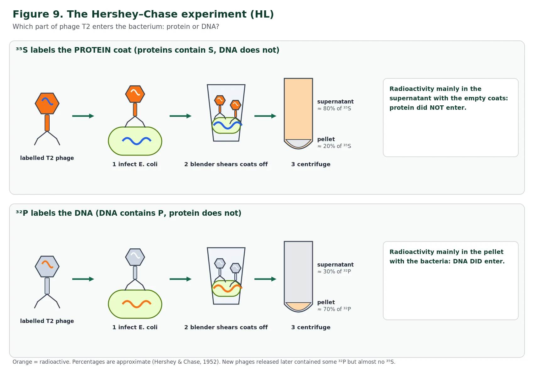
With 35S-labelled phages, most of the radioactivity (about 80%) was found in the supernatant with the empty coats, so the protein had stayed outside the bacteria. With 32P-labelled phages, most of the radioactivity (roughly 70%) was in the pellet with the bacteria, so the DNA had entered the cells. In addition, the new phages produced inside the bacteria contained some of the 32P but almost none of the 35S. Since the material that enters the cell and is passed to the next generation of phages must be the genetic material, the results supported the conclusion that genes are made of DNA, not protein.
The results were not perfectly clean. Some 35S ended up in the pellet because the blender did not remove every coat and some supernatant was trapped in the pellet, and some 32P stayed in the supernatant because not every phage injected its DNA and some cells broke open. A good exam answer acknowledges these imperfections and explains why they do not change the conclusion.
Nature of science: technology opens new experiments
The experiment depended on radioisotopes, which only became widely available to biologists in the 1940s. Isotopes of an element have the same number of protons but different numbers of neutrons; for example, ordinary 32S has 16 protons and 16 neutrons and is stable, whereas 35S has 16 protons and 19 neutrons and is unstable, releasing radiation as it decays.
Because chemical behaviour depends on protons and electrons, not neutrons, isotopes of an element behave identically in chemical reactions, so cells build 35S and 32P into molecules exactly as they would the ordinary isotopes, but the radioactive atoms can be detected and measured. A new technology made it possible to “follow” each type of molecule separately, and so to answer a question that had been debated for decades. Radioactive tracers had been pioneered by George de Hevesy, who received the 1943 Nobel Prize in Chemistry for this work.
A1.2.15 Chargaff’s data on the relative amounts of purine and pyrimidine bases HL
Before the structure of DNA was known, a popular idea called the tetranucleotide hypothesis (proposed in the early twentieth century) held that DNA was made of a short sequence of the four nucleotides repeated over and over. If that were true, the four bases would be present in equal amounts, 25% each, in every DNA sample, and DNA could carry little or no information. This was one reason many scientists assumed proteins must be the genetic material.
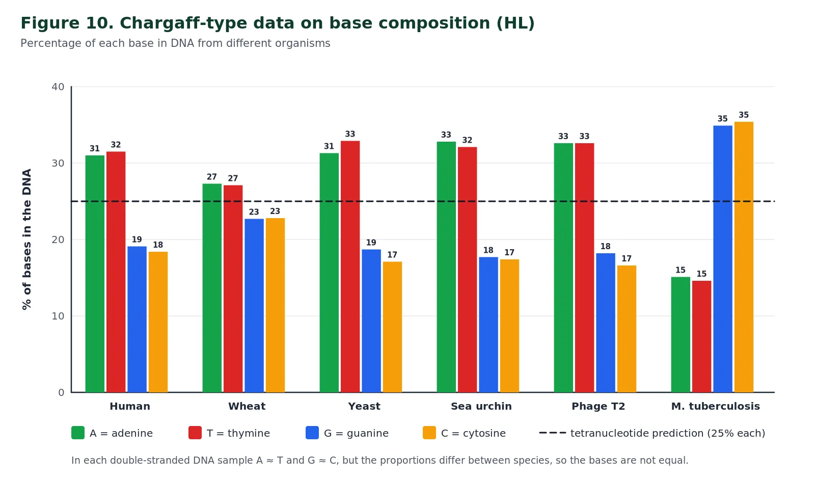
In the late 1940s Erwin Chargaff and his colleagues used newly developed paper chromatography to measure the proportions of the four bases in DNA from many different species. Their results, and those of others who repeated the work, are summarised below. Two clear patterns emerged.
| Source of DNA | Type of organism | % A | % T | % G | % C |
|---|---|---|---|---|---|
| Human | Mammal | 31.0 | 31.5 | 19.1 | 18.4 |
| Wheat | Plant | 27.3 | 27.1 | 22.7 | 22.8 |
| Yeast | Fungus | 31.3 | 32.9 | 18.7 | 17.1 |
| Sea urchin | Invertebrate | 32.8 | 32.1 | 17.7 | 17.4 |
| Bacteriophage T2 | Virus (double-stranded DNA) | 32.6 | 32.6 | 18.2 | 16.6 |
| Mycobacterium tuberculosis | Bacterium | 15.1 | 14.6 | 34.9 | 35.4 |
First, the four bases are not present in equal amounts, and the proportions differ from one species to another: human DNA is about 62% A + T, whereas the DNA of Mycobacterium tuberculosis is about 70% G + C. Second, within each sample the amount of adenine is very close to the amount of thymine, and the amount of guanine is very close to the amount of cytosine.
It follows that purines (A + G) make up about half of the bases and pyrimidines (T + C) the other half. These regularities are known as Chargaff’s rules. Chargaff could not explain the second pattern; it made sense only when Watson and Crick showed that A pairs with T and G with C across the two strands.
Data check: purine to pyrimidine ratio
Using the human data in the table, calculate the ratio (A + G) ÷ (T + C) to two decimal places.
Show working
A + G = 31.0 + 19.1 = 50.1; T + C = 31.5 + 18.4 = 49.9. Ratio = 50.1 ÷ 49.9 = 1.00. For M. tuberculosis: (15.1 + 34.9) ÷ (14.6 + 35.4) = 50.0 ÷ 50.0 = 1.00. The ratio is close to 1 in every double-stranded DNA sample, even though the A + T content varies widely.
Nature of science: the problem of induction and the certainty of falsification
Scientists generalise from a limited number of observations to a universal claim. This is induction, and its weakness is that the next observation could always be different, so no generalisation can be proved with certainty. Falsification turns this around: a single reliable counter-example is enough to show a hypothesis is false. Chargaff’s measurements were such a counter-example. Because the bases were not equal in amount, the tetranucleotide hypothesis was falsified with certainty, and the way was opened for DNA to be taken seriously as the genetic material.
Nature of science and TOK links SL & HL
A1.2 is rich in examples of how scientific knowledge is built. The structure of DNA was not observed directly; it was inferred by building models that had to fit X-ray diffraction measurements, chemical data and base ratios at the same time. The Hershey–Chase experiment shows how a new technology, radioisotope labelling, made a long-standing question testable. Chargaff’s data show how a single set of careful measurements can falsify a widely held hypothesis. The story of Rosalind Franklin raises questions about recognition, collaboration and competition in science.
TOK questions to discuss
- To what extent can we claim to “know” the structure of a molecule that no one has ever seen directly?
- Hershey and Chase’s results were not 100% clear-cut. How much uncertainty can an experiment contain and still provide convincing evidence?
- Is keeping research data secret ever justified in science, and how does the sharing of data affect who receives credit?
- Linking question: what makes RNA more likely than DNA to have been the first genetic material? (Think about RNA’s ability both to store information and to act as a catalyst.)
Common mistakes and examiner tips SL & HL
✗ Saying a nucleotide is a sugar, a phosphate and an amino acid
✓ A nucleotide = pentose sugar + phosphate + nitrogenous base. Amino acids are the monomers of proteins.
✗ Joining bases together to make the backbone
✓ The backbone is alternating sugar and phosphate joined by covalent bonds. Bases project from it and pair with the other strand by hydrogen bonds.
✗ Drawing both strands the same way up
✓ Antiparallel strands point in opposite directions; draw one strand’s pentagons upside down and, at HL, label the 5′ and 3′ ends.
✗ Writing that RNA contains thymine
✓ RNA has uracil instead of thymine. Thymine is found in DNA.
✗ Saying deoxyribose has no oxygen
✓ Deoxyribose has one oxygen atom fewer than ribose: H instead of OH on carbon 2′.
✗ Calling the bonds between bases covalent
✓ Base pairs are held by hydrogen bonds, which is why strands can separate for replication without breaking the backbone.
✗ Pairing A with G because both are purines
✓ Complementary pairs are always one purine + one pyrimidine: A–T (A–U in RNA) and G–C.
✗ Applying A = T and G = C to RNA or single-stranded DNA
✓ Chargaff’s pairing rule applies only to double-stranded DNA. Single-stranded genomes need not follow it.
✗ Saying Hershey–Chase showed protein entered the cell
✓ 35S (protein) stayed mostly in the supernatant; 32P (DNA) entered the cells, so DNA is the genetic material.
✗ Writing that DNA is made 3′ to 5′
✓ New nucleotides are added to the 3′ end, so strands are built 5′→3′; the template is read 3′→5′.
✗ Saying hydrogen bonds join the nucleotides in a strand
✓ Nucleotides in a strand are joined by covalent phosphodiester bonds (3′–OH to 5′ phosphate). Hydrogen bonds only join bases on opposite strands.
✗ Calling the sugar–base link a peptide or phosphodiester bond
✓ The base is joined to C1′ of the sugar by an N-glycosidic bond; peptide bonds join amino acids and phosphodiester bonds join nucleotides.
✗ Describing purines as one-ring bases
✓ Purines (A, G) have two fused rings; pyrimidines (C, T, U) have one ring, even though “pyrimidine” is the longer word.
Key terms flip cards SL & HL
Tap or press Enter on a card to see the definition. Try to say the definition aloud before you flip.
NucleotideFlip
The monomer of nucleic acids: a pentose sugar, a phosphate group and a nitrogenous base joined by covalent bonds.
Sugar–phosphate backboneFlip
The chain of alternating sugar and phosphate groups, joined by covalent bonds, that forms the strong spine of each DNA or RNA strand.
Condensation reactionFlip
A reaction that joins two molecules into one with the release of water; used to link nucleotides.
Complementary base pairingFlip
The pairing of A with T (or U) and G with C by hydrogen bonds.
AntiparallelFlip
Describes the two strands of DNA, which run side by side in opposite directions (5′→3′ and 3′→5′).
Double helixFlip
The shape of DNA: two antiparallel strands twisted round each other, about 2 nm in diameter.
Semi-conservative replicationFlip
Replication in which each new DNA molecule contains one original strand and one newly made strand.
CodonFlip
A sequence of three bases in mRNA that codes for one amino acid or a stop signal.
PurineFlip
A base with two fused rings (6 + 5 atoms, four N): adenine or guanine. Bonds to the sugar at N9.
PyrimidineFlip
A base with one six-membered ring (two N): cytosine, thymine or uracil. Bonds to the sugar at N1.
Nucleosome (HL)Flip
DNA wrapped round a core of eight histone proteins, held by an additional H1 histone, joined to neighbours by linker DNA.
Tetranucleotide hypothesis (HL)Flip
The falsified idea that DNA is a repeating sequence of the four nucleotides in equal amounts.
N-glycosidic bondFlip
The covalent bond joining C1′ of the pentose to a ring nitrogen of the base (N9 of a purine, N1 of a pyrimidine), formed by condensation.
Phosphodiester bondFlip
The covalent link between nucleotides: the 3′ carbon of one sugar is joined through a phosphate to the 5′ carbon of the next (C3′–O–P–O–C5′).
NucleosideFlip
A base bonded to a pentose sugar, with no phosphate. Adding phosphate to C5′ makes a nucleotide.
Methyl group in thymineFlip
The –CH₃ on carbon 5 that distinguishes thymine (DNA) from uracil (RNA).
Self-check: multiple-choice questions SL & HL
Questions 1–14 cover SL and HL content; questions 15–20 are HL only. Choose an answer to get instant feedback, or answer them all and press Check all.
Exam-style and data-based questions SL & HL
Attempt each question on paper before revealing the model answer. Ticks (✔) show where a mark would be awarded; you need one valid point per mark. Questions 10 and 11 use published experimental data.
1. [SL & HL] Draw and label a diagram of a single DNA nucleotide. [3]
Show model answer (markscheme style)
Circle labelled phosphate, pentagon labelled deoxyribose (pentose sugar) and rectangle labelled base ✔; phosphate linked to the sugar on the C5′ side and base linked to C1′ on the opposite side ✔; all parts joined by lines representing covalent bonds ✔.
Do not credit “ribose” or an unattached base.
2. [SL & HL] Compare and contrast the structure of DNA and RNA. [4]
Show model answer (markscheme style)
Similarities (any one): both are polymers of nucleotides ✔; both have a sugar–phosphate backbone with covalent bonds ✔; both contain A, C and G ✔. Differences (any three): DNA usually double-stranded, RNA usually single-stranded ✔; DNA contains deoxyribose, RNA ribose ✔; DNA contains thymine, RNA uracil ✔; DNA strands form a double helix, RNA does not ✔.
“Compare and contrast” needs at least one similarity and one difference.
3. [SL & HL] Explain how complementary base pairing allows DNA to be copied accurately. [4]
Show model answer (markscheme style)
The two strands separate as hydrogen bonds between bases are broken ✔; each strand acts as a template ✔; free nucleotides pair with complementary bases on the template, A with T and G with C ✔; pairing depends on hydrogen bonding between specific bases ✔; each new strand has the same sequence as the strand previously paired with the template, giving two identical molecules ✔; replication is semi-conservative ✔.
4. [SL & HL] A sample of double-stranded DNA contains 21% guanine. Calculate the percentage of adenine. Show your working. [2]
Show model answer (markscheme style)
G = C = 21%, so G + C = 42% ✔; A + T = 58%, and A = T, so A = 29% ✔.
5. [SL & HL] Draw a labelled diagram to show how two nucleotides are linked in a strand of RNA, and name the bonds within and between the nucleotides. [4]
Show model answer (markscheme style)
Two nucleotides drawn as circle–pentagon–rectangle with the phosphate of the second nucleotide linked to the sugar of the first ✔; label ribose, phosphate and base ✔; base joined to C1′ of each sugar by an N-glycosidic bond ✔; nucleotides joined by a phosphodiester bond between the 3′ carbon of one sugar and the 5′ phosphate of the next ✔; formed by condensation with loss of water (accept as an alternative mark) ✔.
6. [SL & HL] Distinguish between the structure of purines and pyrimidines, and explain why a DNA base pair always contains one of each. [4]
Show model answer (markscheme style)
Purines (adenine, guanine) have two fused rings, a six-membered ring and a five-membered ring ✔; pyrimidines (cytosine, thymine, uracil) have one six-membered ring ✔; (purines attach to the sugar at N9, pyrimidines at N1 ✔); a purine + pyrimidine pair is always the same width / a purine–purine pair would be too wide and a pyrimidine–pyrimidine pair too narrow ✔; so the backbones stay a constant distance apart and the helix has a uniform diameter of about 2 nm ✔.
7. [HL] Outline the significance of the directionality of nucleic acids for DNA replication and transcription. [4]
Show model answer (markscheme style)
Strands have a 5′ end (free phosphate) and a 3′ end (free sugar –OH) ✔; nucleotides can only be added to the 3′ end of a growing strand ✔; so DNA and RNA are synthesised in the 5′→3′ direction ✔; polymerases can only bind the strand in one orientation ✔; template strands are read 3′→5′, and because DNA strands are antiparallel replication differs on the two strands ✔; in transcription only one strand is used as the template ✔.
8. [HL] Explain how purine-to-pyrimidine bonding contributes to the stability of the DNA double helix. [3]
Show model answer (markscheme style)
Adenine and guanine are purines with two rings; cytosine and thymine are pyrimidines with one ring ✔; every base pair contains one purine and one pyrimidine ✔; so A–T and C–G pairs have the same length ✔; the distance between the backbones is constant, giving a helix of uniform diameter ✔; so any base sequence can be accommodated without distorting the molecule ✔.
9. [HL] Describe the structure of a nucleosome. [3]
Show model answer (markscheme style)
Core of eight histone proteins ✔ (two each of four types); DNA wrapped around the core about twice ✔; an additional histone (H1) holds the DNA in place ✔; linker DNA connects adjacent nucleosomes ✔; positively charged histones attract negatively charged DNA ✔.
10. [HL, data-based] In the Hershey–Chase experiment, about 80% of the 35S was found in the supernatant and about 70% of the 32P was found in the pellet. (a) Explain how these results support the conclusion that DNA is the genetic material. [3] (b) Suggest why about 20% of the 35S was found in the pellet. [1] [4]
Show model answer (markscheme style)
(a) 35S labels protein and 32P labels DNA ✔; most protein stayed outside the bacteria, in the supernatant with the coats ✔; most DNA entered the bacteria, in the pellet ✔; the material that enters the cell directs production of new phages, so it is the genetic material ✔ (new phages also contained 32P).
(b) Some protein coats were not removed by blending and stayed attached to cells ✔, or some supernatant was trapped in the pellet.
11. [HL, data-based] Base composition (%) of three genomes: human DNA A 31.0, G 19.1, C 18.4, T 31.5; M. tuberculosis DNA A 15.1, G 34.9, C 35.4, T 14.6; poliovirus A 30.4, G 25.4, C 19.5, T 0.0. (a) Calculate (A + G) ÷ (T + C) for M. tuberculosis. [1] (b) Explain how data like these falsified the tetranucleotide hypothesis. [2] (c) Suggest reasons for the composition of the poliovirus genome. [2] [5]
Show model answer (markscheme style)
(a) (15.1 + 34.9) ÷ (14.6 + 35.4) = 50.0 ÷ 50.0 = 1.0 ✔.
(b) The hypothesis predicted equal amounts (25%) of each base ✔; the bases are unequal and the proportions differ between species, for example 31% A in humans but 15% A in M. tuberculosis ✔; so DNA is not a simple repeating unit and could carry information ✔.
(c) No thymine, so the genome is RNA, containing uracil (about 24.7%) ✔; A ≠ U and G ≠ C, so it is single-stranded and the bases are not paired ✔.
References and further reading SL & HL
These authoritative sources support specific facts in this chapter. The explanations, diagrams and questions here are original.
- Hershey, A. D. and Chase, M. (1952). Independent functions of viral protein and nucleic acid in growth of bacteriophage. Journal of General Physiology 36(1): 39–56: the original radioisotope experiment.
- Chargaff, E. (1950). Chemical specificity of nucleic acids and mechanism of their enzymatic degradation. Experientia 6: 201–209: base composition data and the regularities now called Chargaff’s rules.
- Watson, J. D. and Crick, F. H. C. (1953). Molecular structure of nucleic acids: a structure for deoxyribose nucleic acid. Nature 171: 737–738: the double-helix model.
- Luger, K. et al. (1997). Crystal structure of the nucleosome core particle at 2.8 Å resolution. Nature 389: 251–260: histone octamer and about 147 bp of wrapped DNA.
- RCSB Protein Data Bank, entry 1KX5: nucleosome core particle: free 3D structure for the molecular visualisation skill.
- OpenStax Biology 2e, 14.2 DNA Structure and Sequencing: dimensions of the double helix (2 nm diameter, 0.34 nm per base pair).
Nucleic acids IB Biology: frequently asked questions SL & HL
What are nucleic acids?
Nucleic acids are large polymers made of nucleotide monomers. The two types are DNA (deoxyribonucleic acid), which stores hereditary information, and RNA (ribonucleic acid), which helps express that information as proteins.
What are the three parts of a nucleotide?
A nucleotide has a phosphate group, a pentose (five-carbon) sugar and a nitrogenous base. The base is attached to carbon 1′ of the sugar and the phosphate to carbon 5′.
What are the main differences between DNA and RNA?
DNA is usually double-stranded, contains the sugar deoxyribose and uses the base thymine. RNA is usually single-stranded, contains ribose and uses uracil instead of thymine.
Why are the two strands of DNA antiparallel?
Complementary bases can only line up and form hydrogen bonds when the two strands run in opposite directions, so one strand runs 5′ to 3′ while its partner runs 3′ to 5′.
Why does adenine pair with thymine and guanine with cytosine?
The hydrogen-bonding groups on adenine match those on thymine, and those on guanine match cytosine. Each pair also combines a two-ring purine with a one-ring pyrimidine, so every pair is the same width.
Do I need to know the number of hydrogen bonds in each base pair for IB Biology?
No. The IB guide says students are not required to memorise the numbers of hydrogen bonds, although it is useful to know that A–T pairs have two and G–C pairs have three.
Which parts of IB topic A1.2 are HL only?
A1.2.11 to A1.2.15 are additional higher level: directionality of DNA and RNA, purine-to-pyrimidine bonding, nucleosome structure, the Hershey–Chase experiment and Chargaff’s data.
What did the Hershey–Chase experiment prove?
It showed that the DNA of bacteriophage T2, labelled with phosphorus-32, entered bacteria while the protein coat, labelled with sulfur-35, stayed outside. This supported DNA rather than protein as the genetic material.
What bond joins the sugar and the base in a nucleotide?
An N-glycosidic (β-N-glycosidic) bond. It is a covalent bond between carbon 1′ of the pentose and nitrogen 9 of a purine (adenine, guanine) or nitrogen 1 of a pyrimidine (cytosine, thymine, uracil), formed by a condensation reaction that releases water.
What bond is formed when two nucleotides join together?
A phosphodiester bond. It forms by condensation between the hydroxyl group on carbon 3′ of one nucleotide’s sugar and the phosphate on carbon 5′ of the next, releasing a water molecule. Repeated phosphodiester bonds make the sugar–phosphate backbone, which runs from a 5′ end to a 3′ end.
What is the difference between a purine and a pyrimidine?
Purines (adenine and guanine) have two fused rings, a six-membered ring joined to a five-membered ring, with four nitrogen atoms. Pyrimidines (cytosine, thymine and uracil) have a single six-membered ring with two nitrogen atoms. In DNA each base pair contains one purine and one pyrimidine, which keeps the helix about 2 nm wide.
What is the difference between a nucleoside and a nucleotide?
A nucleoside is a nitrogenous base bonded to a pentose sugar. A nucleotide is a nucleoside with one or more phosphate groups attached to carbon 5′ of the sugar. Nucleotides are the monomers of DNA and RNA.
What are Chargaff’s rules?
In double-stranded DNA the amount of adenine equals the amount of thymine and the amount of guanine equals the amount of cytosine, so purines make up half of the bases. The overall base composition varies between species.
What is a nucleosome made of?
A nucleosome is a length of DNA wrapped about twice around a core of eight histone proteins, with an additional H1 histone holding it in place. Linker DNA joins neighbouring nucleosomes.
Why is the genetic code described as universal?
With only minor exceptions, all organisms and viruses use the same 64 codons with the same meanings, which is strong evidence that all life shares a common ancestor.