
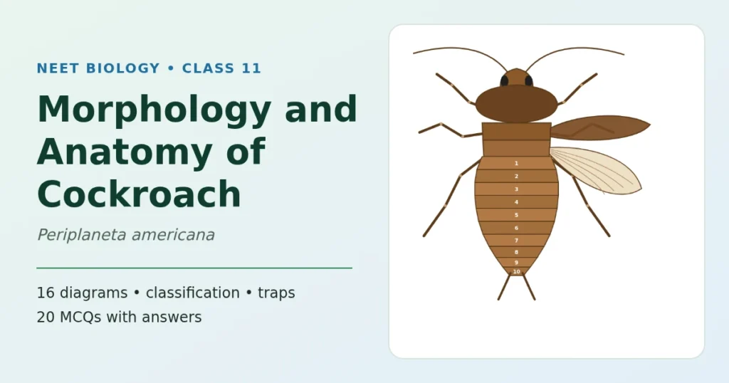
NEET Biology • Class XI • Structural Organisation in Animals
The cockroach, one system at a time
NEET (UG) syllabus: an insect, brief accountThe cockroach, Periplaneta americana, is the insect you study to learn how an arthropod body is built. These notes explain the morphology of cockroach (what you see from outside) and the anatomy of cockroach (the organ systems inside) in simple steps: classification from kingdom to species, head, mouthparts, thorax, legs and wings, the abdomen and how to tell male from female, then the digestive, circulatory, respiratory, excretory, nervous and reproductive systems. Each part has its own clear diagram. At the end you get a numbers table, common traps, 20 MCQs and FAQs.
- 19 diagrams
- Kingdom to species
- All 6 organ systems
- Common traps
- 20 MCQs
Cockroach at a glance
Cockroaches are brown or black insects of the class Insecta, phylum Arthropoda. Bright yellow, red and green cockroaches have also been reported in tropical regions. Their size ranges from about ¼ inch to 3 inches (0.6–7.6 cm). They have long antennae, long legs and a flat extension of the upper body wall that hides the head from above.
Cockroaches are nocturnal omnivores: they come out at night and eat almost anything. They live in damp places throughout the world. Many have become residents of human homes, which is why they are serious pests and carry several diseases.
The species you study is the common or American cockroach, Periplaneta americana. Adults are about 34–53 mm long, and in males the wings extend beyond the tip of the abdomen.
| Feature | Cockroach |
|---|---|
| Body plan | Segmented (metameric), bilaterally symmetrical, dorsoventrally flattened |
| Body regions | Head, thorax (3 segments), abdomen (10 segments) |
| Covering | Hard, brown, chitinous exoskeleton made of plates called sclerites |
| Body cavity | Haemocoel (filled with haemolymph) |
| Circulation | Open type |
| Breathing | Tracheae opening by 10 pairs of spiracles |
| Excretion | Malpighian tubules; uricotelic |
| Sexes | Separate (dioecious) |
| Development | Indirect, paurometabolous (egg → nymph → adult) |
Is the cockroach still in the NEET syllabus?
The earlier NCERT Class XI textbook taught the cockroach in detail in Chapter 7 (section 7.4). The current rationalised textbook no longer has that detailed section. The NEET (UG) 2026 syllabus, however, still asks for the “morphology, anatomy and functions of different systems (digestive, circulatory, respiratory, nervous and reproductive) of an insect”, as a brief account. The cockroach is the standard insect for this, and the NCERT Class XI lab manual (Exercise 14) still uses it. So keep this topic in your revision, at the level of these notes.
Classification of cockroach: kingdom to species
Classification places the cockroach in groups that get narrower at each step. Learn the seven ranks in order, and learn why it belongs to each one, because NEET asks the reasons too.
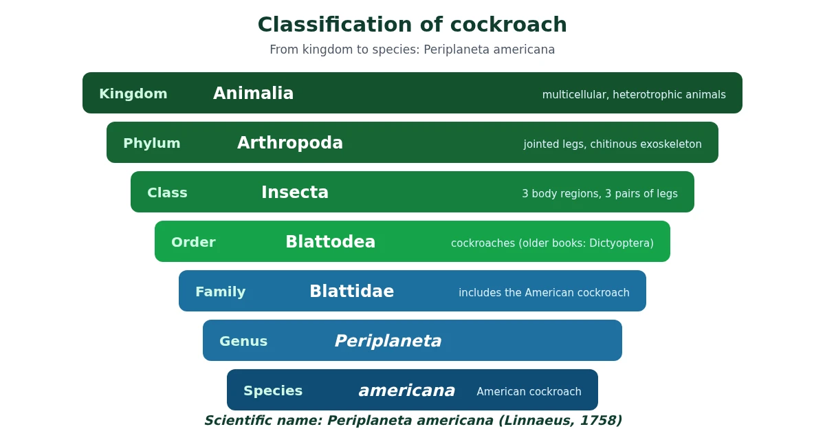
| Rank | Name | Why the cockroach belongs here |
|---|---|---|
| Kingdom | Animalia | Multicellular, eukaryotic and heterotrophic; no cell wall |
| Phylum | Arthropoda | Jointed appendages, chitinous exoskeleton, segmented body, open circulation |
| Class | Insecta (Hexapoda) | Body in head, thorax and abdomen; 3 pairs of legs; 1 pair of antennae; usually 2 pairs of wings; tracheal breathing |
| Order | Blattodea | The cockroach order (older Indian books: Dictyoptera) |
| Family | Blattidae | Includes the American cockroach |
| Genus | Periplaneta | Written with a capital letter, in italics |
| Species | americana | Written in small letters, in italics |
The scientific name is written as Periplaneta americana (Linnaeus, 1758): genus first with a capital letter, species second in small letters, both in italics (or underlined when handwritten).
Order: Blattodea or Dictyoptera?
Many older Indian reference books place the cockroach in the order Dictyoptera (some very old books say Orthoptera). Current taxonomic databases, ITIS and GBIF, list it in the order Blattodea. Neither NCERT text names the order, so if an exam option shows only one of these names, choose the one given.
Morphology of cockroach: body plan and exoskeleton
Morphology means the external form. The body of the cockroach is segmented and divided into three distinct regions: head, thorax and abdomen.
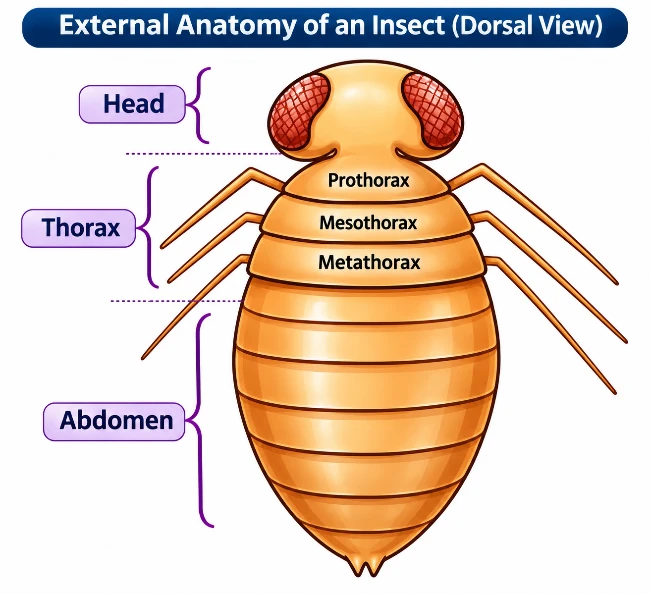
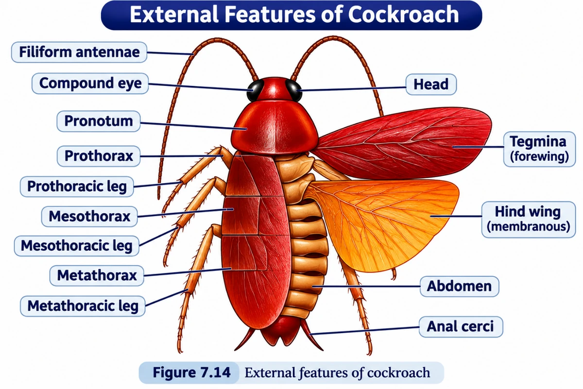
The entire body is covered by a hard, brown chitinous exoskeleton. In each segment, the exoskeleton has hardened plates called sclerites. The dorsal plates are tergites (terga), the ventral plates are sternites (sterna) and the lateral plates are pleurites (pleura). The plates are joined to each other by a thin, flexible articular membrane (arthrodial membrane).
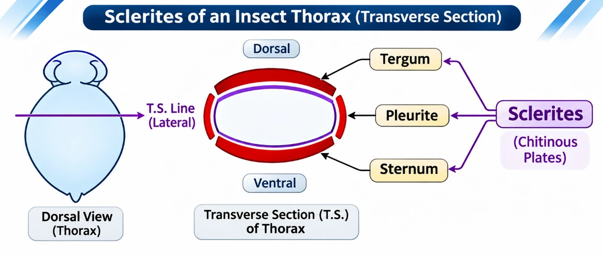
Why both? Hard plates protect the body and give muscles something firm to pull on. The soft membranes between them let the body bend and the segments move. The tergum of the prothorax is especially large and is called the pronotum; it is the shield that partly covers the head when you look from above. The terga of the mesothorax and metathorax are the mesonotum and metanotum.
Head of cockroach and its mouthparts
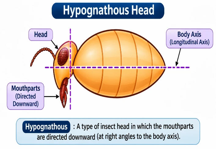
The head is triangular and lies at the front of the body at right angles to the long body axis, with the mouthparts pointing downward. This kind of head is called hypognathous. The head is formed by the fusion of six segments and can move in all directions because of a flexible neck, which is a short extension of the prothorax.
What the head bears
- A pair of compound eyes on the dorsal surface of the head.
- A pair of thread-like (filiform) antennae arising from membranous sockets in front of the eyes. Antennae carry sensory receptors that help the cockroach monitor its surroundings.
- Mouthparts at the front end, of the biting and chewing type.
The five mouthparts
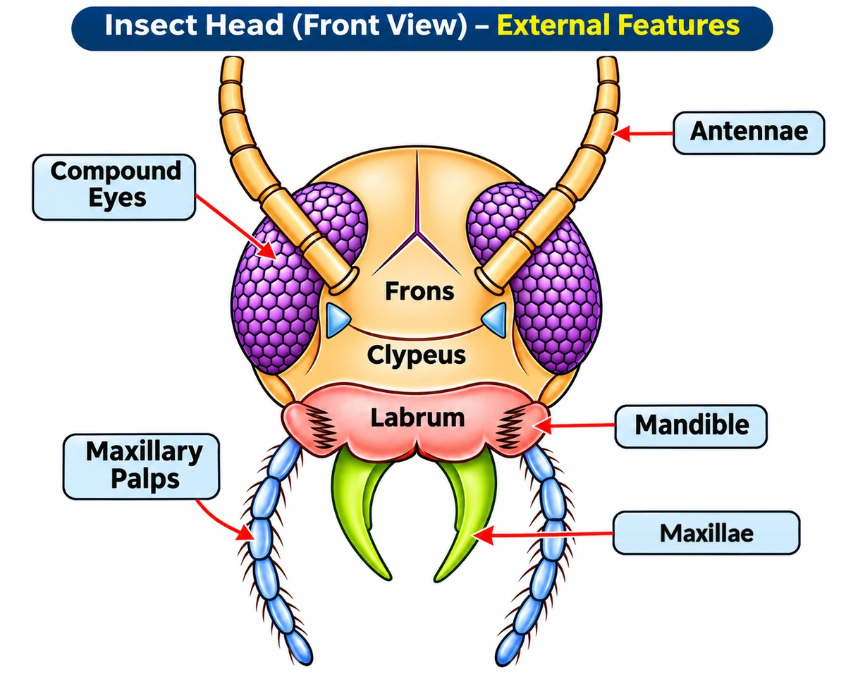
| Mouthpart | Number | What it is and does |
|---|---|---|
| Labrum | 1 | Upper lip; covers the mouthparts from the front |
| Mandibles | 1 pair | Hard jaws with an incising (cutting) region and a grinding region; they bite and crush food |
| Maxillae | 1 pair | Hold food and push it towards the mouth; each bears a jointed maxillary palp, a sense organ |
| Labium | 1 | Lower lip, formed by the fusion of the second pair of maxillae; bears a pair of labial palps |
| Hypopharynx | 1 | A median flexible lobe that acts as the tongue; it lies inside the cavity enclosed by the other mouthparts |
The front view shows the labrum, the mandibles beneath it, the maxillae and the maxillary palps. The labium and hypopharynx lie behind these and are hidden from the front.
Insect mouthparts are modified appendages, so the mouthparts of different insects (biting in a cockroach, piercing and sucking in a mosquito) are homologous: same basic parts, different jobs.
Thorax: legs and wings
The thorax has three segments: prothorax, mesothorax and metathorax. The head is joined to the thorax by the neck, a short extension of the prothorax. Each thoracic segment bears one pair of walking legs, so a cockroach has three pairs of legs, as every insect does.
Leg
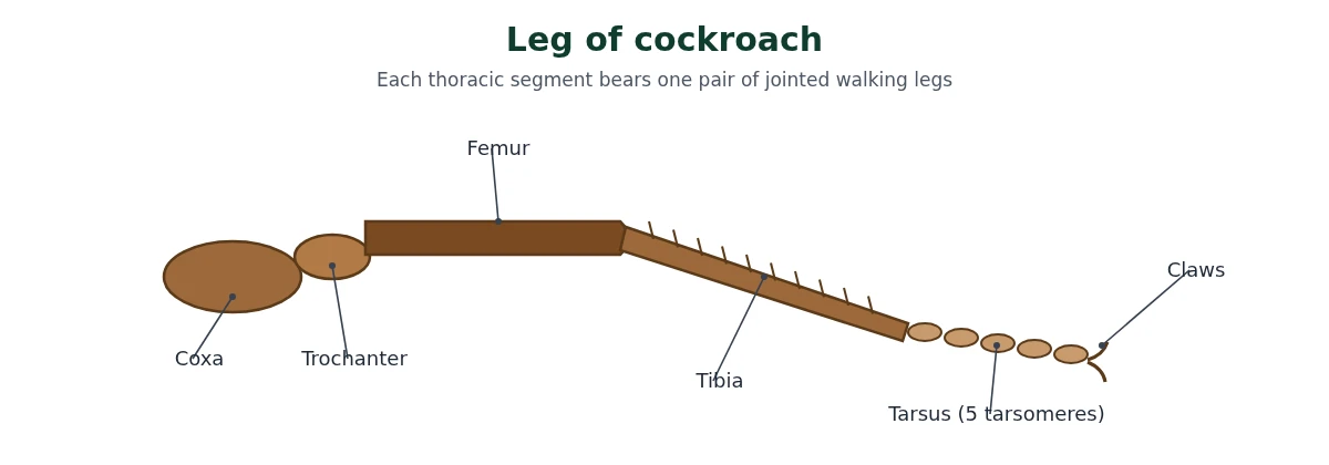
Each leg is jointed and has five main parts: coxa, trochanter, femur, tibia and tarsus. The tarsus is made of five small segments called tarsomeres. The legs are named after the segment they come from: prothoracic, mesothoracic and metathoracic legs.
Wings
There are two pairs of wings, and this is where many students slip:
| Wing | Comes from | Texture | Job |
|---|---|---|---|
| Forewings (tegmina, singular tegmen) | Mesothorax | Opaque, dark and leathery | Cover and protect the hind wings at rest |
| Hind wings | Metathorax | Transparent and membranous | Used in flight |
The prothorax bears no wings; it bears only the first pair of legs and the large pronotum.
Abdomen: how to tell a male from a female
The abdomen has 10 segments in both males and females. The difference between the sexes (sexual dimorphism) is seen at the hind end.
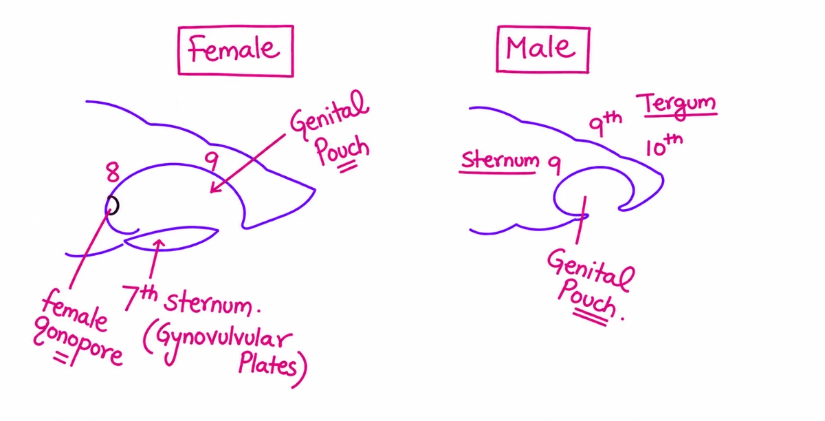
In females, the 7th sternum is boat-shaped. Together with the 8th and 9th sterna it forms a brood or genital pouch. The front part of this pouch contains the female gonopore, the spermathecal pores and the openings of the collateral glands. The lobes of the boat-shaped 7th sternum are also called gynovulvular plates.
In males, the genital pouch or chamber lies at the hind end of the abdomen. It is bounded dorsally by the 9th and 10th terga and ventrally by the 9th sternum. It contains the dorsal anus, the ventral male genital pore and the gonapophyses. Males also bear a pair of short, thread-like anal styles, which are absent in females.
In both sexes, the 10th segment bears a pair of jointed, filamentous anal cerci.
| Feature | Male | Female |
|---|---|---|
| Wings | Extend beyond the tip of the abdomen | Do not extend beyond the abdomen as in males |
| Anal styles | Present (one pair) | Absent |
| Anal cerci | Present (10th segment) | Present (10th segment) |
| 7th sternum | Normal | Boat-shaped; forms the brood pouch with the 8th and 9th sterna |
| Genital pouch | Bounded by 9th and 10th terga and 9th sternum | Formed by 7th, 8th and 9th sterna |
Anatomy of cockroach: digestive system
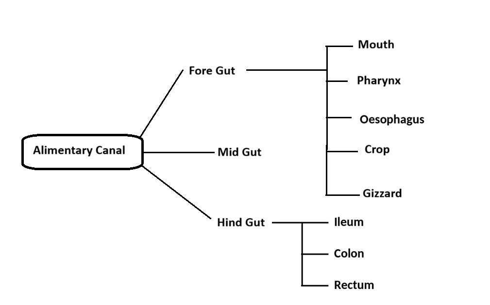
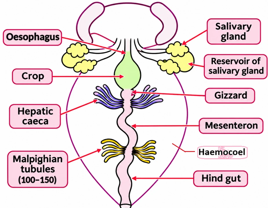
The alimentary canal lies in the body cavity and has three regions: foregut, midgut and hindgut.
Foregut
The mouth opens into a short tubular pharynx, which leads into a narrow tube, the oesophagus. A pair of salivary glands with salivary reservoirs pours saliva near the mouth. The oesophagus opens into a sac-like crop, which stores food. The crop is followed by the gizzard or proventriculus.
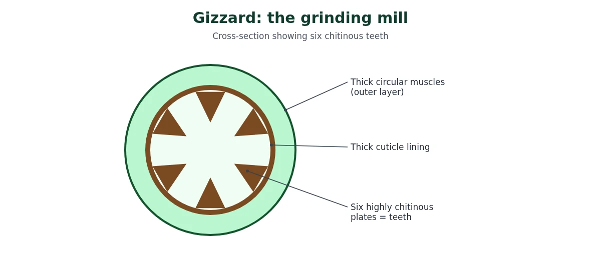
The gizzard has an outer layer of thick circular muscles and a thick inner cuticle that forms six highly chitinous plates called teeth. It grinds the food particles. The whole foregut is lined by cuticle, which protects it from hard bits of food.
Midgut
A ring of 6–8 blind tubules called hepatic or gastric caeca is present at the junction of the foregut and midgut. They secrete digestive juice. The midgut (mesenteron) is where most digestion and absorption happen. It has no cuticle lining, so its cells secrete a thin peritrophic membrane around the food, which protects the delicate midgut wall from hard food particles.
Hindgut
At the junction of the midgut and hindgut is another ring of 100–150 yellow, thin, filamentous Malpighian tubules. They remove excretory products from the haemolymph; they are not digestive glands. The hindgut is broader than the midgut and is differentiated into the ileum, colon and rectum. The rectum opens out through the anus.
| Part | Region | Job |
|---|---|---|
| Crop | Foregut | Stores food |
| Gizzard (proventriculus) | Foregut | Grinds food with 6 chitinous teeth |
| Hepatic (gastric) caeca, 6–8 | Foregut–midgut junction | Secrete digestive juice |
| Midgut (mesenteron) | Midgut | Digestion and absorption; no cuticle |
| Malpighian tubules, 100–150 | Midgut–hindgut junction | Excretion (not digestion) |
| Ileum, colon, rectum | Hindgut | Carry and expel undigested food; rectum opens by the anus |
Circulatory (blood vascular) system
The blood vascular system of the cockroach is of the open type. Blood vessels are poorly developed and open into spaces called the haemocoel. The internal organs lying in the haemocoel are directly bathed in blood, called haemolymph.
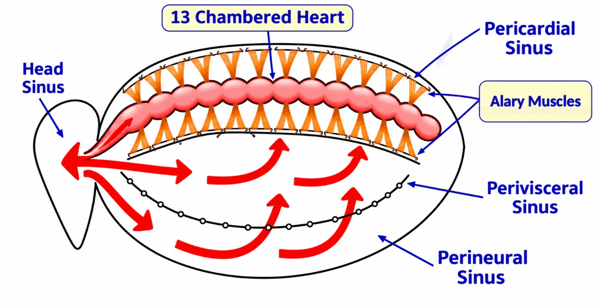
- Haemolymph is made of colourless plasma and blood cells called haemocytes. It has no respiratory pigment, so it does not carry oxygen; the tracheae do that job.
- Heart: an elongated muscular tube lying along the mid-dorsal line of the thorax and abdomen. It is divided into 13 funnel-shaped chambers, each with a pair of openings called ostia, one on either side.
- Alary muscles: paired, fan-shaped (triangular) muscles attached to the heart region. They help the heart fill and pump.
- Sinuses: the haemocoel is divided by the dorsal and ventral diaphragms into the pericardial sinus (dorsal, around the heart), the perivisceral sinus (middle, around the gut) and the perineural sinus (ventral, around the nerve cord).
Path of haemolymph
The heart contracts from back to front, so haemolymph flows from posterior to anterior inside the heart. It leaves through the anterior aorta into the head sinus. From there it spreads back through the perineural and perivisceral sinuses, bathing the organs, then passes into the pericardial sinus. Finally it re-enters the heart through the ostia, and the cycle repeats.
Respiratory system of cockroach
The cockroach breathes through a network of air tubes called tracheae. They open to the outside through 10 pairs of small holes called spiracles on the lateral sides of the body: 2 pairs in the thorax and 8 pairs in the abdomen.
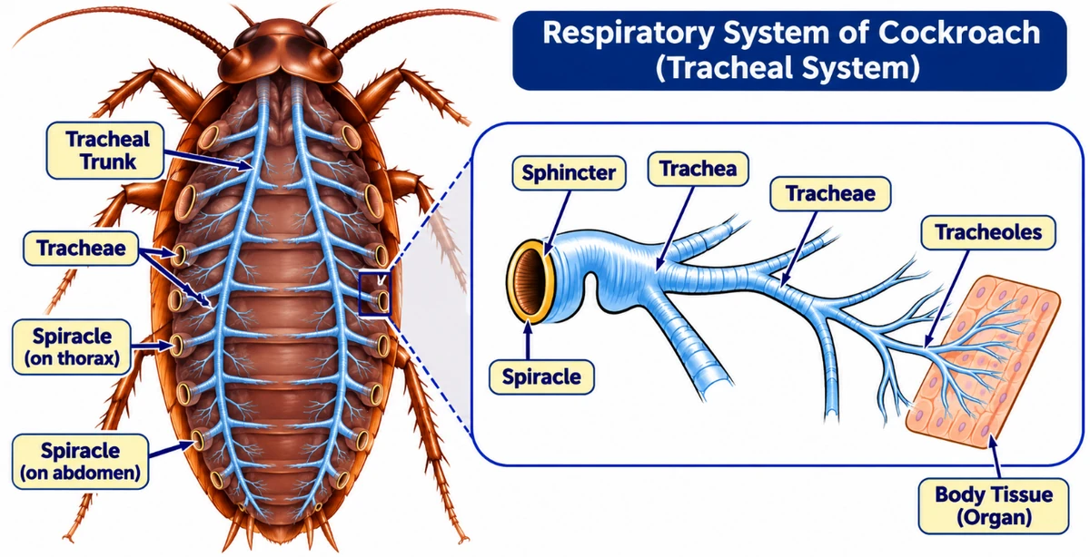
- Thin branching tubes (tracheal tubes, subdivided into tracheoles) carry oxygen from the air straight to all parts of the body.
- The opening of the spiracles is regulated by sphincters.
- The exchange of gases takes place at the tracheoles by diffusion.
The key idea: in a cockroach the respiratory system is separate from the circulatory system. Air goes spiracle → trachea → tracheole → cell. This is why the haemolymph can be colourless and carry no oxygen.
Excretory system of cockroach
Excretion is performed by the Malpighian tubules. Each tubule is lined by glandular and ciliated cells. The tubules lie free in the haemolymph, absorb nitrogenous waste products from it and convert them into uric acid, which is passed out through the hindgut along with undigested food. Because its main nitrogenous waste is uric acid, the cockroach is called uricotelic.
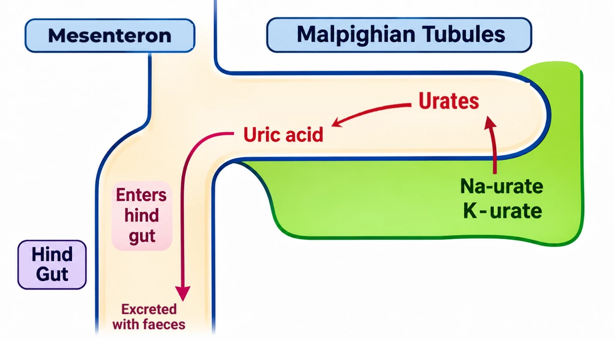
Nitrogenous wastes reach the tubules from the haemolymph as soluble sodium and potassium urates. Inside the tubule they are changed into insoluble uric acid, while water and the sodium and potassium are taken back into the haemolymph. The uric acid then moves into the hindgut and leaves with the faeces. Uric acid needs very little water to remove, which suits an animal living on land.
Three other structures also help in excretion:
- Fat body
- Nephrocytes
- Urecose glands
Nervous system and sense organs
The nervous system of the cockroach is a series of fused, segmentally arranged ganglia joined by paired longitudinal connectives on the ventral side. Three ganglia lie in the thorax and six in the abdomen.
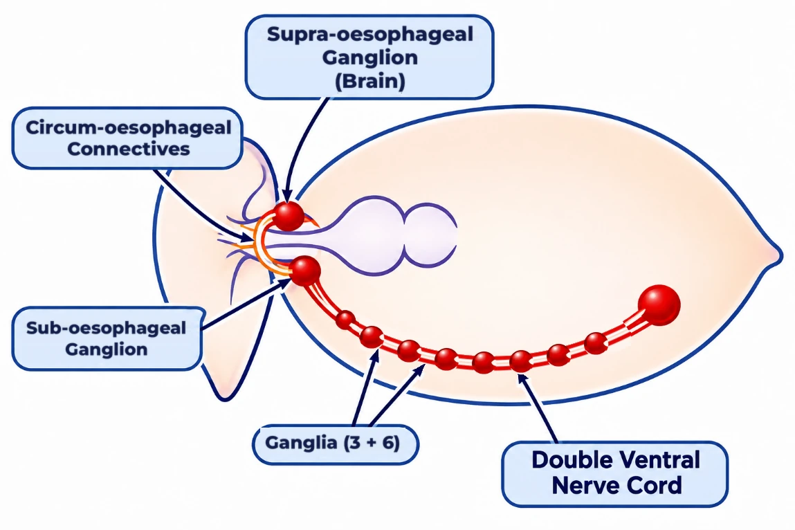
- In the head, the brain is the supra-oesophageal ganglion, which lies above the oesophagus and supplies nerves to the antennae and compound eyes.
- The brain is joined by circum-oesophageal connectives to the sub-oesophageal ganglion below the oesophagus. Together they form a nerve ring around the oesophagus.
- From the sub-oesophageal ganglion, the double ventral nerve cord runs back along the belly side, carrying the thoracic and abdominal ganglia.
The nervous system is spread throughout the body. The head holds only a small part of it, while the rest lies along the ventral side. That is why a cockroach can live for as long as one week after its head is cut off.
Sense organs
The sense organs of the cockroach are the antennae, eyes, maxillary palps, labial palps and anal cerci, among others.
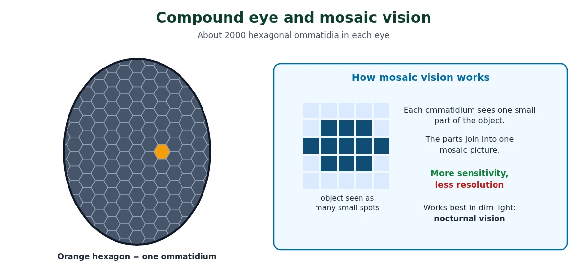
The compound eyes lie on the dorsal surface of the head. Each eye has about 2000 hexagonal ommatidia (singular: ommatidium). With several ommatidia, a cockroach receives several images of an object, one part from each. This is mosaic vision, which gives more sensitivity but less resolution. It works best at night, so it is also called nocturnal vision.
Reproductive system of cockroach
Cockroaches are dioecious: the sexes are separate, and both have well-developed reproductive organs.
Male reproductive system
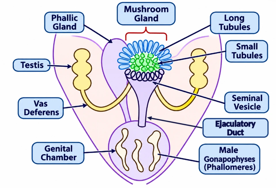
- Testes: one pair, one on each lateral side in the 4th–6th abdominal segments.
- Vas deferens: a thin duct arises from each testis and opens into the ejaculatory duct through the seminal vesicle.
- Ejaculatory duct: opens into the male gonopore, which lies ventral to the anus.
- Mushroom-shaped gland: a characteristic accessory reproductive gland in the 6th–7th abdominal segments, made of long and short tubules.
- External genitalia: the male gonapophyses or phallomeres, chitinous asymmetrical structures surrounding the male gonopore.
The sperms are stored in the seminal vesicles and are glued together in bundles called spermatophores, which are discharged during copulation. Each spermatophore has three coats: the innermost is made by the mushroom gland, the middle one by the ejaculatory duct and the outermost by the phallic gland.
Female reproductive system
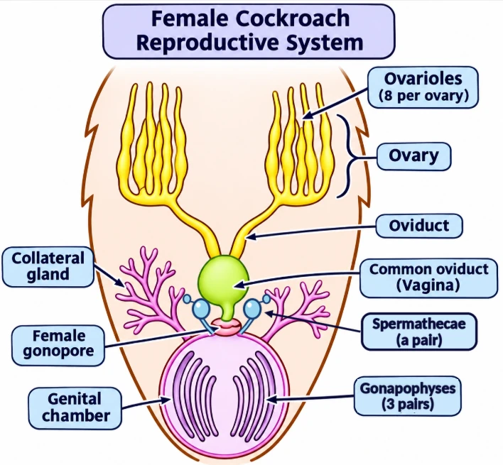
- Ovaries: two large ovaries lie laterally in the 2nd–6th abdominal segments.
- Ovarioles: each ovary is a group of eight ovarian tubules or ovarioles, each containing a chain of developing ova.
- Oviducts: the oviducts of the two ovaries unite into a single median oviduct, also called the vagina, which opens into the genital chamber.
- Spermatheca: a pair is present in the 6th segment and opens into the genital chamber. It stores the sperms received during mating.
- Collateral glands: open into the genital chamber; their secretion forms the egg case (ootheca).
Mating and fertilisation
During copulation the male uses his phallomeres to deliver spermatophores into the female, and she stores the sperms in her spermathecae. One mating can serve for a long time: the female keeps the stored sperms and uses them to fertilise batch after batch of eggs. Eggs released from the ovarioles reach the genital chamber, where they are fertilised by the stored sperms. The fertilised eggs are then encased in a capsule made by the collateral glands.
Ootheca and development
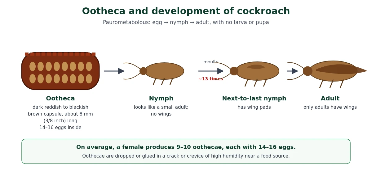
The fertilised eggs are encased in capsules called oothecae (singular: ootheca). An ootheca is a dark reddish to blackish brown capsule about 3/8 inch (8 mm) long. Oothecae are dropped or glued to a suitable surface, usually in a crack or crevice of high relative humidity near a food source. On average, a female produces 9–10 oothecae, each containing 14–16 eggs.
The development of Periplaneta americana is paurometabolous: it passes through a nymphal stage, with no larva and no pupa. The nymphs look very much like adults. A nymph grows by moulting about 13 times to reach the adult form. The next-to-last nymphal stage has wing pads, but only adult cockroaches have wings.
Economic importance of cockroach
Many species of cockroaches are wild and are of no economic importance. A few species live in and around human homes, and these are pests because:
- they destroy food;
- they contaminate food with their smelly excreta;
- they can transmit a variety of bacterial diseases by contaminating food material.
Numbers to remember
NEET loves numbers in this topic. Revise this table the night before the exam.
| What | Number |
|---|---|
| Size range of cockroaches | ¼–3 inches (0.6–7.6 cm) |
| Length of adult P. americana | 34–53 mm |
| Segments fused to form the head | 6 |
| Thoracic segments / pairs of legs / pairs of wings | 3 / 3 / 2 |
| Abdominal segments (both sexes) | 10 |
| Teeth in the gizzard | 6 |
| Hepatic (gastric) caeca | 6–8 |
| Malpighian tubules | 100–150 |
| Chambers in the heart | 13 |
| Pairs of spiracles | 10 (2 thoracic + 8 abdominal) |
| Ganglia on the ventral nerve cord | 3 thoracic + 6 abdominal |
| Ommatidia in each compound eye | About 2000 |
| Testes lie in | 4th–6th abdominal segments |
| Mushroom gland lies in | 6th–7th abdominal segments |
| Ovaries lie in | 2nd–6th abdominal segments |
| Ovarioles per ovary | 8 |
| Spermatheca lies in | 6th segment |
| Oothecae per female (average) | 9–10 |
| Eggs per ootheca | 14–16 |
| Length of ootheca | About 8 mm (3/8 inch) |
| Moults of the nymph | About 13 |
| Survival after the head is cut off | Up to about one week |
Common traps in cockroach questions
These are the points where students lose marks most often.
- The tegmina come from the mesothorax and the hind wings from the metathorax. The prothorax has no wings.
- Anal styles are only in males; anal cerci are in both sexes. Styles are on the 9th sternum, cerci on the 10th segment.
- Hepatic caeca (6–8) are at the foregut–midgut junction; Malpighian tubules (100–150) are at the midgut–hindgut junction. Do not swap the numbers or the positions.
- Malpighian tubules are excretory, not digestive. Digestive juice comes from the hepatic caeca (and saliva).
- The crop stores food; the gizzard grinds it. Gizzard = proventriculus.
- The foregut and hindgut are lined by cuticle; the midgut is not.
- Haemolymph is colourless and has no respiratory pigment. Oxygen reaches the cells through the tracheae, not through blood.
- There are 10 pairs of spiracles (20 spiracles in all), not 10 spiracles.
- Gas exchange happens at the tracheoles, not at the spiracles.
- Blood flows from back to front in the heart, and enters the heart through the ostia.
- Ganglia: 3 thoracic + 6 abdominal. The brain is the supra-oesophageal ganglion, not the sub-oesophageal one.
- Mosaic vision means more sensitivity and less resolution, not the other way round.
- Testes: 4th–6th segments. Ovaries: 2nd–6th. Mushroom gland: 6th–7th. Spermatheca: 6th.
- Development is paurometabolous: nymphs, no larva, no pupa. Only adults have wings; the next-to-last nymph has only wing pads.
20 cockroach MCQs with answers
Original practice questions: 14 single-answer (including a statement count), 3 assertion-reason, 2 match-the-column and 2 diagram-based. Try each one before you show the answer.
Q1. Which is the correct classification of the American cockroach?
- (a) Arthropoda → Arachnida → Blattidae → Periplaneta
- (b) Arthropoda → Insecta → Blattidae → Periplaneta
- (c) Annelida → Insecta → Blattidae → Periplaneta
- (d) Arthropoda → Crustacea → Blattidae → Periplaneta
Answer: (b) Phylum Arthropoda, class Insecta, family Blattidae, genus Periplaneta, species americana.
Q2. The forewings of cockroach, called tegmina, arise from the:
- (a) prothorax
- (b) mesothorax
- (c) metathorax
- (d) first abdominal segment
Answer: (b) Tegmina are mesothoracic. The membranous hind wings come from the metathorax; the prothorax has no wings.
Q3. The head of a cockroach is formed by the fusion of:
- (a) three segments
- (b) four segments
- (c) six segments
- (d) eight segments
Answer: (c) The triangular head is formed by the fusion of six segments and moves freely because of the flexible neck.
Q4. Which mouthpart of cockroach acts as the tongue?
- (a) Labrum
- (b) Labium
- (c) Hypopharynx
- (d) Maxilla
Answer: (c) The hypopharynx is a median flexible lobe inside the cavity enclosed by the mouthparts.
Q5. In a cockroach, food is ground mainly in the:
- (a) crop
- (b) gizzard
- (c) hepatic caeca
- (d) rectum
Answer: (b) The gizzard (proventriculus) has thick circular muscles and six chitinous teeth. The crop only stores food.
Q6. Malpighian tubules of cockroach are present at the junction of:
- (a) crop and gizzard
- (b) foregut and midgut
- (c) midgut and hindgut
- (d) ileum and colon
Answer: (c) 100–150 Malpighian tubules lie at the midgut–hindgut junction. The 6–8 hepatic caeca lie at the foregut–midgut junction.
Q7. How many pairs of spiracles does a cockroach have?
- (a) 8
- (b) 10
- (c) 12
- (d) 6
Answer: (b) 10 pairs: 2 thoracic and 8 abdominal.
Q8. In cockroach, exchange of gases takes place at the:
- (a) spiracles
- (b) tracheae
- (c) tracheoles
- (d) haemolymph
Answer: (c) Gases diffuse at the tracheoles. Spiracles are only openings, regulated by sphincters.
Q9. A cockroach can live for up to a week after its head is cut off because:
- (a) its brain lies in the abdomen
- (b) most of its nervous system lies along the ventral side of the body
- (c) it breathes through its haemolymph
- (d) it has no nervous system in the head
Answer: (b) The head holds only a bit of the nervous system; the ganglia of the thorax and abdomen lie along the ventral nerve cord.
Q10. The male cockroach can be identified by the presence of:
- (a) anal cerci
- (b) anal styles
- (c) a boat-shaped 7th sternum
- (d) spermathecae
Answer: (b) Anal styles occur only in males. Anal cerci are present in both sexes; the boat-shaped 7th sternum and spermathecae are female features.
Q11. In a female cockroach, each ovary is made of:
- (a) 4 ovarioles
- (b) 6 ovarioles
- (c) 8 ovarioles
- (d) 10 ovarioles
Answer: (c) Each ovary has eight ovarian tubules (ovarioles), each with a chain of developing ova.
Q12. On average, an ootheca of Periplaneta americana contains:
- (a) 4–6 eggs
- (b) 9–10 eggs
- (c) 14–16 eggs
- (d) 30–40 eggs
Answer: (c) Each ootheca has 14–16 eggs. The 9–10 figure is the number of oothecae a female produces on average.
Q13. Assertion (A): The haemolymph of cockroach is colourless.
Reason (R): In cockroach, oxygen is carried to the tissues by the tracheal system and not by blood.
- (a) Both A and R are true, and R is the correct explanation of A
- (b) Both A and R are true, but R is not the correct explanation of A
- (c) A is true, but R is false
- (d) A is false, but R is true
Answer: (a) Haemolymph has no respiratory pigment because it does not transport oxygen; the tracheae deliver air directly to the cells.
Q14. Assertion (A): Cockroach is uricotelic.
Reason (R): Its Malpighian tubules convert nitrogenous wastes into uric acid, which is excreted through the hindgut.
- (a) Both A and R are true, and R is the correct explanation of A
- (b) Both A and R are true, but R is not the correct explanation of A
- (c) A is true, but R is false
- (d) A is false, but R is true
Answer: (a) Uric acid is the main nitrogenous waste, so the cockroach is uricotelic.
Q15. Assertion (A): Compound eyes of cockroach give high resolution.
Reason (R): Each compound eye has about 2000 ommatidia.
- (a) Both A and R are true, and R is the correct explanation of A
- (b) Both A and R are true, but R is not the correct explanation of A
- (c) A is true, but R is false
- (d) A is false, but R is true
Answer: (d) Mosaic vision gives more sensitivity but less resolution, so A is false. R is true.
Q16. Match the structure with its location in the cockroach.
A. Testes B. Mushroom gland C. Ovaries D. Spermatheca
(i) 2nd–6th segments (ii) 6th segment (iii) 4th–6th segments (iv) 6th–7th segments
- (a) A-iii, B-iv, C-i, D-ii
- (b) A-i, B-iv, C-iii, D-ii
- (c) A-iii, B-ii, C-i, D-iv
- (d) A-iv, B-iii, C-ii, D-i
Answer: (a) Testes 4th–6th, mushroom gland 6th–7th, ovaries 2nd–6th, spermatheca 6th.
Q17. Match the part with its function.
A. Crop B. Hepatic caeca C. Malpighian tubules D. Collateral glands
(i) Excretion (ii) Form the ootheca (iii) Store food (iv) Secrete digestive juice
- (a) A-iii, B-i, C-iv, D-ii
- (b) A-iii, B-iv, C-i, D-ii
- (c) A-iv, B-iii, C-i, D-ii
- (d) A-ii, B-iv, C-i, D-iii
Answer: (b) Crop stores food, hepatic caeca secrete digestive juice, Malpighian tubules excrete and collateral glands form the ootheca.
Q18. Diagram-based: In the alimentary canal diagram on this page, the ring of 6–8 blind tubules just behind the gizzard is the:
- (a) Malpighian tubules
- (b) salivary glands
- (c) hepatic (gastric) caeca
- (d) colon
Answer: (c) The hepatic caeca sit at the foregut–midgut junction, right after the gizzard.
Q19. Diagram-based: In the circulation diagram on this page, the sinus in which the 13-chambered heart and alary muscles lie is the:
- (a) pericardial sinus
- (b) perivisceral sinus
- (c) perineural sinus
- (d) head sinus
Answer: (a) The heart lies in the dorsal pericardial sinus. The perivisceral sinus surrounds the gut and the perineural sinus surrounds the ventral nerve cord.
Q20. How many of these statements about cockroach are correct?
1. Development is paurometabolous.
2. The nymph moults about 13 times.
3. Nymphs have fully formed wings.
4. The heart has 13 chambers.
- (a) One
- (b) Two
- (c) Three
- (d) Four
Answer: (c) Statements 1, 2 and 4 are correct. Only adults have wings; the next-to-last nymph has just wing pads.
Cockroach FAQs
What is the classification of cockroach from kingdom to species?
Kingdom Animalia, Phylum Arthropoda, Class Insecta, Order Blattodea (older Indian books say Dictyoptera), Family Blattidae, Genus Periplaneta, Species americana. The scientific name is Periplaneta americana.
Is the cockroach in the NEET syllabus?
The current rationalised NCERT Class XI textbook dropped the detailed cockroach section, but the NEET (UG) 2026 syllabus still asks for the morphology, anatomy and functions of the digestive, circulatory, respiratory, nervous and reproductive systems of an insect, as a brief account. The cockroach is the standard insect for this, so it is worth revising.
How many spiracles does a cockroach have?
A cockroach has 10 pairs of spiracles on the sides of its body: 2 pairs in the thorax and 8 pairs in the abdomen. Their opening is controlled by sphincters.
How many chambers are there in the heart of a cockroach?
The heart of a cockroach is a long muscular tube along the mid-dorsal line, divided into 13 funnel-shaped chambers. Each chamber has ostia on either side through which haemolymph enters from the sinuses.
How do you tell a male cockroach from a female?
Males have a pair of short, thread-like anal styles and wings that extend beyond the tip of the abdomen. Females have no anal styles, a boat-shaped 7th sternum that, with the 8th and 9th sterna, forms the brood pouch. Both sexes have anal cerci on the 10th segment.
Why can a cockroach live without its head for about a week?
Its nervous system is spread through the body. The head holds only a small part of it, while most of the ganglia lie along the ventral nerve cord in the thorax and abdomen. It also breathes through spiracles along its body, not through its head.
Keep going
Phylum Arthropoda
Features, classes and examples of arthropods, the phylum the cockroach belongs to.
Read the Arthropoda notesAnimal Kingdom hub
Basis of classification, every phylum in one table, flowcharts and MCQs.
Animal Kingdom NEET notes