
Biology › Human physiology › Gas Exchange and the Respiratory System
Gas Exchange and the Respiratory System
How does oxygen get from the air into a muscle cell, and how does carbon dioxide get out? These notes cover gas exchange IB Biology students need for B3.1 and the NCERT chapter Breathing and Exchange of Gases: airways, the mechanism of breathing, alveoli, partial pressures, the oxygen dissociation curve, CO2 transport, control of breathing, respiratory volumes, lung diseases, and gas exchange in fish, insects and leaves.
Intro What is gas exchange?
Syllabus link for gas exchange IB Biology and NCERT: IB B3.1.1 to B3.1.3; NCERT Ch 14 introduction (Breathing and Exchange of Gases)
AHL = additional higher level (IB HL only) NCERT = extra detail for NEET / CBSE students
Every living cell respires all the time, so every organism must take in oxygen and get rid of carbon dioxide. Gas exchange (see the Wikipedia overview of gas exchange) is the movement of these gases between an organism and its surroundings, by diffusion, across a gas-exchange surface. This chapter covers gas exchange IB Biology students need for topic B3.1 (and older topics 6.4 and D.6), together with the full NCERT Class XI chapter Breathing and Exchange of Gases used for CBSE and NEET.
Three words are often mixed up, and examiners like to test the difference:
- Ventilation (breathing) is the physical, muscle-driven movement of air into and out of the lungs. It is a mechanical process, not a chemical one.
- Gas exchange is the diffusion of O2 and CO2 across a surface: alveoli and blood in the lungs, and blood and cells in the tissues.
- Cell respiration is the chemical release of energy (ATP) from glucose and other foods inside cells. Aerobic respiration uses O2 and makes CO2.
So breathing serves gas exchange, and gas exchange serves cell respiration. NCERT lists five steps: breathing, diffusion of gases across the alveolar membrane, transport of gases by blood, diffusion between blood and tissues, and use of O2 by cells.
Properties of gas-exchange surfaces
Whether it is the skin of a flatworm, the gill of a fish or the alveolus of a human, an efficient exchange surface shares the same features (IB B3.1.2). They follow directly from Fick’s law: the rate of diffusion is proportional to surface area times concentration difference, divided by the thickness of the barrier.
| Property | Why it matters | Human lung example |
|---|---|---|
| Permeable | Gases must be able to cross the membranes | O2 and CO2 are small and pass through phospholipid bilayers |
| Thin | Short diffusion distance speeds diffusion | Respiratory membrane well under 1 µm thick |
| Moist | Gases dissolve in water before crossing cells | Film of fluid and surfactant lining each alveolus |
| Large surface area | More area allows more diffusion per second | About 300 to 500 million alveoli, roughly 70 to 100 m2 in total |
Keeping the concentration gradients steep
Diffusion only continues while a gradient exists. Animals maintain gradients at exchange surfaces in three ways (IB B3.1.3): dense networks of blood vessels remove O2 and bring CO2 as soon as they cross; continuous blood flow keeps replacing the blood; and ventilation brings fresh air (or, in fish, fresh water) to the surface. In fish the countercurrent arrangement of gills improves this further (see the section on other organisms).
Key points for gas exchange IB Biology
- Gas exchange is passive diffusion; ventilation is active and needs muscle contraction.
- Exchange surfaces are permeable, thin, moist and large.
- Gradients are maintained by ventilation, blood flow and dense capillary networks.
1 Structure of the human respiratory system
Syllabus link for gas exchange IB Biology and NCERT: IB B3.1.4; NCERT 14.1.1 Human respiratory system
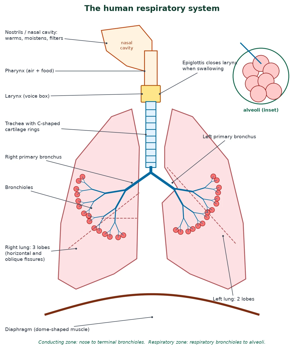
In humans the system begins at the external nostrils and ends at the alveoli. For gas exchange IB Biology answers, learn the route air takes in this order:
Nostrils → nasal cavity → pharynx (nasopharynx, oropharynx, laryngopharynx) → larynx → trachea → primary bronchi → secondary and tertiary bronchi → bronchioles → terminal bronchioles → respiratory bronchioles → alveolar ducts → alveoli
Nose and pharynx
The nasal cavity is divided by a septum into two chambers. Each has three regions: the vestibule near the nostril, with hairs that filter large particles; the respiratory region, where mucus from goblet cells traps dust, cilia sweep it towards the throat, and a rich blood supply warms and moistens the air (three bony ridges, the nasal conchae, add surface area and make the air swirl); and the olfactory region in the roof, which detects smells and has no part in gas exchange. Behind the internal nares lies the pharynx, a common passage for food and air.
Larynx, trachea and bronchi
The larynx (voice box) is a cartilage box at the top of the trachea. It has nine cartilages: three unpaired (thyroid, the largest, which forms the Adam’s apple; cricoid, the only complete ring; and the epiglottis) and three paired (arytenoid, corniculate and cuneiform). During swallowing the larynx rises and the epiglottis covers the glottis so that food goes into the oesophagus. The vocal folds stretch between the thyroid and arytenoid cartilages and vibrate as air passes out, producing sound.
The trachea (windpipe, about 10 to 12 cm long) runs down the neck in front of the oesophagus. It is held open by 16 to 20 C-shaped rings of hyaline cartilage; the open part of each C faces the oesophagus, so swallowed food can expand it. It is lined with ciliated pseudostratified epithelium and goblet cells that form a mucus escalator. At about the level of the fifth thoracic vertebra it divides into the right and left primary bronchi. These branch again and again, into secondary (lobar) and tertiary (segmental) bronchi and then bronchioles. NCERT notes that cartilage rings support the trachea, bronchi and initial bronchioles; the smaller bronchioles have none and are kept open by smooth muscle and elastic tissue.
Lungs, pleura and the thorax
The two lungs sit in the thoracic cavity, an air-tight chamber bounded by the vertebral column, sternum, ribs and the dome-shaped diaphragm. The right lung is larger and has three lobes; the left lung has two lobes and a notch for the heart. Each lung is covered by a double pleural membrane with a thin layer of pleural fluid between the layers, which reduces friction and links the lungs to the chest wall, so that any change in the volume of the thorax changes the volume of the lungs.
Functionally, the passages from the nostrils to the terminal bronchioles form the conducting zone: they filter, warm and humidify air but no exchange occurs there. The respiratory bronchioles, alveolar ducts and alveoli form the respiratory (exchange) zone.
NCERT Respiratory organs across animals
Sponges, cnidarians and flatworms exchange gases over the whole body surface; earthworms use their moist skin; insects have tracheal tubes; most aquatic arthropods, molluscs and fish use gills; and land vertebrates use lungs. Amphibians such as frogs can also exchange gases through their moist skin.
Test yourself: gas exchange IB Biology recall (click a question to reveal the answer)
Why are the cartilage rings of the trachea C-shaped rather than complete?
The open gap faces the oesophagus, so the oesophagus can bulge forward as food is swallowed, while the cartilage still stops the trachea collapsing.
Where does the conducting zone end?
At the terminal bronchioles. Gas exchange begins in the respiratory bronchioles.
2 Ventilation and the mechanism of breathing
Syllabus link for gas exchange IB Biology and NCERT: IB B3.1.5; NCERT 14.2 Mechanism of breathing
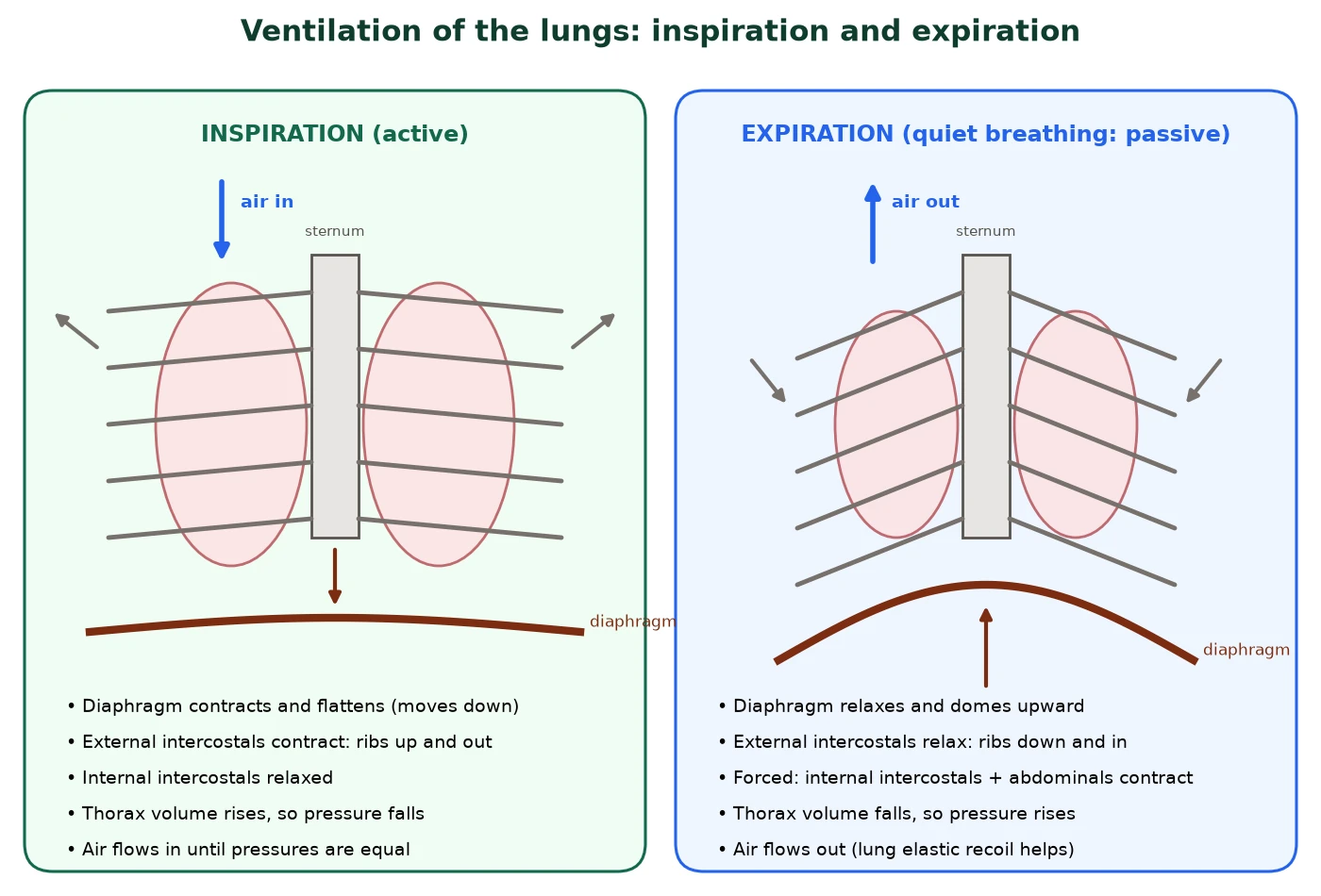
The lungs have no muscle of their own that can expand them. Instead, the mechanism of breathing relies on muscles that change the volume of the thorax. Because pressure and volume of a gas are inversely related (Boyle’s law), a bigger thorax means lower pressure inside the lungs, and air flows in from the atmosphere. Ventilation keeps alveolar air high in O2 and low in CO2, which is why it matters so much for gas exchange IB Biology questions.
Inspiration
- The diaphragm contracts and flattens, moving down and increasing the vertical size of the thorax.
- The external intercostal muscles contract, pulling the ribs and sternum up and out and increasing the front-to-back and side-to-side size.
- Thoracic and lung volume increase, so the pressure in the lungs falls below atmospheric pressure.
- Air flows in through the airways until the pressures are equal.
In quiet breathing the diaphragm does most of the work (often quoted as roughly three-quarters of the volume change). During forced inspiration, accessory muscles such as the sternocleidomastoid and scalenes lift the ribs further.
Expiration
Quiet expiration is passive: the diaphragm and external intercostals relax, the elastic lungs recoil, the diaphragm domes upwards and the ribs move down and in. Volume falls, pressure rises above atmospheric, and air flows out. Forced expiration is active: the internal intercostal muscles pull the ribs down and in, and the abdominal muscles push the abdominal organs up against the diaphragm.
Gas exchange IB Biology exam tip: antagonistic muscles
The IB guide expects you to name antagonistic pairs: external versus internal intercostals, and diaphragm versus abdominal wall muscles. Always describe the chain muscle → volume → pressure → air flow; do not write that air flowing in makes the lungs expand.
Test yourself: gas exchange IB Biology recall (click a question to reveal the answer)
Which muscles contract during quiet expiration?
None. Quiet expiration is passive; it relies on muscle relaxation and elastic recoil of the lungs.
Why does air enter the lungs when the diaphragm contracts?
The thorax volume increases, so the pressure in the lungs falls below atmospheric pressure; air moves down the pressure gradient.
3 Structure and function of alveoli
Syllabus link for gas exchange IB Biology and NCERT: IB B3.1.4 Adaptations of mammalian lungs; NCERT 14.1.1, 14.3
Alveoli are the structural and functional units of the lung. Each is a tiny air sac, about 0.2 mm across, surrounded by a dense network of capillaries. The adaptations below are the core of gas exchange IB Biology students must explain:
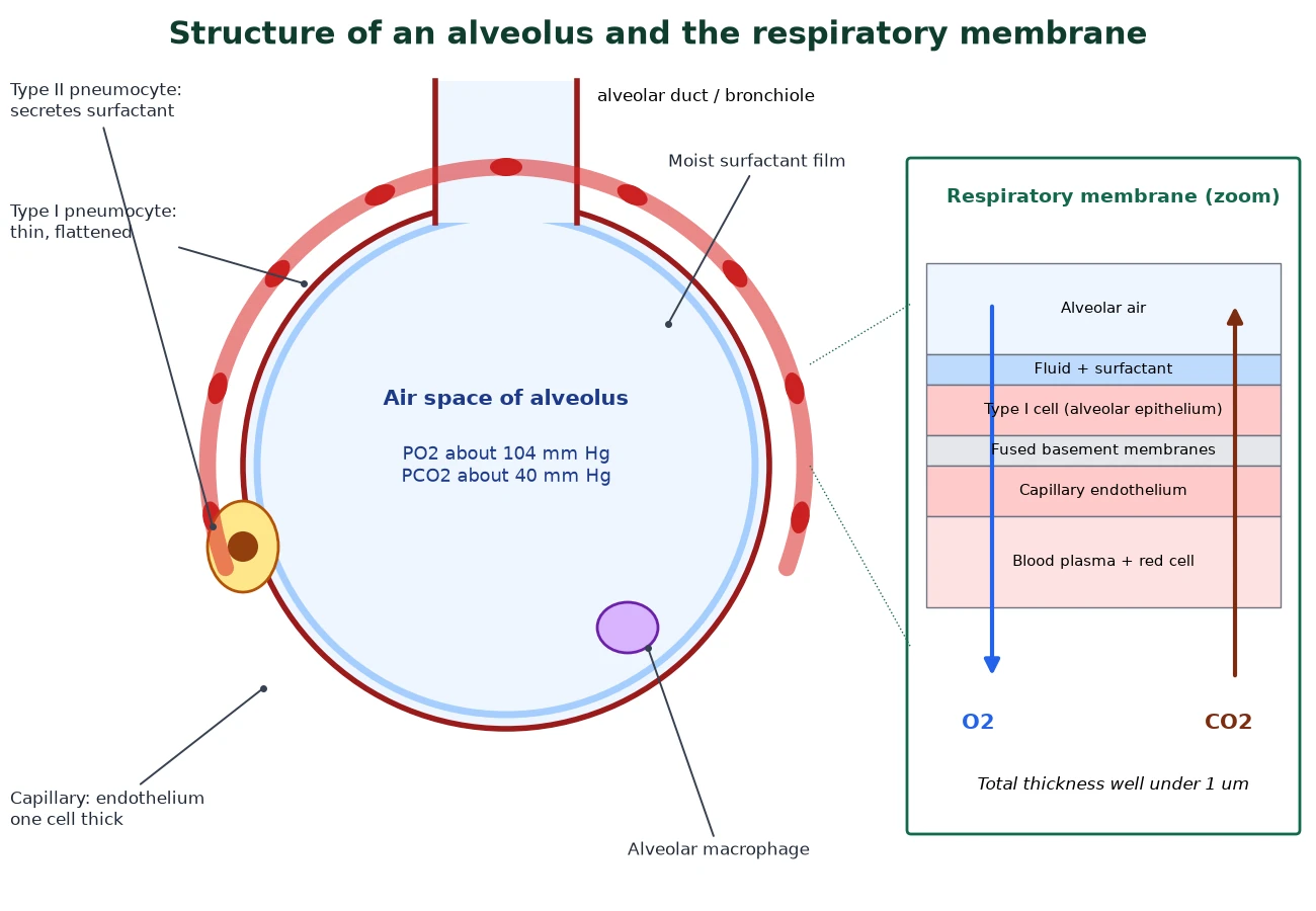
- Huge number and surface area: hundreds of millions of alveoli give a surface roughly the size of a badminton or tennis court.
- Type I pneumocytes: extremely thin, flattened cells forming most of the wall, giving a short diffusion distance.
- Type II pneumocytes: rounded cells that secrete surfactant (mainly phospholipids with proteins). Surfactant lowers the surface tension of the moisture film, so alveoli do not stick together and collapse when air leaves them (collapse is called atelectasis). Premature babies often lack surfactant, causing infant respiratory distress syndrome.
- Moist lining: gases dissolve before diffusing.
- Dense capillary bed: capillary walls are one cell thick and red blood cells are squeezed close to the wall.
- Alveolar macrophages: engulf dust and microorganisms that reach the alveoli.
- Elastic fibres: allow alveoli to stretch on inspiration and recoil on expiration.
The respiratory membrane that a gas crosses has three main layers: the thin squamous epithelium of the alveolus, the endothelium of the capillary, and the fused basement substance between them. NCERT notes that the total thickness is much less than a millimetre; it is actually only around 0.2 to 0.6 µm in most places.
Drawing tip for gas exchange IB Biology (B3.1.4)
When asked to annotate an alveolus, label type I and type II pneumocytes, surfactant, the capillary and a macrophage, and add a function to each label, for example “type I pneumocyte: thin, so short diffusion distance”.
4 Diffusion and exchange of gases
Syllabus link for gas exchange IB Biology and NCERT: IB B3.1.2 to B3.1.4; NCERT 14.3 Exchange of gases
The amount of a gas in a mixture is described by its partial pressure: the share of total pressure due to that gas. Air at sea level is at about 760 mm Hg and about 21% O2, so PO2 is about 0.21 × 760 = 159 mm Hg. Each gas diffuses from a region of higher to lower partial pressure, independently of the other gases. This idea is the heart of breathing and exchange of gases in NCERT and of gas exchange IB Biology Paper 1 questions.
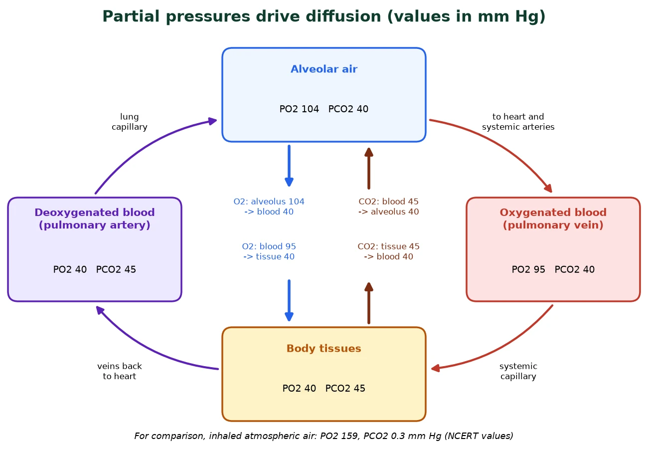
| Gas (mm Hg) | Atmospheric air | Alveoli | Deoxygenated blood | Oxygenated blood | Tissues |
|---|---|---|---|---|---|
| PO2 | 159 | 104 | 40 | 95 | 40 |
| PCO2 | 0.3 | 40 | 45 | 40 | 45 |
Alveolar PO2 (104) is lower than in inhaled air because each breath mixes with air already in the lungs and humidified air contains water vapour. The O2 gradient (104 to 40) is much larger than the CO2 gradient (45 to 40), yet CO2 still crosses easily because it is about 20 to 25 times more soluble in water than O2. The diffusing capacity of the membrane for CO2 is therefore far higher.
Factors that affect the rate of diffusion are the partial pressure gradient, the solubility of the gas, the thickness of the membrane and its surface area. In humans the exchange is so fast that blood is almost fully loaded with O2 in the first third of its journey through a lung capillary.
Test yourself: gas exchange IB Biology recall (click a question to reveal the answer)
Why can CO2 diffuse fast even though its partial pressure gradient is only 5 mm Hg?
CO2 is about 20 to 25 times more soluble than O2, so its rate of diffusion per mm Hg of gradient is much greater.
5 Transport of O2 and CO2
Syllabus link for gas exchange IB Biology and NCERT: IB B3.1.11 to B3.1.13 (AHL); old IB D.6; NCERT 14.4 Transport of gases
Blood transport links the lungs to every respiring cell, and it is the most heavily examined HL part of gas exchange IB Biology.
Transport of oxygen
About 97% of O2 in blood is carried by haemoglobin in red blood cells and only about 3% is dissolved in plasma. Haemoglobin is a protein of four polypeptide chains (two alpha and two beta in adults), each with a haem group containing an iron(II) ion that binds one O2 molecule. So one haemoglobin molecule carries up to four O2 molecules (Hb + 4O2 → Hb(O2)4, oxyhaemoglobin). Binding O2 is oxygenation, not oxidation: the iron stays Fe2+.
Each gram of haemoglobin can bind about 1.34 mL of O2. With about 15 g per 100 mL of blood, fully saturated blood carries about 20 mL O2 per 100 mL. NCERT states that every 100 mL of oxygenated blood delivers about 5 mL of O2 to the tissues at rest; during heavy exercise much more is unloaded.
The oxygen dissociation curve AHL
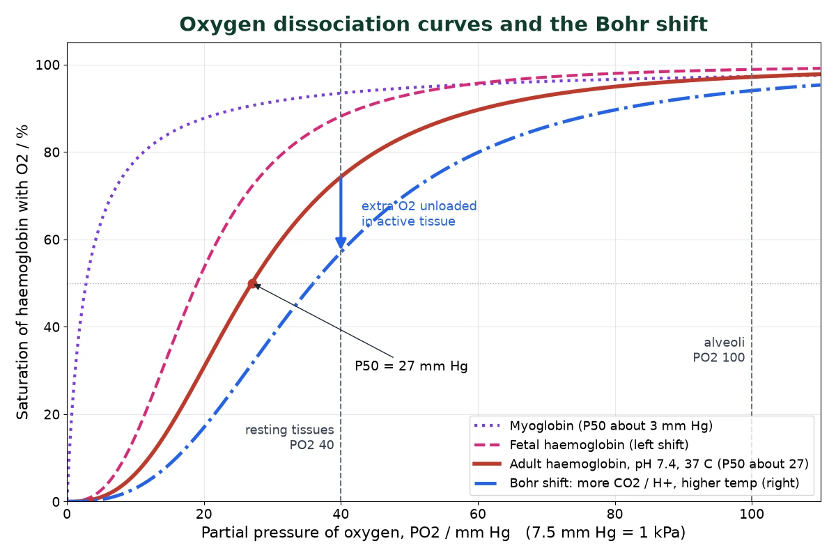
The oxygen dissociation curve plots percentage saturation of haemoglobin against PO2. It is sigmoid (S-shaped) because of cooperative binding: once the first O2 binds, the shape of the protein changes and the remaining haem groups bind O2 more easily. Three regions matter:
- Flat top (alveoli, PO2 about 100 mm Hg): haemoglobin is about 97 to 98% saturated, and small falls in PO2 (for example at moderate altitude) barely reduce loading.
- Steep middle (tissues, PO2 20 to 40 mm Hg): a small drop in PO2 releases a large amount of O2, exactly where respiring cells need it.
- P50: the PO2 at which haemoglobin is 50% saturated, about 26 to 27 mm Hg (3.5 kPa) for adult blood. A higher P50 means lower affinity.
The Bohr shift AHL
In actively respiring tissue, CO2 rises and the pH falls (more H+). CO2 and H+ bind to haemoglobin at sites away from the haem (allosteric sites) and lower its affinity for O2. The curve moves to the right: at the same PO2, haemoglobin is less saturated, so more O2 is unloaded exactly where respiration is fastest. Higher temperature and higher 2,3-BPG (made by red cells at altitude) also shift the curve right. In the lungs, CO2 is lost, pH rises and the curve shifts back to the left, helping loading. The mirror effect, in which oxygenation of haemoglobin makes it release CO2 and H+ in the lungs, is the Haldane effect.
Fetal haemoglobin and myoglobin AHL
Fetal haemoglobin (HbF) has two alpha and two gamma chains. It binds 2,3-BPG less strongly and so has a higher affinity for O2 than adult haemoglobin: its curve lies to the left. At the low PO2 of the placenta, fetal blood can therefore take O2 from the mother’s blood. After birth HbF is gradually replaced by adult haemoglobin. Myoglobin, the O2 store in muscle, has one chain and one haem, so it cannot bind cooperatively; its curve is hyperbolic and far to the left. It releases O2 only when muscle PO2 is very low, such as in intense exercise.
Transport of carbon dioxide
CO2 travels in three forms (NCERT values): about 7% dissolved in plasma, about 20 to 25% as carbaminohaemoglobin (bound to amino groups of haemoglobin, not to the haem), and about 70% as hydrogencarbonate (bicarbonate) ions.
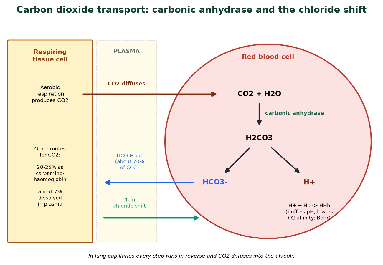
- CO2 diffuses from tissues into plasma and into red blood cells.
- The enzyme carbonic anhydrase, abundant in red blood cells, speeds up CO2 + H2O → H2CO3 enormously.
- Carbonic acid dissociates: H2CO3 → H+ + HCO3–.
- H+ binds to haemoglobin, forming HHb. This buffers the blood pH and lowers O2 affinity (Bohr shift).
- HCO3– leaves the red cell into the plasma through a carrier, and Cl– moves in to keep the charge balanced: the chloride shift (Hamburger phenomenon).
- In lung capillaries, where PCO2 is low, every step reverses and CO2 diffuses into the alveoli. NCERT gives about 4 mL of CO2 delivered to the alveoli by every 100 mL of deoxygenated blood.
Test yourself: gas exchange IB Biology recall (click a question to reveal the answer)
What is the effect of a right shift on P50?
P50 increases: a higher PO2 is needed to half-saturate haemoglobin, so affinity is lower and O2 is unloaded more easily.
Why is carbon monoxide so dangerous?
CO binds to haem over 200 times more strongly than O2, forming carboxyhaemoglobin, which blocks O2 binding and also makes the remaining sites hold O2 more tightly (a left shift), so tissues are starved of O2.
6 Regulation of breathing and respiratory volumes
Syllabus link for gas exchange IB Biology and NCERT: IB B3.1.6; old IB D.6; NCERT 14.2.1, 14.5
How breathing is controlled
Breathing is normally involuntary. A respiratory rhythm centre in the medulla oblongata sets the basic rhythm; its dorsal group of neurons drives inspiration, while its ventral group is used mainly in forced breathing. The pneumotaxic centre in the pons limits how long inspiration lasts, so a stronger pneumotaxic signal gives shorter, faster breaths.
- Central chemoreceptors next to the rhythm centre in the medulla are very sensitive to CO2 and H+. A rise in PCO2 (and so a fall in the pH of cerebrospinal fluid) is the strongest stimulus to breathe.
- Peripheral chemoreceptors in the aortic and carotid bodies detect rises in CO2 and H+ and large falls in O2. They signal through the vagus (from aortic bodies) and glossopharyngeal nerves (from carotid bodies).
- The rhythm centre increases the rate and depth of breathing through nerves to the diaphragm and intercostal muscles, removing CO2 and restoring blood pH. This is negative feedback.
The role of O2 in normal regulation is small; PO2 must fall a long way before it drives breathing. Higher brain centres allow voluntary control, such as holding your breath or speaking. Control of breathing comes from old IB D.6 and NCERT, but it is useful context for gas exchange IB Biology data questions on exercise.
Respiratory volumes and capacities
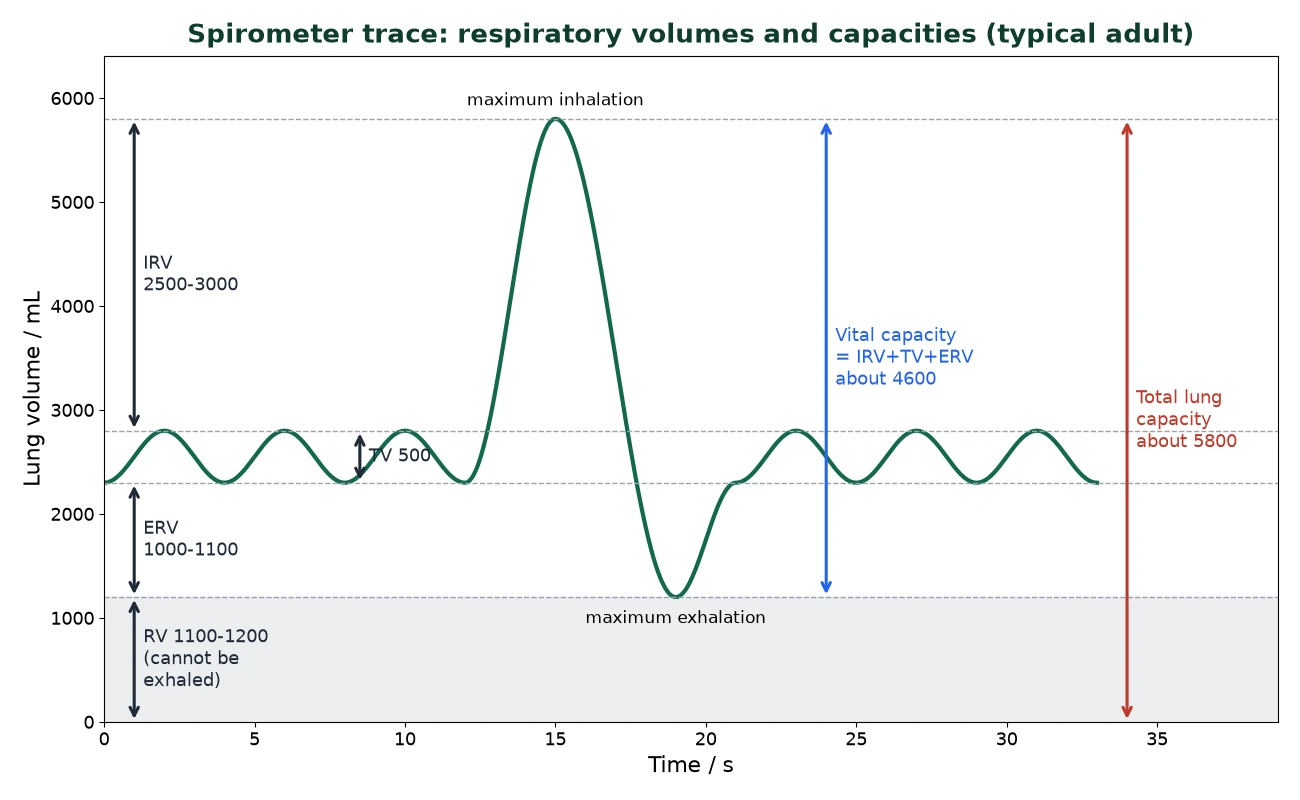
Lung volumes are measured with a spirometer (IB B3.1.6 also expects simple methods such as a bell jar or a lung-volume bag). Typical respiratory volumes for a healthy adult, from NCERT, are:
| Volume or capacity | Definition | Typical value |
|---|---|---|
| Tidal volume (TV) | Air breathed in or out in one quiet breath | About 500 mL (6000 to 8000 mL per minute) |
| Inspiratory reserve volume (IRV) | Extra air that can be forcibly inhaled after a normal inhalation | 2500 to 3000 mL |
| Expiratory reserve volume (ERV) | Extra air that can be forcibly exhaled after a normal exhalation | 1000 to 1100 mL |
| Residual volume (RV) | Air left in the lungs after the most forceful exhalation | 1100 to 1200 mL |
| Inspiratory capacity (IC) | TV + IRV | About 3000 to 3500 mL |
| Functional residual capacity (FRC) | ERV + RV: air left after a normal exhalation | About 2100 to 2300 mL |
| Vital capacity (VC) | ERV + TV + IRV: maximum air breathed out after a maximum breath in | About 4000 to 4600 mL |
| Total lung capacity (TLC) | VC + RV | About 5100 to 5800 mL |
Of each 500 mL tidal breath, roughly 150 mL stays in the conducting airways (anatomical dead space) and only about 350 mL reaches the alveoli. That is why slow, deep breathing ventilates the alveoli better than fast, shallow breathing at the same minute volume.
Gas exchange IB Biology exam tip
Ventilation rate = breaths per minute; pulmonary ventilation = tidal volume × ventilation rate. For example, 0.5 L × 12 min-1 = 6 L min-1.
7 Asthma, emphysema and occupational respiratory disorders
Syllabus link for gas exchange IB Biology and NCERT: Old IB 6.4 (lung cancer, emphysema); NCERT 14.6 Disorders of respiratory system
Lung diseases make good gas exchange IB Biology data questions, because each one changes the surface area, the diffusion distance or the gradient.
| Disorder | Cause | What goes wrong | Effect on gas exchange |
|---|---|---|---|
| Asthma | Allergens, cold air, exercise, smoke in sensitive people | Inflammation of bronchi and bronchioles, extra mucus, and contraction of the smooth muscle (bronchoconstriction), partly due to histamine and other mediators | Narrow airways raise resistance; wheezing and breathlessness. Treated mainly with inhaled bronchodilators and corticosteroids |
| Emphysema | Mainly cigarette smoking; rarely alpha-1 antitrypsin deficiency | Phagocytes release elastase that digests elastic fibres; alveolar walls break down into a few large air sacs | Much smaller surface area, loss of elastic recoil, air trapping; low blood O2. Part of chronic obstructive pulmonary disease (COPD) |
| Lung cancer | Smoking (main cause), radon, asbestos, air pollution | Mutations cause uncontrolled cell division; tumours block airways | Less functional lung tissue, coughing blood, breathlessness |
| Silicosis and asbestosis | Long-term inhalation of silica or asbestos dust (stone cutting, mining, glass, construction) | Macrophages cannot digest the particles; inflammation and fibrosis (scar tissue) thicken lung tissue | Thicker, stiffer membrane: longer diffusion distance and lower lung compliance |
NCERT describes occupational respiratory disorders in industries such as grinding and stone-breaking, where so much dust is produced that the lung’s defences cannot cope; long exposure causes inflammation and fibrosis. Silicosis and asbestosis are the best-known examples. Prevention includes reducing dust at source, wet drilling, ventilation of workplaces and protective masks for workers.
Hypoxia, carbon monoxide and decompression sickness
- Hypoxic hypoxia: low PO2 in inhaled air, for example at high altitude.
- Anaemic hypoxia: normal PO2 but too little functional haemoglobin, for example anaemia or carbon monoxide poisoning.
- Ischaemic (stagnant) hypoxia: poor blood flow to a tissue.
- Histotoxic hypoxia: cells cannot use O2, for example cyanide blocking cytochrome oxidase.
- Decompression sickness (the bends): at depth, more nitrogen dissolves in a diver’s blood; rising too fast lets it form bubbles in blood and tissues. Divers ascend slowly, and deep divers breathe gas mixtures with helium, which is less soluble and less narcotic than nitrogen.
8 Gas exchange in other animals
Syllabus link for gas exchange IB Biology and NCERT: IB B3.1.1 to B3.1.3 (comparative); NCERT 14.1 Respiratory organs
Comparing organisms shows that gas exchange IB Biology questions always come back to the same rules of surface area, distance and gradient.
| Organism | Exchange surface | Key adaptation |
|---|---|---|
| Unicellular organisms, sponges, cnidarians, flatworms | Whole body surface | Small size or flat shape gives a high surface area to volume ratio and short distances |
| Earthworm | Moist skin | Mucus keeps the skin wet; dense capillaries and haemoglobin in the blood |
| Insects | Tracheal system | Spiracles lead to chitin-ringed tracheae and fine tracheoles that deliver air directly to cells |
| Bony fish | Gills (under an operculum) | Gill filaments with many lamellae; countercurrent flow; one-way flow of water |
| Frog | Lungs, moist skin and buccal lining | Cutaneous exchange is important under water and in hibernation |
| Birds | Lungs with air sacs | One-way air flow through parabronchi gives very efficient exchange |
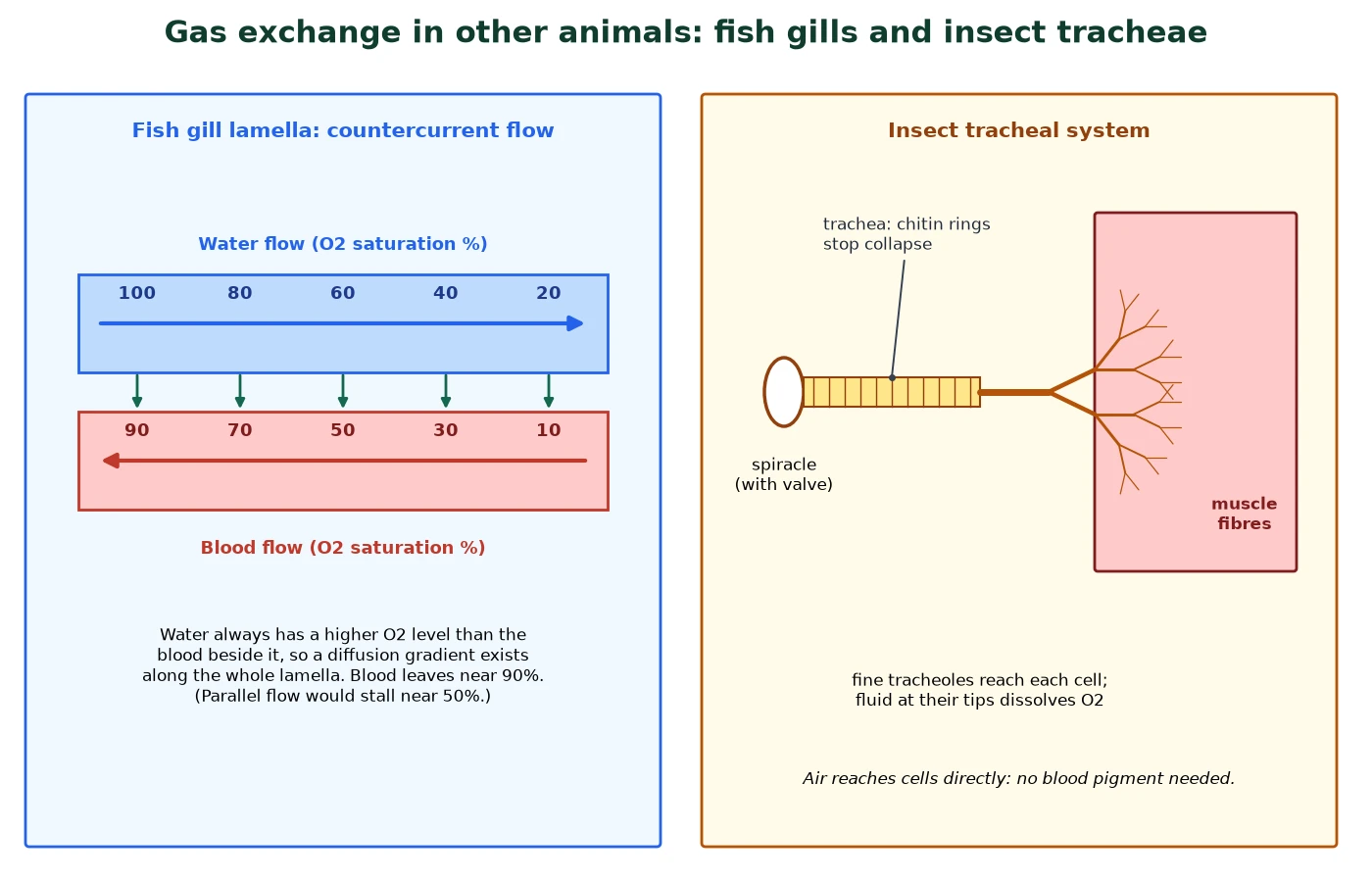
Countercurrent flow in gills: water flows over the lamellae in the opposite direction to blood. Along the whole lamella, the water next to any part of the blood always has more O2 than that blood, so diffusion continues all the way, and blood can leave almost as oxygenated as the incoming water. With parallel (concurrent) flow the two would equalise at about half saturation.
Insect tracheae: air enters through spiracles, which can close to reduce water loss. The tracheae branch into tracheoles less than 1 µm wide that end inside or next to muscle fibres. Fluid at the tips moves out when muscles work hard, pulling air closer to the cells. Large insects ventilate by pumping movements of the abdomen.
9 Gas exchange in plants and leaves
Syllabus link for gas exchange IB Biology and NCERT: IB B3.1.7 to B3.1.10
Plants respire all the time, and in light they also photosynthesise. In bright light, photosynthesis is faster than respiration, so a leaf takes in CO2 and releases O2; in darkness the direction reverses. Leaves are adapted for gas exchange IB Biology students must be able to annotate:
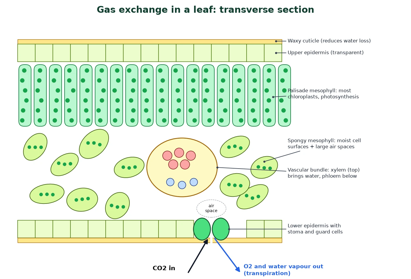
- Waxy cuticle: reduces water loss from the upper surface.
- Epidermis: transparent, letting light reach the mesophyll.
- Palisade mesophyll: tightly packed columnar cells with many chloroplasts for photosynthesis.
- Spongy mesophyll: loosely packed cells with large, connected air spaces and moist cell walls, giving a large internal surface for gases to dissolve and diffuse.
- Stomata and guard cells: pores, mostly on the lower surface, that open and close to control the exchange of CO2 and the loss of water vapour.
- Vascular bundle (vein): xylem brings water to replace evaporation; phloem carries sugars away.
Transpiration as a consequence of gas exchange
The same open stomata that let CO2 in also let water vapour out. Water evaporates from the moist mesophyll walls into the air spaces and diffuses out through the stomata: this is transpiration. Rates rise with higher temperature, light intensity and wind, and fall with higher humidity. Plants in dry habitats balance the two needs, for example by closing stomata at midday.
Stomatal density (practical skill)
Stomatal density is the number of stomata per unit area, usually per mm2. Paint clear nail varnish on the leaf surface, peel the dried layer with clear tape, mount it on a slide and count stomata in a field of view of known area (area = πr2). Repeat in several fields and on several leaves, then calculate a mean. For example, 24 stomata in a circular field of radius 0.2 mm (area 0.126 mm2) gives about 190 stomata per mm2.
Test yourself: gas exchange IB Biology recall (click a question to reveal the answer)
Why is transpiration described as a consequence of gas exchange?
Stomata must open for CO2 to enter for photosynthesis, and water vapour inevitably diffuses out through the same pores.
10 Quick check
Eight questions on gas exchange IB Biology and NEET style. Try them before opening the answers.
-
Where are the respiratory rhythm centre and central chemoreceptors found?
- A. Cerebellum
- B. Hypothalamus
- C. Medulla oblongata
- D. Pons only
-
Which muscles contract during quiet inspiration?
- A. Diaphragm and external intercostals
- B. Diaphragm and internal intercostals
- C. Abdominal muscles only
- D. Internal intercostals only
-
About what percentage of CO2 is carried as hydrogencarbonate ions?
- A. 7%
- B. 20 to 25%
- C. 97%
- D. 70%
-
Which cells secrete surfactant?
- A. Type I pneumocytes
- B. Type II pneumocytes
- C. Goblet cells
- D. Alveolar macrophages
-
A right shift of the oxygen dissociation curve means haemoglobin has
- A. higher affinity for O2
- B. no change in affinity
- C. lower affinity for O2
- D. stopped binding CO2
-
Vital capacity equals
- A. TV + IRV
- B. ERV + RV
- C. TV + IRV + ERV + RV
- D. TV + IRV + ERV
-
Explain why the oxygen dissociation curve is sigmoid.
-
Explain how emphysema reduces the rate of gas exchange.
Show answers
- C. The medulla oblongata; the pons holds the pneumotaxic centre.
- A. Diaphragm and external intercostals.
- D. About 70%.
- B. Type II pneumocytes.
- C. Lower affinity, so more unloading.
- D. TV + IRV + ERV.
- Cooperative binding: binding of each O2 changes haemoglobin’s shape and raises the affinity of the remaining haem groups, so the curve is steep in the middle.
- Alveolar walls are destroyed, so surface area falls; loss of elastic fibres means the lungs do not recoil and stale air is trapped, reducing the concentration gradient.
Practice MCQs: Breathing and Exchange of Gases MCQs
11 Glossary
Key terms for gas exchange IB Biology and NEET revision.
© competitorspoint.com · Biology notes. Diagrams and text are original to competitorspoint.com.
Ready to practise? Download the Gas Exchange IB-style practice questions with full markscheme (100 marks, free for signed-up members).
⬇ Download practice questions (PDF)