
IB Biology › Cell Biology › Membrane Transport
Membrane Transport
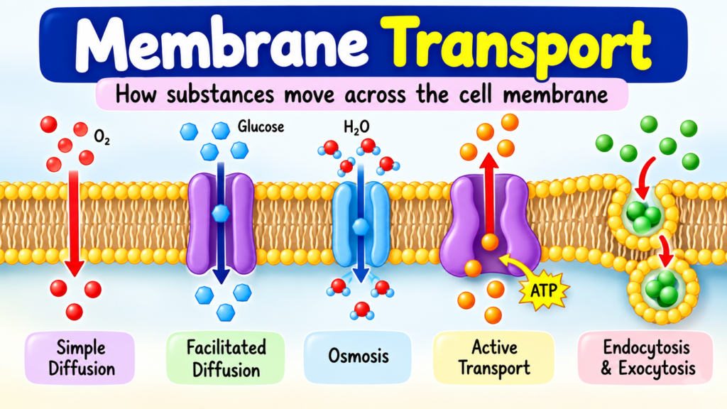
Every cell has to let some things in, keep other things out and send products away. This IB Biology chapter on membrane transport shows how the plasma membrane manages that traffic, from simple diffusion of oxygen to ATP-powered pumps and vesicles that carry whole packages of material.
Intro Why membranes need transport
Syllabus link: B2.1 lipid bilayers as barriers; selectivity in membrane permeability
A cell is a watery space wrapped in a very thin film: the plasma membrane. Inside, the cell must keep the right amounts of water, ions, sugars and enzymes. Outside, conditions keep changing. Membrane transport is the set of methods a cell uses to control what crosses this boundary.
Quick recap: the phospholipid bilayer
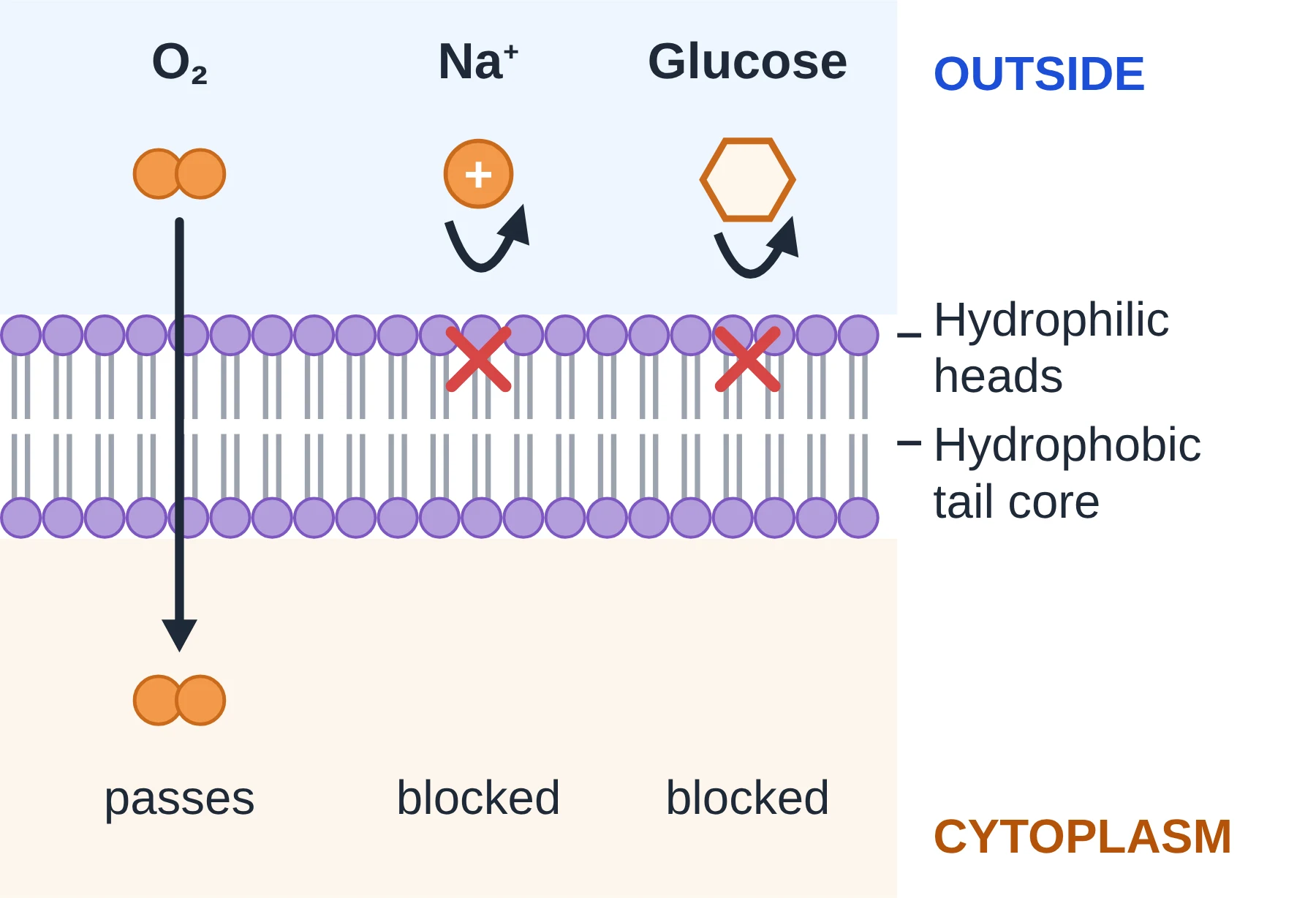
- Each phospholipid has a hydrophilic (water-loving) phosphate head and two hydrophobic (water-fearing) fatty acid tails.
- In water, phospholipids arrange themselves into a bilayer: heads face the watery fluid on each side, tails point inwards and form an oily core.
- That hydrophobic core is the key barrier. Ions and large polar molecules (such as glucose) are attracted to water and cannot easily pass through it.
- Proteins sit in the bilayer. Integral proteins are embedded in it (many span it completely); peripheral proteins sit on one surface. Channels, carriers and pumps are integral proteins.
- Membranes are fluid: the phospholipids and many proteins drift sideways, which lets the membrane bend, fuse and reseal.
Selective permeability
A membrane is selectively permeable (also called partially permeable): some substances cross easily, others slowly, and some only with help. Two things decide this:
- The particle itself. Small, non-polar particles cross the bilayer quickly; ions and large polar molecules hardly cross at all.
- The proteins present. Channel, carrier and pump proteins are specific, so each cell controls exactly which ions and molecules get through by choosing which proteins to make.
Transport is either passive (no energy from the cell; particles move down a concentration gradient) or active (uses energy, usually from ATP, and can move particles against a gradient).
Key points
- The hydrophobic core of the bilayer is a barrier to ions and polar molecules.
- Simple diffusion is not selective: it depends only on particle size and polarity.
- Membrane proteins make permeability selective.
- Passive transport needs no ATP; active transport does.
Common mistakes
- Saying the membrane is ‘semi-permeable’ and stopping there. Explain why: the hydrophobic core blocks charged and polar particles.
- Thinking that cells ‘want’ or ‘need’ particles to move. Particles move randomly; the cell controls the route, not the particles.
1 Diffusion
Syllabus link: B2.1 simple diffusion across membranes
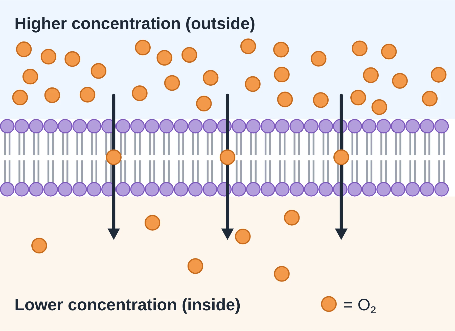
Diffusion is the net movement of particles from a region of higher concentration to a region of lower concentration. It happens because particles are always moving randomly. More particles on one side means more of them happen to cross in that direction, so there is a net flow until the concentrations even out.
When particles cross a membrane by passing straight between the phospholipids, it is called simple diffusion. No protein and no ATP are involved, so it is a form of passive transport.
What crosses by simple diffusion?
- Oxygen (O2): enters cells for aerobic respiration, for example from alveoli into blood capillaries.
- Carbon dioxide (CO2): leaves respiring cells and enters photosynthesising leaf cells.
- Other small non-polar molecules, such as steroid hormones and small fat-soluble substances, which dissolve in the hydrophobic core.
- Very small polar molecules (water, urea) can slip through slowly, but much more slowly than O2.
Factors that affect the rate of diffusion
- Concentration gradient: a bigger difference in concentration across the membrane gives a faster net rate. Cells keep gradients steep, e.g. blood flow carries O2 away from the alveoli.
- Temperature: warmer particles have more kinetic energy, move faster and diffuse faster.
- Surface area: a larger area means more particles can cross at once (think microvilli and alveoli).
- Distance or thickness: the shorter the path, the faster the diffusion. Gas exchange surfaces are often only one cell thick.
- Size and polarity of the particle: small, non-polar particles cross fastest; large or charged particles barely cross the bilayer at all.
A handy summary (Fick’s law, in words): the rate of diffusion increases with surface area × concentration difference and decreases with thickness of the surface.
Key points
- Diffusion is net movement down a concentration gradient, caused by random motion.
- Simple diffusion through the bilayer needs no protein and no ATP.
- O2, CO2 and small non-polar molecules cross this way.
- Rate depends on gradient, temperature, surface area, distance and particle size and polarity.
Common mistakes
- Writing ‘particles move from high to low’ without saying concentration. Always name the gradient.
- Forgetting the word net: particles still move both ways, but more move down the gradient.
- Saying diffusion stops at equilibrium. Random movement continues; only the net movement stops.
2 Osmosis
Syllabus link: B2.1 osmosis and aquaporins; D2.3 movement of water from less to more concentrated solutions
Osmosis is the net movement of water across a partially permeable membrane from a solution with a lower solute concentration to one with a higher solute concentration. It is passive: no ATP is used.
Why does water move?
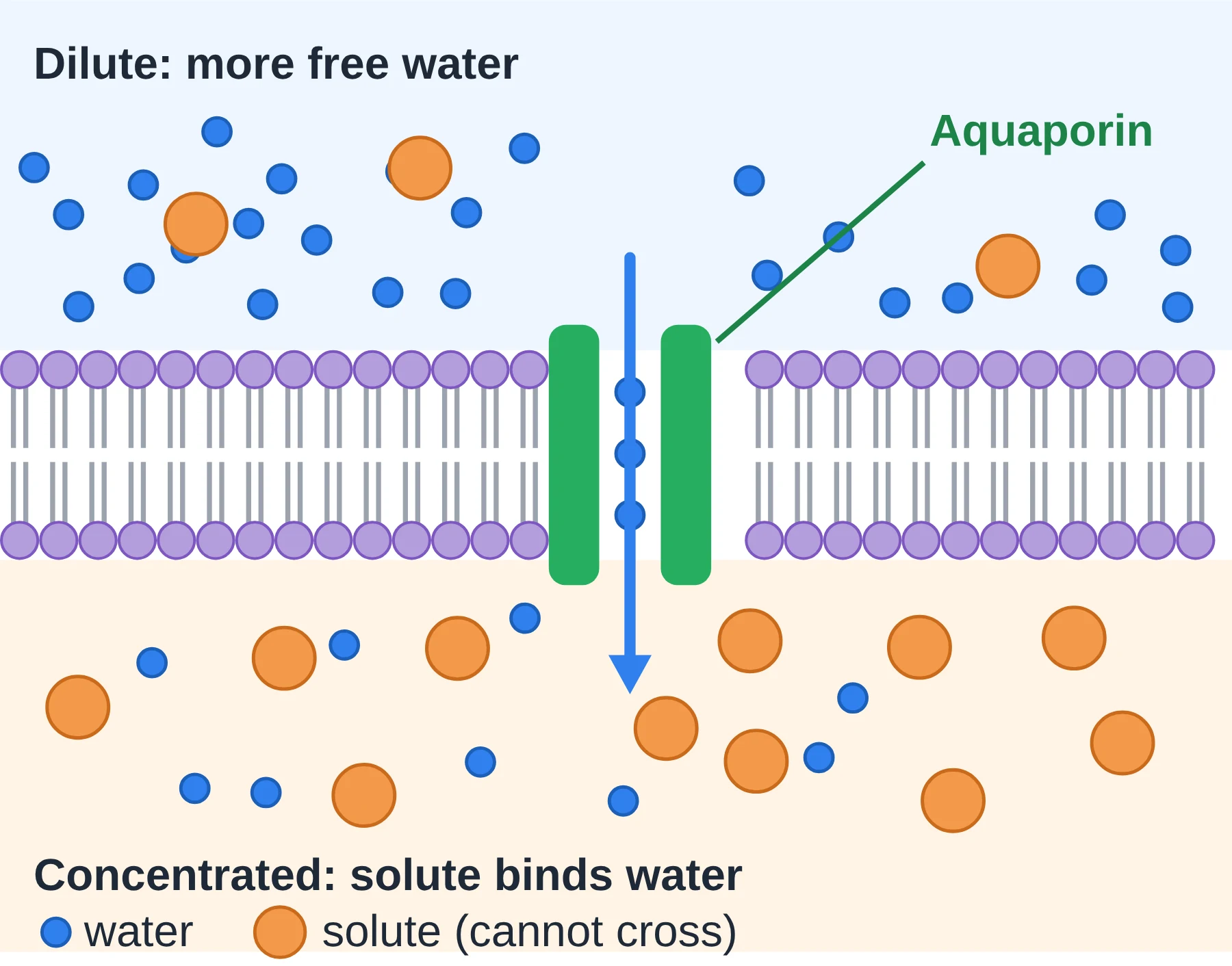
- Water is a polar molecule, so it forms hydrogen bonds and clusters around dissolved solute particles such as ions and sugars.
- Water molecules held around solutes are less free to move. A concentrated solution therefore has fewer free water molecules.
- More free water molecules hit the membrane from the dilute side, so more cross from dilute to concentrated than the other way: a net flow.
- The solute particles themselves cannot cross (the membrane is impermeable to them), so only water moves.
Aquaporins
A few water molecules squeeze through the bilayer, but this is slow. Many cells add aquaporins: integral channel proteins with a narrow pore that lets water molecules pass in single file, very quickly. The pore is lined so that ions (even tiny H+ ions) are excluded. Cells that move lots of water, such as kidney collecting duct cells and plant root cells, have many aquaporins.
Osmosis is really diffusion of water, so the same factors (gradient, temperature, surface area, distance) affect its rate. The number of aquaporins also matters.
AHL At HL, you describe this using water potential: water moves from higher to lower water potential. See the water potential notes in the Tonicity section.
Key points
- Osmosis = net movement of water across a partially permeable membrane.
- Direction: from lower solute concentration to higher solute concentration.
- Aquaporins are channel proteins that greatly speed up water movement.
- Osmosis is passive.
Common mistakes
- Saying ‘water moves from high concentration to low concentration’ is confusing. Talk about solute concentration (or water potential at HL).
- Saying solutes move during osmosis. In osmosis it is water that moves.
- Calling aquaporins ‘pumps’. They are channels and use no ATP.
3 Facilitated Diffusion
Syllabus link: B2.1 channel proteins for facilitated diffusion; AHL gated ion channels in neurons
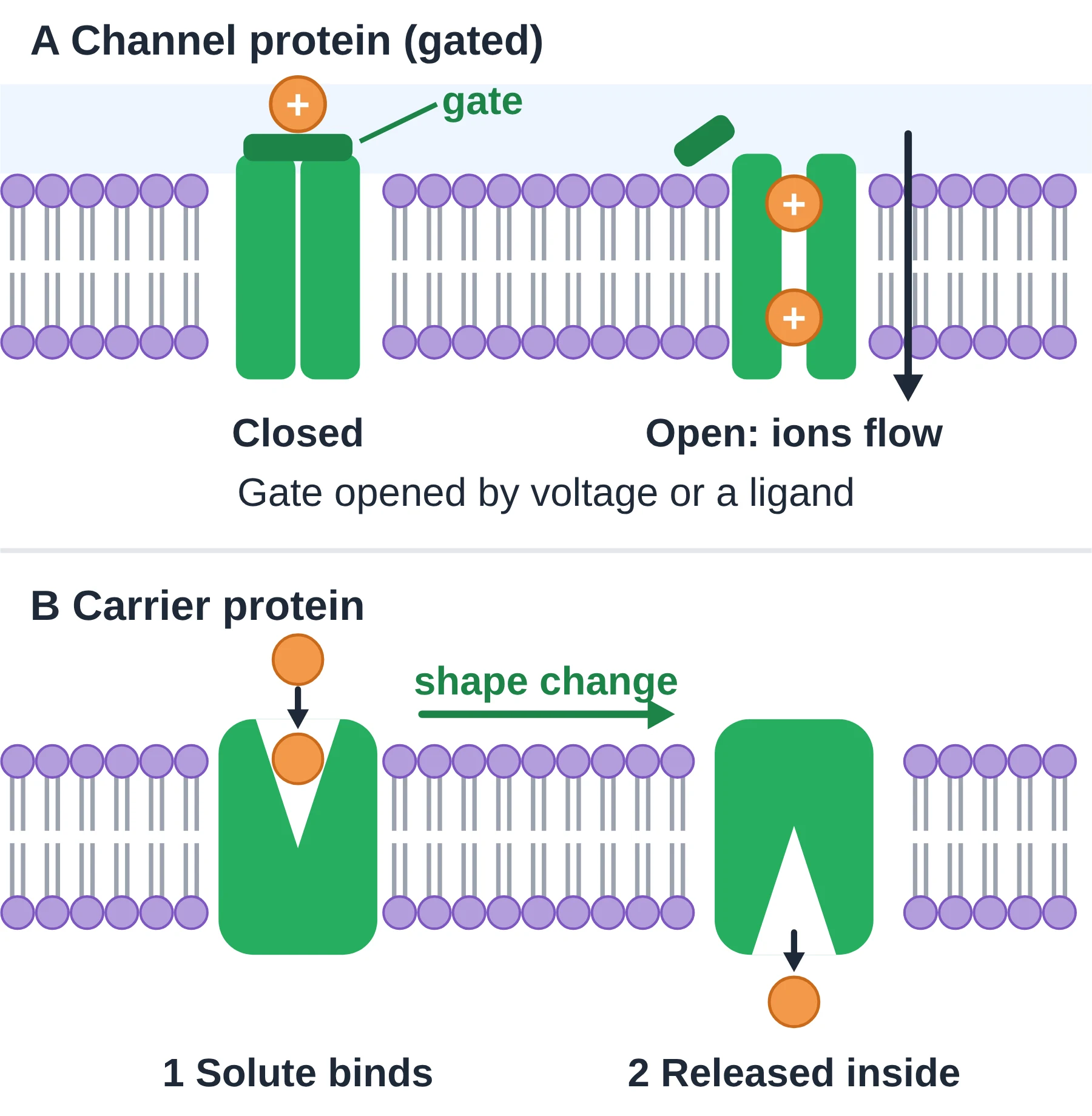
Ions and large polar molecules cannot get through the hydrophobic core, but they can still move down their concentration gradient if a protein gives them a route. This is facilitated diffusion: passive transport through a specific channel protein or carrier protein. No ATP is used.
Channel proteins
- Form a water-filled pore right through the membrane.
- The diameter of the pore and the charges lining it decide which particle fits. A potassium channel, for example, lets K+ through but not Na+.
- Very fast: millions of ions per second can pass through one open channel.
- Many are gated: they open or close, so the cell can switch permeability on and off.
Gated channels AHL
- Voltage-gated channels open or close when the voltage (membrane potential) across the membrane changes. Voltage-gated Na+ and K+ channels in axons produce nerve impulses.
- Ligand-gated channels open when a specific chemical (a ligand) binds. The nicotinic acetylcholine receptor at synapses opens when acetylcholine binds, and Na+ flows into the next neuron or muscle fibre.
Carrier proteins
- Have a binding site that fits one specific solute, for example glucose.
- When the solute binds, the protein changes shape (a conformational change) and releases the solute on the other side.
- Slower than channels, because each cycle moves only one or a few particles.
- Example: GLUT glucose transporters let glucose diffuse into most body cells.
Why the rate levels off
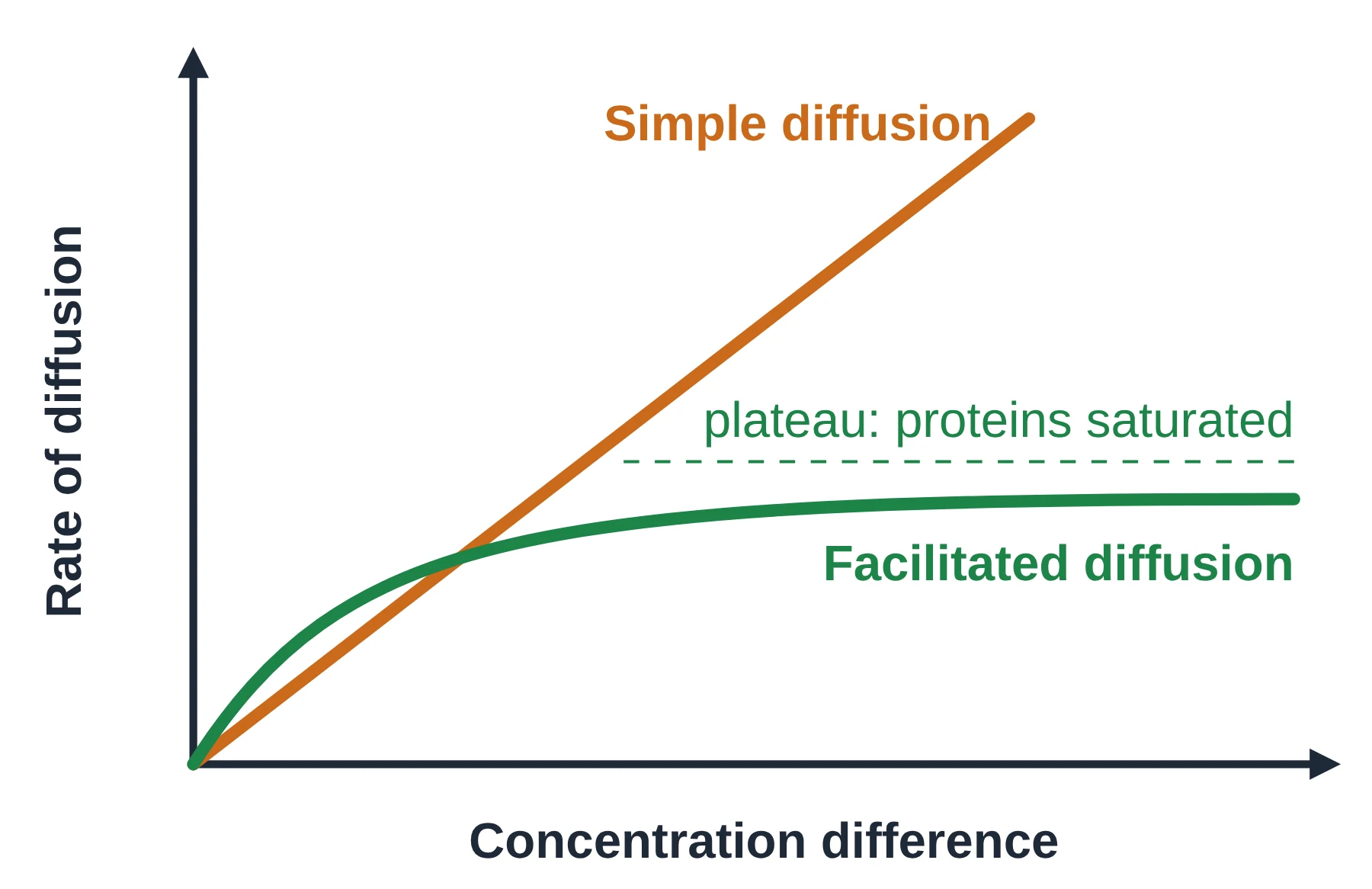
In simple diffusion, doubling the concentration difference roughly doubles the rate. In facilitated diffusion the rate rises at first, then reaches a plateau: every channel or carrier is already in use, so adding more solute cannot speed things up. The only way to go faster is to put more transport proteins into the membrane.
Key points
- Facilitated diffusion is passive and moves particles down their gradient.
- Channel proteins form a pore; carrier proteins bind and change shape.
- Both are specific, so they create selective permeability.
- AHL Gates can be voltage-gated (Na+, K+ channels) or ligand-gated (nicotinic acetylcholine receptor).
Common mistakes
- Writing that carrier proteins always use ATP. Carriers in facilitated diffusion do not; pumps do.
- Forgetting that facilitated diffusion can only go down a concentration gradient.
- Describing a channel protein as ‘changing shape to carry’ the ion. That describes a carrier.
4 Active Transport
Syllabus link: B2.1 pump proteins for active transport; AHL sodium–potassium pumps; sodium-dependent glucose cotransporters
Sometimes a cell needs to move particles against their concentration gradient, from lower to higher concentration. This is active transport. It is carried out by pump proteins and uses energy from ATP (adenosine triphosphate).
- Pumps are specific: each binds only certain ions or molecules.
- ATP is hydrolysed: ATP → ADP + Pi. The phosphate attaches to the pump and makes it change shape.
- Because energy is supplied, particles can be concentrated on one side of the membrane.
- Cells that do lots of active transport (root hair cells, kidney tubule cells) contain many mitochondria to make ATP.
Examples: root hair cells take up mineral ions such as nitrate from soil where they are scarce; stomach lining cells pump H+ into the stomach; muscle cells pump Ca2+ into storage.
The sodium–potassium pump AHL
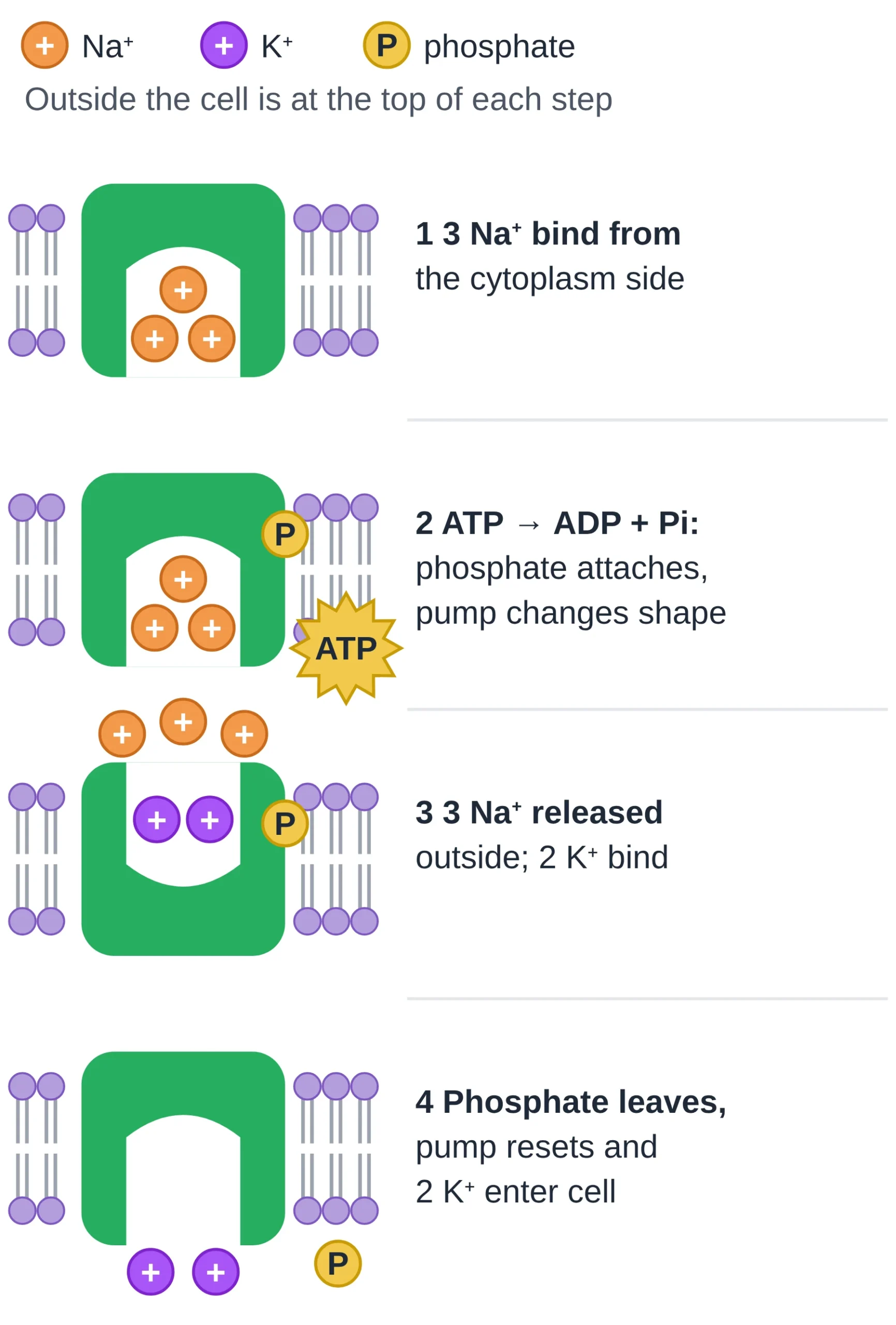
The Na+/K+ pump is found in almost all animal cells. It is an exchange transporter: it moves two ions in opposite directions. In each cycle, one ATP is used to move 3 Na+ out and 2 K+ in.
- Three Na+ ions from the cytoplasm bind to the pump, which is open to the inside.
- ATP transfers a phosphate group to the pump (ATP → ADP + Pi). The pump changes shape and opens to the outside.
- The three Na+ are released outside. Two K+ ions from outside bind.
- The phosphate is released. The pump returns to its original shape and the two K+ are released into the cytoplasm. The cycle repeats.
Why it matters: the pump builds up high Na+ outside and high K+ inside. Because more positive charge leaves than enters, the inside becomes negative. This membrane potential (about −70 mV in a resting neuron) is needed for nerve impulses, and the Na+ gradient powers other transport.
AHL Glucose–sodium cotransport (indirect active transport)
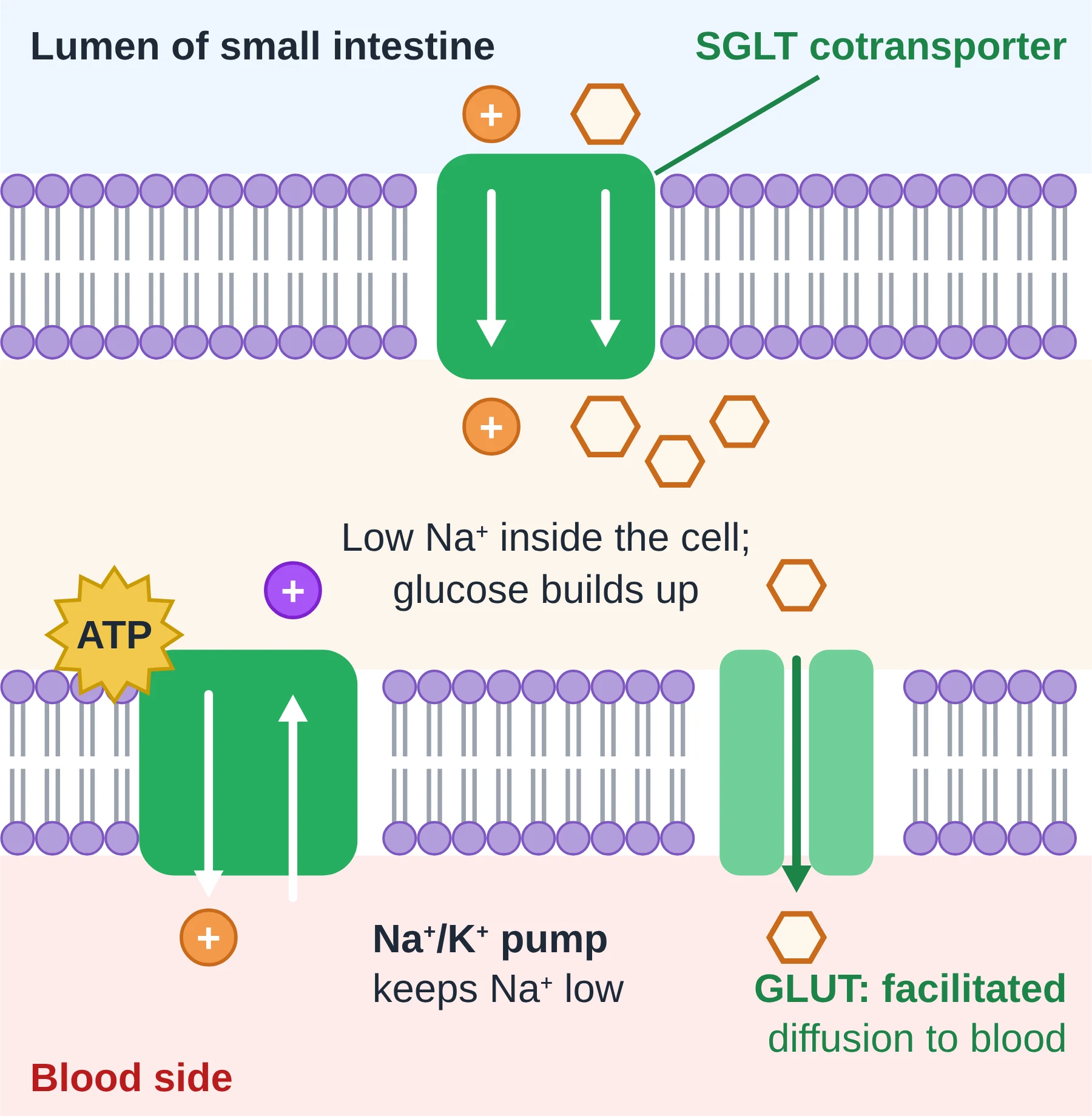
Cells lining the small intestine (and the proximal tubule of the nephron) must take up glucose even when there is more glucose inside the cell than outside. They use a sodium-dependent glucose cotransporter (SGLT).
- Na+/K+ pumps in the other side of the cell use ATP to keep the Na+ concentration inside the cell low.
- Na+ diffuses back into the cell down its gradient, through the SGLT, and glucose must travel with it.
- The energy released by Na+ moving down its gradient drags glucose in against its gradient.
- Glucose then leaves the cell on the blood side by facilitated diffusion through GLUT proteins.
- The SGLT does not use ATP itself; the ATP was used earlier by the pump. That is why this is called indirect active transport.
Key points
- Active transport moves particles against their concentration gradient.
- It uses pump proteins and energy from ATP.
- AHL Na+/K+ pump: 3 Na+ out, 2 K+ in per ATP; it generates the membrane potential.
- AHL SGLT cotransport is indirect active transport powered by the Na+ gradient.
Common mistakes
- Writing ‘3 K+ in and 2 Na+ out’. Remember: three Na out, two K in.
- Saying ATP is ‘made’ by the pump. ATP is used (hydrolysed to ADP + Pi).
- Saying the cotransporter uses ATP directly. It uses the Na+ gradient that ATP created.
5 Endocytosis
Syllabus link: B2.1 AHL membrane fluidity and the fusion and formation of vesicles
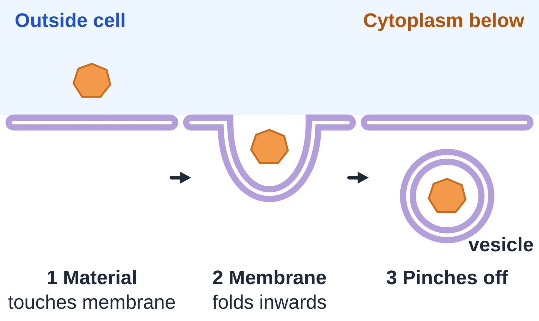
Some materials are far too big for any channel or pump, for example a whole bacterium or a droplet of fluid. Cells move these in bulk, wrapped in a piece of membrane called a vesicle. Taking material in this way is endocytosis. It is a type of bulk transport and uses ATP.
How it works
- Material outside the cell touches the plasma membrane (often binding to receptors).
- The membrane folds inwards, forming a pocket around the material. Proteins of the cytoskeleton help pull it in, using ATP.
- The edges of the pocket meet and fuse, so a vesicle pinches off into the cytoplasm. The plasma membrane reseals behind it.
Two main types
- Phagocytosis (‘cell eating’): large solid particles are engulfed. White blood cells such as macrophages take in bacteria this way; the vesicle then fuses with a lysosome, whose enzymes digest the contents. Amoeba feeds this way too.
- Pinocytosis (‘cell drinking’): small droplets of extracellular fluid, with whatever is dissolved in them, are taken in by small vesicles.
Why membrane fluidity matters AHL
Phospholipids in the bilayer are not fixed in place; they can move sideways. This fluidity lets the membrane bend, break and reseal without leaving a hole. The amount of unsaturated fatty acids and cholesterol in the membrane helps keep fluidity at the right level for vesicles to form.
Key points
- Endocytosis brings material into the cell inside vesicles.
- Phagocytosis takes in solid particles; pinocytosis takes in fluid.
- It needs ATP and depends on membrane fluidity.
- The plasma membrane gets slightly smaller each time a vesicle forms.
Common mistakes
- Saying the material crosses the membrane. It does not pass through the bilayer; it is enclosed by membrane.
- Mixing up endo- (into) and exo- (out of). Think: endo = enter, exo = exit.
6 Exocytosis
Syllabus link: B2.1 AHL membrane fluidity and the fusion and formation of vesicles
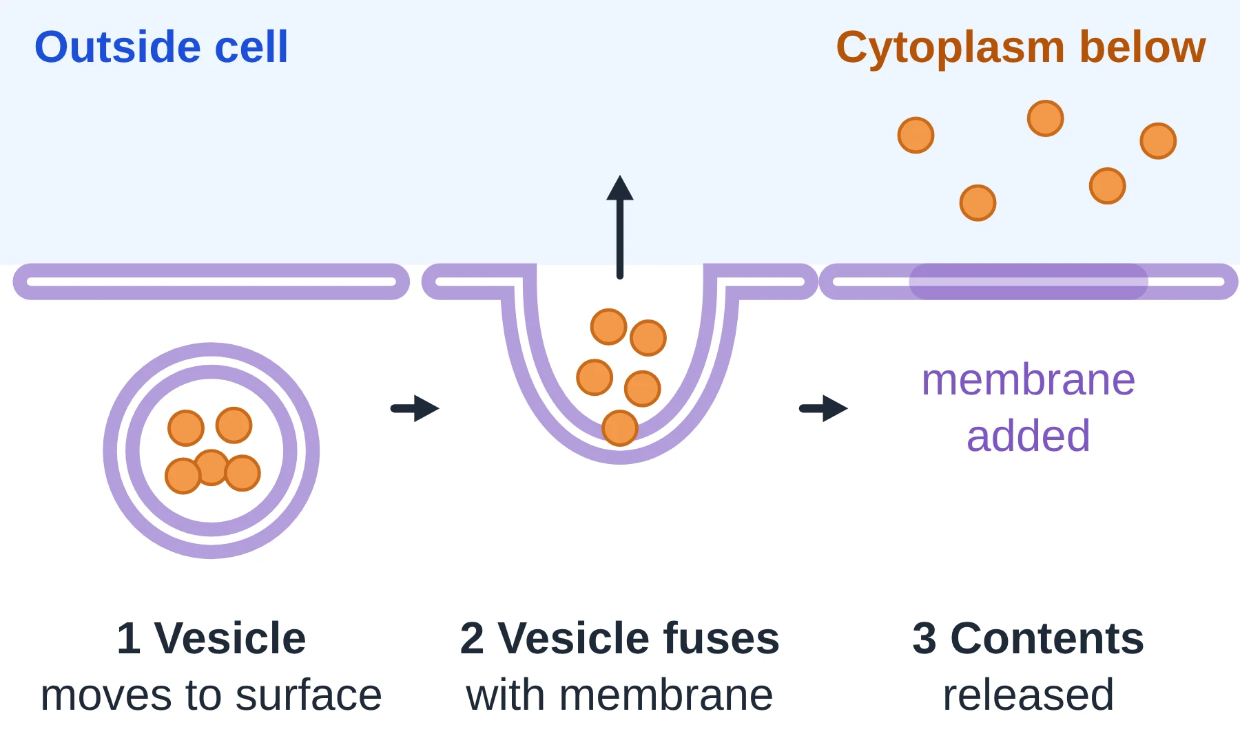
Exocytosis is the reverse of endocytosis: material packaged inside a vesicle is released out of the cell. It is how cells secrete products and get rid of waste. It also uses ATP.
How it works
- Proteins made on the rough endoplasmic reticulum are processed in the Golgi apparatus and packed into vesicles.
- Vesicles are carried to the plasma membrane along the cytoskeleton by motor proteins (using ATP).
- The vesicle membrane fuses with the plasma membrane. Because both are fluid bilayers, they join smoothly.
- The vesicle opens and its contents are released outside. The vesicle membrane becomes part of the plasma membrane.
Examples
- Pancreas cells release digestive enzymes and the hormone insulin.
- Neurons release neurotransmitters such as acetylcholine at synapses.
- Plant cells send cell wall materials out to the growing wall.
- Single-celled organisms expel waste or water (contractile vacuoles work in a similar way).
Endocytosis and exocytosis keep each other in balance: one removes membrane from the surface, the other adds it back.
Key points
- Exocytosis releases vesicle contents out of the cell.
- The vesicle fuses with the plasma membrane, which grows a little.
- Used for secretion (enzymes, hormones, neurotransmitters).
- Needs ATP and a fluid membrane.
Common mistakes
- Saying vesicles ‘burst’ at the membrane. They fuse.
- Forgetting that exocytosis is active (needs ATP), even though no pump protein is involved.
7 Tonicity
Syllabus link: D2.3 effects of water movement on cells with and without a cell wall; medical uses of isotonic solutions; AHL water potential
Tonicity compares the solute concentration of a solution outside a cell with the solute concentration inside the cell, and tells you which way water will move by osmosis. Always describe the outside solution relative to the cell.
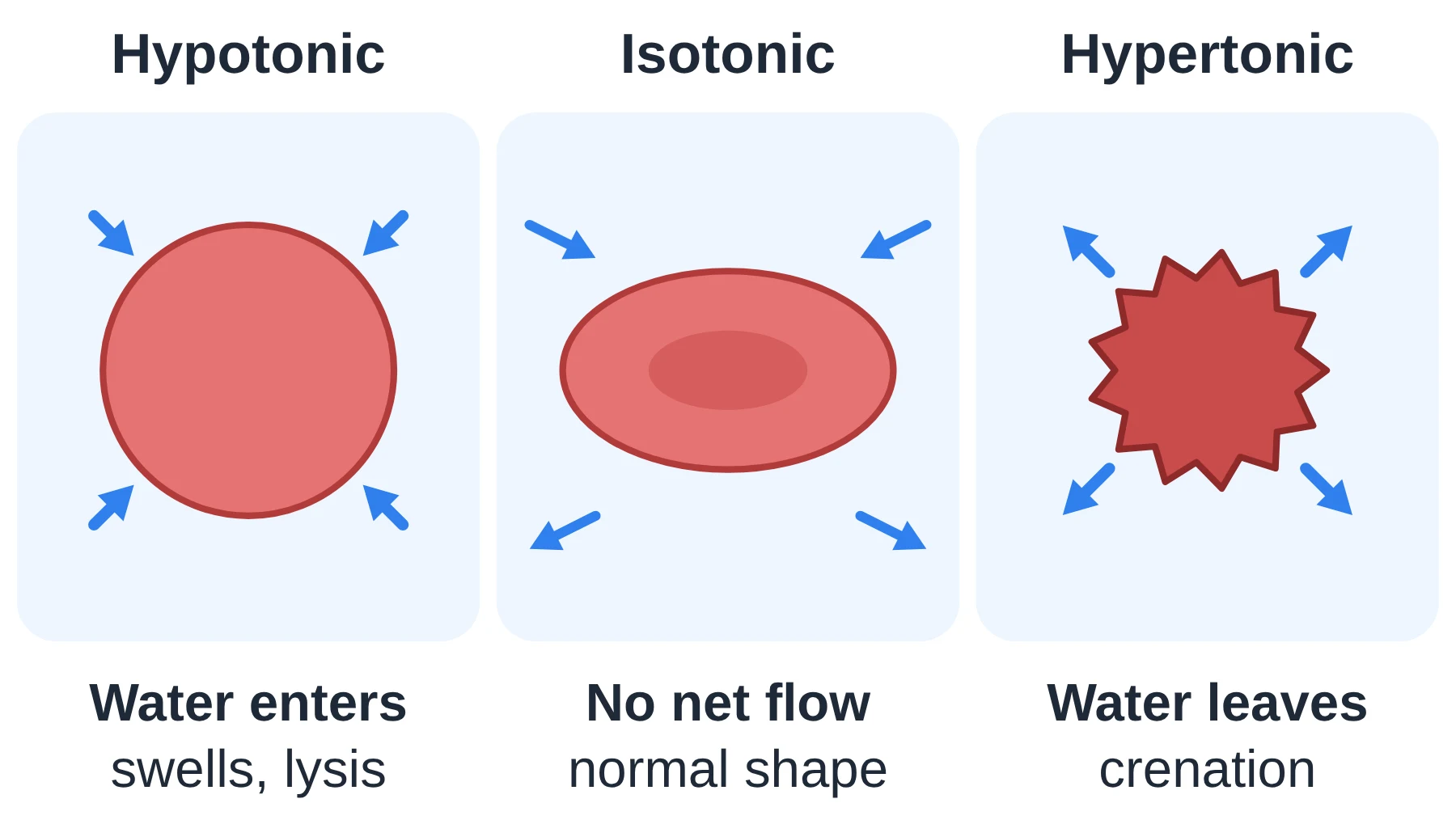
- Hypotonic solution: lower solute concentration than the cell. Water moves into the cell.
- Isotonic solution: same solute concentration as the cell. No net movement of water.
- Hypertonic solution: higher solute concentration than the cell. Water moves out of the cell.
Animal cells (no cell wall)
- In a hypotonic solution, water enters, the cell swells and may burst. This is lysis (for red blood cells, haemolysis).
- In an isotonic solution, the cell keeps its normal shape (a red blood cell stays a biconcave disc).
- In a hypertonic solution, water leaves and the cell shrinks; red blood cells become wrinkled and spiky. This is crenation.
- Freshwater single-celled organisms live in a hypotonic world. They use contractile vacuoles to pump excess water out and avoid bursting.
Plant cells (with a cell wall)
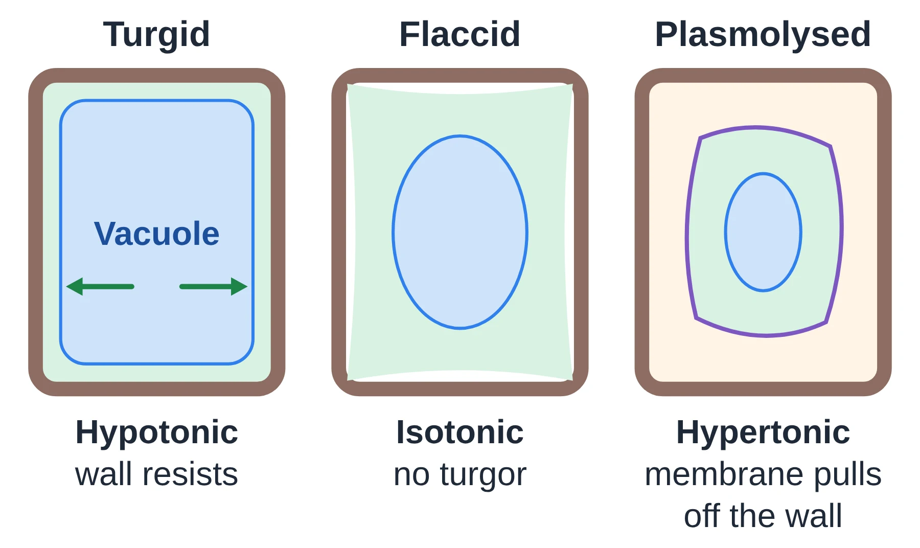
- In a hypotonic solution, water enters the vacuole and the cell swells until the strong cellulose cell wall pushes back. The cell becomes turgid but does not burst. Turgor pressure keeps stems and leaves firm.
- In an isotonic solution, there is no net water movement and no pressure on the wall: the cell is flaccid. Plants with flaccid cells wilt.
- In a hypertonic solution, water leaves the vacuole, the cytoplasm shrinks and the plasma membrane pulls away from the wall. This is plasmolysis. The gap fills with the outside solution, because the wall is fully permeable.
Why medical saline is isotonic
Fluids put directly into the blood (intravenous drips) are usually 0.9% sodium chloride, often called normal saline. This is roughly isotonic with blood plasma, so red blood cells neither take in water and burst nor lose water and crenate. Pure water in a vein would cause haemolysis. For the same reason, organs for transplant are kept in cold isotonic solutions, and contact lens and eye-wash solutions are made isotonic.
AHL Water potential notes
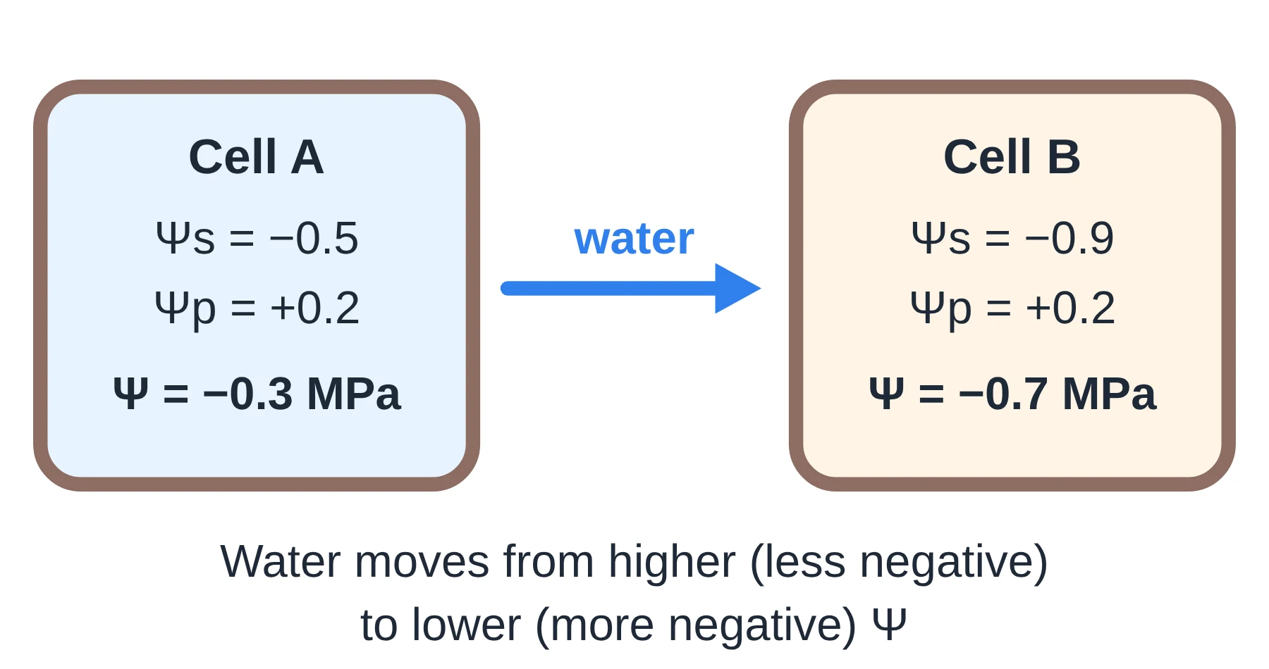
Water potential (symbol Ψ, the Greek letter psi) measures the tendency of water to move. It is measured in kilopascals (kPa) or megapascals (MPa).
- Pure water at atmospheric pressure has Ψ = 0. Most water potentials in cells are negative.
- Water always moves from higher (less negative) Ψ to lower (more negative) Ψ.
- Ψ = Ψs + Ψp
- Solute potential (Ψs): adding solutes lowers it, so it is zero for pure water and negative in any solution.
- Pressure potential (Ψp): positive when a cell wall pushes back on a swollen cell (turgor); zero in a flaccid or plasmolysed cell. In xylem under tension it can be negative.
- To find the water potential of plant tissue, soak samples in a range of sucrose solutions and plot % change in mass. Where the line crosses zero, the tissue is isotonic with that solution.
Key points
- Hypotonic: water in. Isotonic: no net flow. Hypertonic: water out.
- Animal cells: lysis in hypotonic, crenation in hypertonic.
- Plant cells: turgid, flaccid or plasmolysed; the wall stops bursting.
- Medical saline (0.9% NaCl) is isotonic to blood plasma.
- AHL Ψ = Ψs + Ψp; water moves to lower Ψ.
Common mistakes
- Describing the cell as ‘hypertonic’ without saying compared with what. Tonicity compares a solution with the cell.
- Saying plant cells burst in pure water. The cell wall prevents this.
- Saying a plasmolysed cell has an empty gap. The gap is filled with outside solution.
- AHL Thinking a more negative Ψ means more water. It means less free water, so water moves towards it.
8 Passive vs active transport at a glance
| Feature | Passive transport | Active transport |
|---|---|---|
| Includes | Simple diffusion, osmosis, facilitated diffusion | Pumping (e.g. Na+/K+ pump), cotransport, endocytosis, exocytosis |
| Direction | Down the concentration gradient (high to low) | Pumps can move particles against the gradient (low to high) |
| Energy from ATP | Not needed; uses the particles’ kinetic energy | Needed (directly, or indirectly in cotransport) |
| Proteins | None (simple diffusion) or channels and carriers | Pump proteins; vesicles for bulk transport |
| Specific? | Simple diffusion is not; channels and carriers are | Yes, pumps bind specific particles |
| Stops when | Concentrations are equal (no net movement) | ATP runs out (e.g. if respiration is blocked) |
| Examples | O2 into alveolar capillaries; water via aquaporins; glucose via GLUT | Mineral ions into root hairs; 3 Na+ out / 2 K+ in; insulin secretion |
Memory hook: passive = downhill, free; active = uphill, costs ATP.
9 Quick check
Ten questions: six multiple choice and four short answer. Try them before opening the answers.
-
Which of these crosses the phospholipid bilayer most easily by simple diffusion?
- A. Na+ ions
- B. Glucose
- C. Oxygen
- D. Amino acids
-
Aquaporins increase the permeability of a membrane to:
- A. Glucose
- B. Water
- C. Potassium ions
- D. Proteins
-
A red blood cell is placed in a strong salt solution. What happens?
- A. It swells and lyses
- B. It stays the same
- C. It becomes turgid
- D. It shrinks and crenates
-
Which statement about facilitated diffusion is correct?
- A. It uses ATP
- B. It can move ions against their gradient
- C. It needs a channel or carrier protein
- D. It only moves non-polar molecules
-
AHL In one cycle using one ATP, the sodium–potassium pump moves:
- A. 2 Na+ out and 3 K+ in
- B. 3 Na+ out and 2 K+ in
- C. 3 Na+ in and 2 K+ out
- D. 1 Na+ out and 1 K+ in
-
A macrophage engulfing a bacterium is an example of:
- A. Pinocytosis
- B. Exocytosis
- C. Phagocytosis
- D. Osmosis
-
Explain why the rate of facilitated diffusion stops increasing at high concentration differences. (2 marks)
-
Explain why patients are given 0.9% sodium chloride solution in a drip rather than pure water. (3 marks)
-
A strip of onion epidermis is placed in a concentrated sucrose solution. Describe and explain what happens to the cells. (3 marks)
-
AHL Cell X has Ψs = −0.6 MPa and Ψp = +0.2 MPa. Neighbouring cell Y has Ψs = −0.4 MPa and Ψp = +0.1 MPa. Calculate both water potentials and state the direction of water movement. (3 marks)
Show answers
- C, oxygen. O2 is small and non-polar, so it dissolves in the hydrophobic core. Ions and large polar molecules need proteins.
- B, water. Aquaporins are channel proteins that let water pass in single file.
- D. The solution is hypertonic, so water leaves by osmosis and the cell crenates. (Only plant cells become turgid.)
- C. Facilitated diffusion is passive, works down the gradient, and relies on specific membrane proteins.
- B. Three Na+ out and two K+ in.
- C, phagocytosis, a type of endocytosis for solid particles.
- All the channel or carrier proteins are in use (saturated) [1], so the number of proteins becomes the limiting factor and extra solute cannot cross any faster [1].
- 0.9% NaCl is isotonic with blood plasma [1], so there is no net osmosis into or out of red blood cells [1]. Pure water is hypotonic: water would enter the cells, which would swell and burst (haemolysis) [1].
- The solution is hypertonic to the cells [1]. Water leaves the vacuole and cytoplasm by osmosis [1]. The protoplast shrinks and the plasma membrane pulls away from the cell wall: plasmolysis [1].
- X: Ψ = −0.6 + 0.2 = −0.4 MPa [1]. Y: Ψ = −0.4 + 0.1 = −0.3 MPa [1]. Water moves from Y to X, from higher to lower water potential [1].
10 Glossary
© competitorspoint.com · IB Biology notes. Diagrams and text are original to competitorspoint.com.
Want this chapter offline? Download the full Membrane Transport chapter as a PDF (free for signed-up members).
⬇ Download chapter PDF