
Biology › Human physiology › Excretion and osmoregulation
Excretion and Osmoregulation
The human excretory system for IB Biology and NEET: nitrogenous wastes, the kidney and nephron, ultrafiltration and urine formation, the counter-current mechanism, ADH, RAAS and ANF, and kidney disorders, dialysis and transplant. IB-only and NEET-only content is marked.
1 Excretory products: ammonia, urea and uric acid
Syllabus link: NCERT XI Ch 16 (16.0 introduction, 16.1 Human excretory system: introduction to modes of excretion); IB D3.3 Homeostasis (HL: excretion as removal of metabolic waste such as urea)
The human excretory system removes the waste our cells make and keeps the water and salt content of the blood steady. This chapter of Human Physiology covers the whole human excretory system, starting with the wastes themselves.
Excretion is the removal of metabolic waste from the body. It is not the same as egestion (getting rid of undigested food as faeces), because faeces were never made by our cells. Animals have to get rid of ammonia, urea, uric acid, carbon dioxide, water and ions such as Na+, K+, Cl− and phosphate. The main problem is nitrogen: when amino acids and nucleic acids are broken down, the extra amino groups become ammonia, which is very toxic.
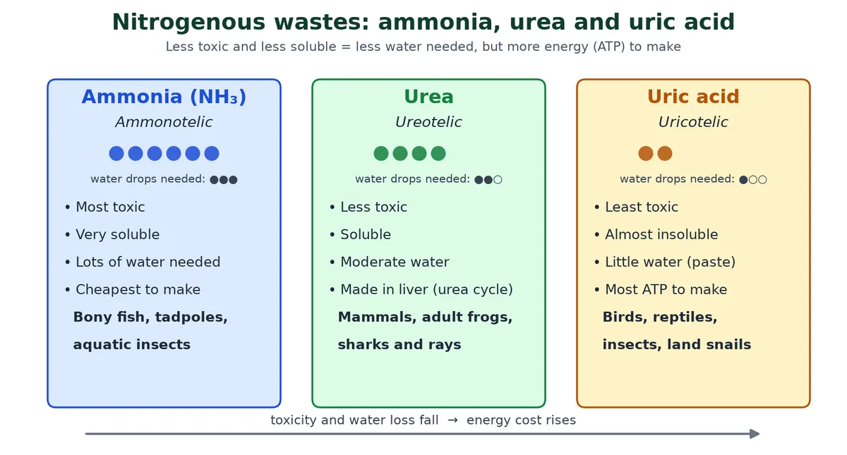
The human excretory system deals mainly with urea, but other animals use other wastes.
Ammonotelic, ureotelic and uricotelic animals
Swipe the table sideways to see every column.
| Mode | Main waste | Toxicity and water need | Examples (NCERT) |
|---|---|---|---|
| Ammonotelic | Ammonia | Most toxic; needs a lot of water; diffuses out across body surface or gills as NH4+ | Many bony fishes, aquatic amphibians (tadpoles), aquatic insects |
| Ureotelic | Urea | Less toxic; moderate water. Made from ammonia in the liver (urea / ornithine cycle) | Mammals, many terrestrial amphibians (adult frog), marine fishes such as sharks |
| Uricotelic | Uric acid | Least toxic; almost insoluble, so it leaves as a paste or pellet with very little water | Reptiles, birds, land snails, insects |
Some urea is kept in the kidney medulla of ureotelic animals to help concentrate urine (see section 5). Humans are ureotelic, but we also make a little uric acid from purines, which is why gout and some kidney stones contain uric acid.
NEET only Excretory organs across the animal kingdom
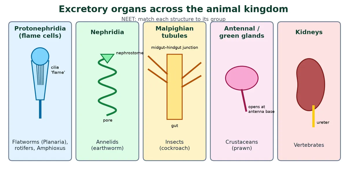
Swipe the table sideways to see every column.
| Structure | Found in | Note |
|---|---|---|
| Protonephridia (flame cells) | Platyhelminthes (Planaria), rotifers, some annelids, Amphioxus | Mainly osmoregulation (ionic and fluid volume), not nitrogen excretion |
| Nephridia | Annelids (earthworm) | Remove nitrogenous waste and keep fluid and ionic balance |
| Malpighian tubules | Most insects, including cockroach | Remove nitrogenous waste (uric acid) and help osmoregulation |
| Antennal glands (green glands) | Crustaceans such as prawn | Excretion |
| Kidneys | Vertebrates | Built from many nephrons; also osmoregulate |
Click to check: which structure is mainly for osmoregulation rather than nitrogen excretion?
Protonephridia (flame cells). In flatworms most nitrogen leaves by diffusion across the body surface.
Types of vertebrate kidney (from the old post, for interest). Three kidneys appear in vertebrate evolution and development: the pronephros (head kidney: functional in larval fishes and amphibians and in adult hagfish and some bony fish as a head kidney), the mesonephros (functional in adult fishes and amphibians, embryonic in reptiles, birds and mammals; its duct is the Wolffian duct) and the metanephros (the adult kidney of reptiles, birds and mammals, with a ureter, cortex and medulla). The human kidney is metanephric; it forms in the embryo after the other two.
Exam tip
- Name the waste and the habitat together: water-rich habitat → ammonia; water-poor → uric acid.
- Tadpoles are ammonotelic but adult frogs are ureotelic: the switch happens at metamorphosis.
- Do not call faeces an excretory product.
2 The human urinary system
Syllabus link: NCERT XI 16.1 Human excretory system; IB D3.3 (HL): role of the kidney in osmoregulation and excretion
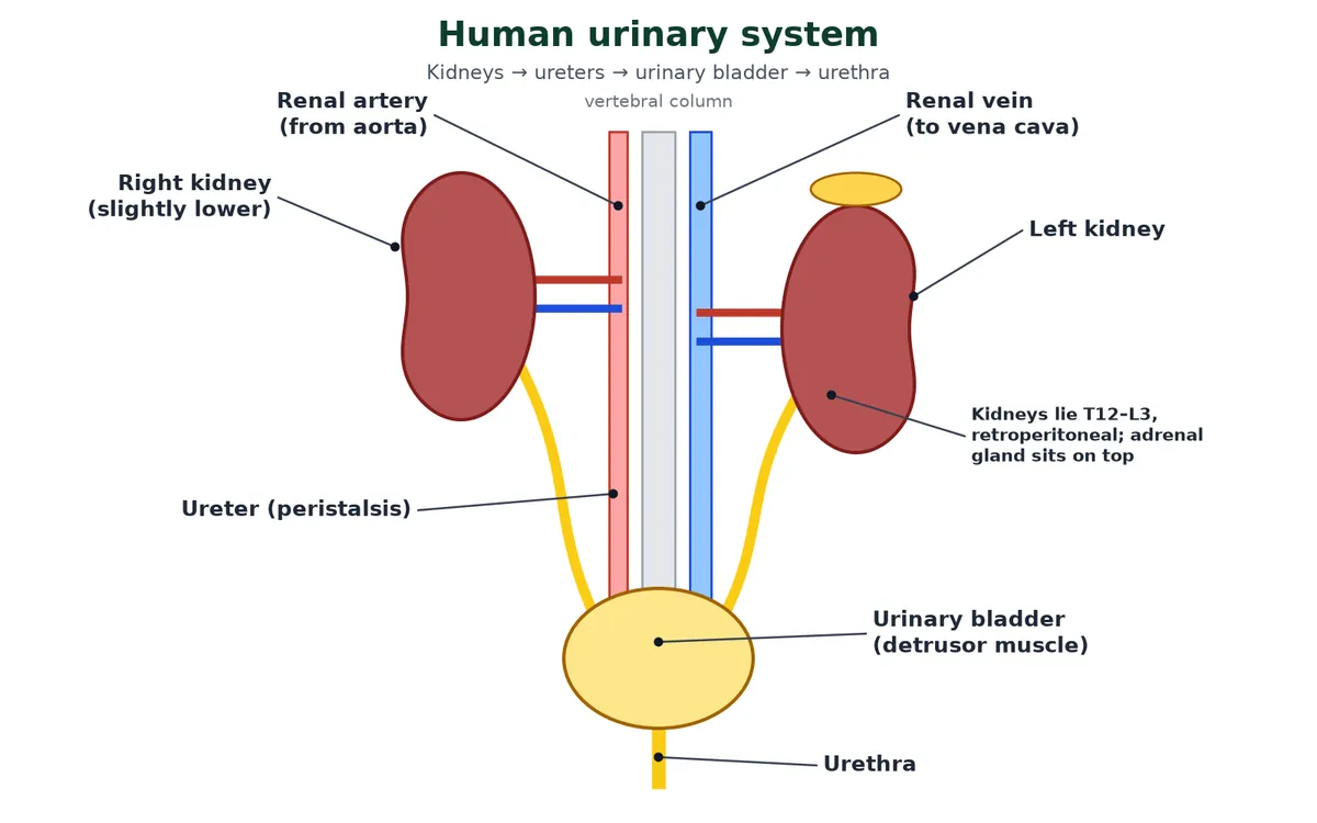
The human excretory system has four parts: a pair of kidneys, a pair of ureters, one urinary bladder and one urethra.
Kidneys
- Reddish-brown, bean-shaped organs between the last thoracic and third lumbar vertebrae (T12 to L3), close to the back wall of the abdomen. The right kidney sits a little lower because the liver is above it.
- They are retroperitoneal: they lie behind the peritoneum, which covers only their front (ventral) surface.
- Each adult kidney is about 10 to 12 cm long, 5 to 7 cm wide, 2 to 3 cm thick and weighs 120 to 170 g.
- The inner, concave side has a notch, the hilum, where the ureter, blood vessels and nerves enter or leave. An adrenal gland sits on top of each kidney.
Ureters, bladder and urethra
Swipe the table sideways to see every column.
| Part | Structure | Job |
|---|---|---|
| Ureter | Muscular tube: transitional epithelium inside (stretches), smooth muscle (longitudinal and circular) in the middle, connective tissue (adventitia) outside | Carries urine from the renal pelvis to the bladder by peristalsis |
| Urinary bladder | Same lining; thick smooth muscle wall called the detrusor. The trigone is a smooth triangular area on its floor between the two ureter openings and the urethral opening | Stores urine. The urge to pass urine starts at about 200 to 300 mL; it can hold up to about 500 to 700 mL |
| Urethra | Tube from bladder to outside; internal sphincter (smooth muscle, involuntary) and external sphincter (skeletal muscle, voluntary) | Carries urine out. Much longer in males, where it also carries semen |
Micturition
Micturition is the passing out of urine. As the bladder fills, stretch receptors in its wall send impulses to the CNS (spinal cord, with control from the brain). The CNS sends motor signals that make the detrusor muscle contract (parasympathetic nerves) and the internal sphincter relax; the voluntary external sphincter is relaxed by reducing signals in the pudendal nerve. While the bladder fills, sympathetic nerves keep the detrusor relaxed and the internal sphincter closed. This is called the micturition reflex. Infants cannot yet override it; adults can delay it until convenient. Incontinence is the loss of this control, so urine leaks.
Key idea
- Kidneys filter; ureters carry; bladder stores; urethra releases.
- An adult passes about 1 to 1.5 L of urine a day (NCERT).
3 Kidney structure and the nephron
Syllabus link: NCERT XI 16.1 (internal structure of kidney, nephron, cortical and juxtamedullary nephrons); IB D3.3 (HL): glomerulus, Bowman’s capsule, tubules and blood supply
Inside the human excretory system, each kidney is packed with about one million tiny tubes called nephrons, the functional units that make urine.
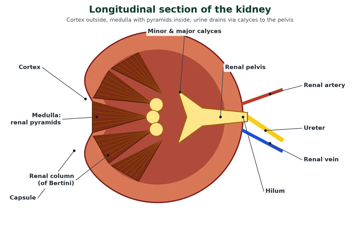
Regions of the kidney
- Capsule: tough fibrous outer layer.
- Cortex: outer zone; contains the glomeruli, PCTs and DCTs.
- Medulla: inner zone divided into conical renal pyramids; contains the loops of Henle and collecting ducts. Cortex extends between the pyramids as renal columns (columns of Bertini).
- The pyramids point into calyces (minor, then major), which open into the funnel-shaped renal pelvis, which continues as the ureter.
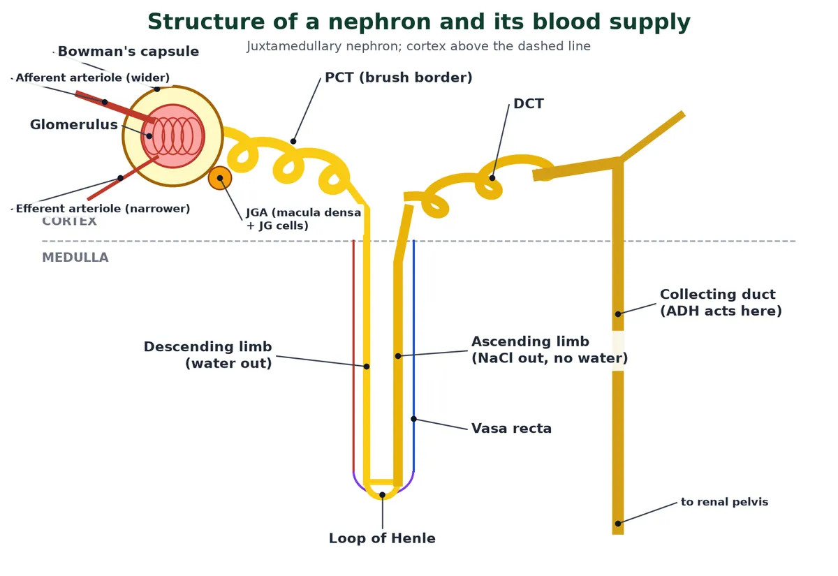
Parts of a nephron
Each nephron has two parts: the glomerulus (a knot of capillaries) and the renal tubule. The tubule starts as the cup-shaped, double-walled Bowman’s capsule around the glomerulus; together they form the Malpighian body or renal corpuscle.
Swipe the table sideways to see every column.
| Part | Lining | Where | Main job |
|---|---|---|---|
| Bowman’s capsule | Outer (parietal) layer: simple squamous; inner (visceral) layer: podocytes with foot processes and filtration slits. The gap between is Bowman’s space | Cortex | Collects the filtrate |
| Proximal convoluted tubule (PCT) | Simple cuboidal with a brush border (microvilli) and many mitochondria | Cortex | Reabsorbs most useful substances |
| Loop of Henle | Thin parts: squamous; thick ascending part: cuboidal | Dips into medulla | Builds the salt gradient in the medulla |
| Distal convoluted tubule (DCT) | Cuboidal, fewer microvilli | Cortex | Fine-tunes Na+, K+, H+ and pH |
| Collecting duct | Cuboidal; many join into ducts of Bellini that open at the tip of a pyramid | Cortex to medulla | Final water reabsorption under ADH |
Every function of the human excretory system described below happens in these segments. Strictly, the collecting duct is not part of the nephron, because many nephrons drain into one duct.
Blood supply
Blood reaches the glomerulus by a wide afferent arteriole and leaves by a narrower efferent arteriole. The efferent arteriole then forms the peritubular capillaries around the tubule; a branch running beside the loop of Henle in a U shape is the vasa recta. Blood finally drains to the renal vein.
Cortical and juxtamedullary nephrons
Swipe the table sideways to see every column.
| Cortical nephrons | Juxtamedullary nephrons | |
|---|---|---|
| Share | About 85% | About 15% |
| Loop of Henle | Short, barely enters the medulla | Long, runs deep into the medulla |
| Vasa recta | Absent or highly reduced | Well developed |
| Role | Routine urine formation | Producing concentrated urine when water must be saved |
NEET only Juxtaglomerular apparatus (JGA)
Where the DCT touches the afferent arteriole of its own glomerulus, three cell types form the JGA: macula densa (tall, packed DCT cells that sense NaCl in the filtrate), juxtaglomerular (JG) cells (modified smooth muscle in the afferent arteriole wall that secrete renin and sense stretch, like baroreceptors) and extraglomerular mesangial (lacis) cells between them, which pass signals. Its role in controlling GFR and blood pressure is covered in section 6.
Click to check: why is the PCT lined by cells with microvilli and many mitochondria?
Microvilli give a huge surface for reabsorption, and mitochondria supply ATP for the active transport (Na+/K+ pumps) that drives most of it.
4 Ultrafiltration and urine formation
Syllabus link: NCERT XI 16.2 Urine formation (glomerular filtration, GFR, reabsorption, secretion); IB D3.3 (HL): ultrafiltration in the glomerulus and Bowman’s capsule, selective reabsorption in the PCT
Urine is made in three steps in every nephron of the human excretory system: glomerular filtration, tubular reabsorption and tubular secretion.
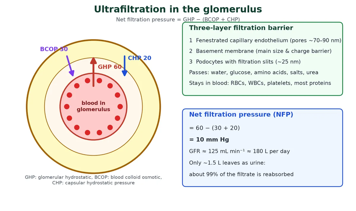
1. Glomerular filtration (ultrafiltration)
Blood in the glomerulus is under high pressure, because the efferent arteriole is narrower than the afferent arteriole. This pressure forces fluid through a three-layer filtration membrane:
- Fenestrated endothelium of the glomerular capillaries: pores about 70 to 90 nm wide stop blood cells.
- Basement membrane: a gel of glycoproteins with negative charge; the main barrier that holds back proteins by size and by charge.
- Podocytes of the inner wall of Bowman’s capsule: their foot processes leave very fine filtration slits (about 25 nm).
Filtration is passive and depends on size (and charge), so useful and harmful small molecules pass alike. The filtrate is like plasma without blood cells and almost no proteins: water, glucose, amino acids, ions, vitamins, urea, uric acid, creatinine and many drugs. Albumin, the main plasma protein, is just too big and too negative to cross in any amount.
Net filtration pressure
Swipe the table sideways to see every column.
| Pressure | Value (typical) | Effect |
|---|---|---|
| Glomerular hydrostatic pressure (GHP) | 60 mm Hg | Pushes fluid out of the capillaries |
| Blood colloid osmotic pressure (BCOP) | 30 mm Hg | Plasma proteins pull water back in |
| Capsular hydrostatic pressure (CHP) | 20 mm Hg | Fluid already in Bowman’s space pushes back |
| Net filtration pressure | 60 − (30 + 20) = 10 mm Hg | Enough to filter about 125 mL per minute |
GFR and renal blood flow
- Glomerular filtration rate (GFR) is the volume of filtrate formed by both kidneys per minute: about 125 mL min−1, or 180 L a day (NCERT).
- The kidneys receive 1100 to 1200 mL of blood a minute, about one-fifth of cardiac output. The plasma part (renal plasma flow) is about 600 to 700 mL min−1.
- Filtration fraction = GFR ÷ renal plasma flow = 125 ÷ 650 ≈ 0.19, so about 16 to 20% of the plasma reaching the glomeruli is filtered.
- With about 3 L of plasma in the body, 180 L a day means the whole plasma volume is filtered about 60 times a day. Only about 1.5 L leaves as urine, so about 99% of the filtrate is reabsorbed.
NEET only How the kidney keeps GFR steady (autoregulation)
- Myogenic response: when blood pressure rises, the afferent arteriole is stretched and its smooth muscle contracts, limiting the rise in GFR.
- Tubuloglomerular feedback: if GFR rises, more NaCl reaches the macula densa, which signals the afferent arteriole to constrict. If GFR falls, the macula densa triggers vasodilation of the afferent arteriole and renin release from the JG cells.
- Sympathetic nerves (noradrenaline) constrict the afferent arteriole during stress or blood loss, lowering GFR to save fluid.
2. Tubular reabsorption
Reabsorption is where the human excretory system saves what the body needs. Useful substances are taken back from the filtrate into the blood in the peritubular capillaries. Glucose, amino acids and Na+ are reabsorbed actively (Na+/K+ pumps, with co-transport for glucose and amino acids); water and urea follow passively. Section 5 follows this segment by segment.
Swipe the table sideways to see every column.
| Type | Examples | What happens |
|---|---|---|
| High-threshold substances | Glucose, amino acids, Na+, Cl− | Almost completely reabsorbed until the carriers are saturated. The renal threshold for glucose is about 180 mg dL−1 of blood; above this, glucose appears in urine |
| Low-threshold substances | Urea, uric acid | Only partly reabsorbed |
| Non-threshold substances | Creatinine, inulin, hippuric acid | Not reabsorbed (creatinine is even secreted a little) |
3. Tubular secretion
Tubule cells move substances from the blood into the filtrate: H+, K+ and ammonia (NH3/NH4+), plus some creatinine and drugs such as penicillin. This happens in the PCT, DCT and collecting duct and keeps blood pH and ionic balance right.
Try it: follow a drop of filtrate through the nephron
Step 1 of 6: Glomerular filtration Glomerulus → Bowman’s capsule
Blood pressure pushes water and small solutes out of the glomerulus. Cells and most proteins stay behind. About 180 L of filtrate a day.
Step 2 of 6: Selective reabsorption PCT
70 to 80% of the filtrate is taken back: all glucose and amino acids, most Na+, Cl−, K+, HCO3− and water. H+ and ammonia are secreted in. Volume falls; concentration does not change.
Step 3 of 6: Descending limb Loop of Henle (going down)
Permeable to water, almost impermeable to salts. Water leaves into the salty medulla, so the filtrate becomes more concentrated.
Step 4 of 6: Ascending limb Loop of Henle (coming up)
Impermeable to water. Na+ and Cl− leave (passively in the thin part, actively in the thick part), so the filtrate becomes dilute and the medulla stays salty.
Step 5 of 6: Distal convoluted tubule DCT
Conditional reabsorption of Na+ (aldosterone) and some water; reabsorbs HCO3−; secretes H+, K+ and NH3 to keep blood pH and K+ balanced. Ca2+ reabsorption is boosted by parathyroid hormone.
Step 6 of 6: Collecting duct Collecting duct → pelvis
With ADH, aquaporins open and water leaves into the medulla, so urine becomes concentrated (up to 4 times plasma). Some urea leaks into the inner medulla. Secretes H+ and K+.
Click to check: glucose is filtered, so why is none in normal urine?
Because all of it is reabsorbed in the PCT by Na+-glucose co-transporters. Glucose appears only when blood glucose is above the renal threshold (about 180 mg dL−1), as in uncontrolled diabetes mellitus.
5 Reabsorption, water balance and the counter-current mechanism
Syllabus link: NCERT XI 16.3 Function of the tubules, 16.4 Mechanism of concentration of the filtrate; IB D3.3 (HL): loop of Henle and the high solute concentration of the medulla
Each part of the tubule in the human excretory system has its own permeability, and that is how the kidney can make urine more dilute or more concentrated than blood.
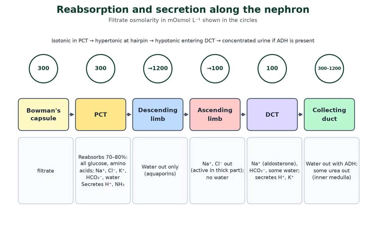
Swipe the table sideways to see every column.
| Segment | Reabsorbed | Secreted | Filtrate becomes |
|---|---|---|---|
| PCT | 70 to 80% of filtrate: all glucose and amino acids, vitamins, most Na+, Cl−, K+, HCO3−, water; a little urea | H+, NH3 | Isotonic (much less volume). Water reabsorption here is obligatory: it follows solutes whatever the body’s needs |
| Descending limb | Water only | None | Hypertonic |
| Ascending limb | Na+, Cl− (also K+, Ca2+, Mg2+); no water | None | Hypotonic (about 100 mOsmol L−1) |
| DCT | Na+ (aldosterone), HCO3−, Ca2+ (PTH), some water | H+, K+, NH3 | Adjusted |
| Collecting duct | Water (ADH: facultative reabsorption), Na+; urea in the inner medulla | H+, K+ | Urine: dilute or up to 4 times plasma |
Why the medulla is salty
Mammals can make urine about four times as concentrated as plasma (up to about 1200 mOsmol L−1). The trick is a gradient of solute in the medulla interstitium, rising from about 300 at the cortex to about 1200 mOsmol L−1 deep in the medulla. It is set up by the loop of Henle and kept by the vasa recta.
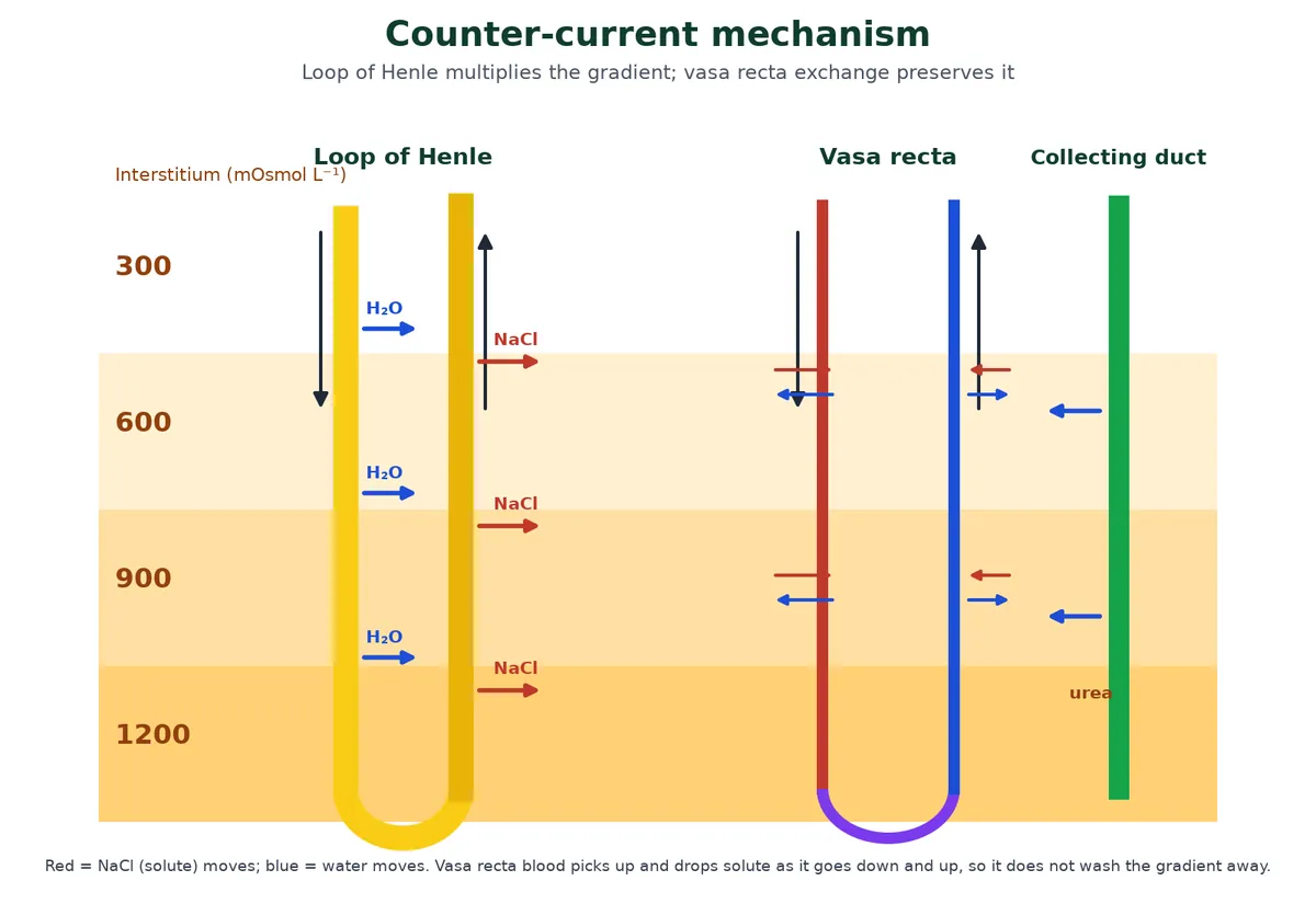
The counter-current mechanism, step by step
- The filtrate flows in opposite directions in the two limbs of the loop of Henle; blood in the two limbs of the vasa recta also flows in a counter-current. That is where the name comes from.
- The thick ascending limb actively pumps Na+ and Cl− out into the medulla but does not let water follow. The interstitium becomes salty and the filtrate becomes dilute.
- The salty interstitium draws water out of the descending limb by osmosis, so the filtrate going down becomes more concentrated. That concentrated fluid then gives up even more NaCl as it goes up. Each pass adds to the gradient: the loop is a counter-current multiplier.
- Urea leaks out of the inner medullary collecting duct; some enters the thin ascending limb and is passed back round (urea recycling). Urea provides about half of the inner medulla’s solute.
- The vasa recta are a counter-current exchanger: blood going down picks up NaCl and loses water; going up it gives NaCl back and takes water. So the blood removes reabsorbed water without washing the gradient away.
- With ADH, the collecting duct runs down through this gradient and water leaves it by osmosis, giving a small volume of concentrated urine.
Two consequences: animals with longer loops of Henle (such as desert rodents) can make more concentrated urine, and a diet rich in protein (more urea) slightly raises the medulla’s concentrating power. The collecting ducts of many nephrons join into ducts of Bellini, which open at the tip of each pyramid into a minor calyx, then a major calyx, the pelvis and the ureter.
IB only HL Exam focus: loop of Henle
- Ascending limb: active transport of Na+ out, impermeable to water.
- This keeps a high solute concentration in the medulla, so water can leave the collecting duct by osmosis.
- The maximum concentration of urine is set by how much Na+ the loop can pump, and by loop length.
What urine contains
Urine is the final product of the human excretory system, and its make-up tells doctors a lot.
- About 1 to 1.5 L a day; pale yellow (from urochrome, a pigment from the breakdown of haemoglobin’s haem), watery and slightly acidic (pH about 6, range roughly 4.5 to 8).
- About 95% water. On average 25 to 30 g of urea is excreted a day, plus creatinine, uric acid, ammonia, Na+, K+, Cl−, phosphate and sulphate.
- Urine analysis helps in diagnosis. Abnormal constituents include:
Swipe the table sideways to see every column.
| Condition | What appears in urine | Usual cause |
|---|---|---|
| Glycosuria | Glucose | Diabetes mellitus (blood glucose above the renal threshold) |
| Ketonuria | Ketone bodies | Diabetes mellitus, starvation (fat being used for energy) |
| Proteinuria / albuminuria | Protein, mainly albumin | Damaged filtration membrane, e.g. glomerulonephritis |
| Haematuria | Red blood cells | Kidney stones, infection, injury, tumours |
| Haemoglobinuria | Free haemoglobin | Haemolysis (red cells breaking up in the blood) |
| Pyuria | Pus cells (dead neutrophils) | Urinary tract infection |
| Xanthinuria (rare, genetic) | Xanthine | Lack of xanthine oxidase in purine breakdown; can cause xanthine stones |
Click to check: which part of the nephron produces a hypotonic filtrate?
The ascending limb of the loop of Henle, because it removes NaCl but is impermeable to water. The filtrate entering the DCT is about 100 mOsmol L−1.
6 Osmoregulation and hormones: ADH, RAAS and ANF
Syllabus link: NCERT XI 16.5 Regulation of kidney function; IB D3.3 (HL): osmoregulation by ADH and aquaporins in the collecting duct; distinction between excretion and osmoregulation
Osmoregulation is the control of the water and solute concentration of body fluids. In the human excretory system it is run by three hormone systems working through the kidney.
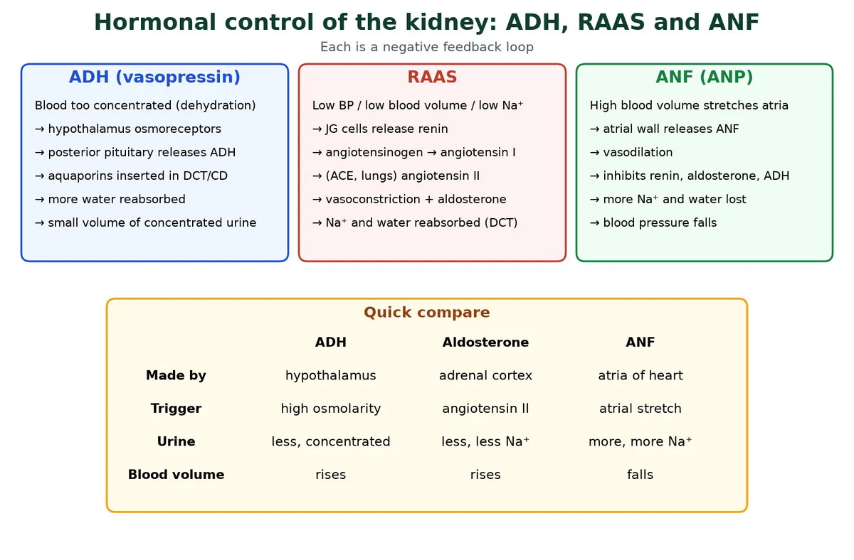
ADH (antidiuretic hormone, vasopressin)
ADH is made in the hypothalamus and released from the posterior pituitary. Normal plasma osmolarity is about 290 to 300 mOsmol L−1. When it rises (dehydration, salty food) or blood volume falls, more ADH is released. ADH makes the late DCT and the collecting duct permeable to water by inserting aquaporins, so more water is reabsorbed and urine is concentrated. ADH also causes some vasoconstriction, which raises blood pressure. When the body has too much water, ADH release is switched off. Alcohol and caffeine reduce ADH release, which is why they increase urine output.
Try it: what does ADH do when you are…
Dehydrated After exercise or sweating; no drinks
Blood osmolarity rises above about 290 to 300 mOsmol L−1. Osmoreceptors in the hypothalamus detect it and you feel thirsty. The posterior pituitary releases more ADH. ADH binds receptors on collecting duct cells, and vesicles carrying aquaporins fuse with the membrane. More water leaves the duct into the salty medulla, so blood osmolarity falls back: negative feedback.
Normal Balanced intake
A steady, moderate level of ADH keeps a moderate number of aquaporins in the collecting duct membranes. Urine volume roughly matches water intake minus water lost in sweat, breath and faeces.
Too much water Drank a lot quickly
Blood becomes too dilute. Osmoreceptors stop stimulating ADH release. Aquaporins are taken back into vesicles, the collecting duct becomes nearly impermeable to water, and dilute urine (as low as about 100 mOsmol L−1) carries the extra water away: diuresis.
Renin-angiotensin-aldosterone system (RAAS)
- A fall in blood pressure, blood volume or GFR is sensed by the JG cells (stretch), the macula densa (less NaCl reaching it) and sympathetic nerves.
- JG cells release the enzyme renin into the blood.
- Renin converts angiotensinogen, a plasma protein made by the liver, into angiotensin I.
- Angiotensin-converting enzyme (ACE), mostly on the lining of lung capillaries, converts it to angiotensin II.
- Angiotensin II is a powerful vasoconstrictor: it raises peripheral resistance and so blood pressure, and raises GFR. It also triggers thirst and ADH release.
- Angiotensin II makes the adrenal cortex (zona glomerulosa) release aldosterone. Aldosterone acts on the principal cells of the late DCT and collecting duct: more Na+ channels in the luminal membrane and more Na+/K+ pumps in the basolateral membrane. Na+ and water are reabsorbed and K+ is secreted. Blood volume and pressure rise, which switches renin release off.
The RAAS has its own page with an example of what happens after heavy bleeding: renin-angiotensin-aldosterone system (RAAS) explained step by step.
ANF (atrial natriuretic factor)
The heart also talks to the human excretory system.
When blood volume and pressure are high, the stretched walls of the atria release ANF (also called ANP). It causes vasodilation, increases GFR, and inhibits renin, aldosterone and ADH. More Na+ (natriuresis) and water (diuresis) are lost and blood pressure falls. ANF is a check on the RAAS. The heart side of this story is in Blood, Heart and Circulation.
Compare: the three systems
Swipe the table sideways to see every column.
| ADH | RAAS / aldosterone | ANF | |
|---|---|---|---|
| Released by | Posterior pituitary (made in hypothalamus) | Kidney (renin), liver, lungs, adrenal cortex | Atrial walls |
| Main trigger | High blood osmolarity | Low blood pressure or volume | High blood volume (atrial stretch) |
| Action on kidney | Water reabsorption in DCT and collecting duct | Na+ (and water) reabsorption; K+ secretion | Less Na+ and water reabsorbed |
| Urine | Less, more concentrated | Less, less Na+ | More, more Na+ |
Disorder link: too little ADH, or kidneys that do not respond to it, causes diabetes insipidus: large volumes of dilute urine and great thirst.
IB only Osmoregulators and osmoconformers
Swipe the table sideways to see every column.
| Osmoconformers | Osmoregulators | |
|---|---|---|
| Body fluid concentration | Matches the surroundings | Kept constant whatever the surroundings |
| Energy | Little spent on osmoregulation | Energy needed (active transport, kidneys) |
| Examples | Most marine invertebrates (e.g. jellyfish, starfish, mussels); sharks and rays are close to it, using urea in the blood | Humans and other mammals, birds, bony fish, freshwater animals |
- Freshwater bony fish gain water by osmosis: they hardly drink, produce lots of dilute urine and actively take up salts at the gills.
- Marine bony fish lose water by osmosis: they drink sea water, pump out excess salt at the gills and make a little urine.
- Distinguish the terms: excretion removes metabolic waste (urea); osmoregulation controls water and solute concentration. The kidney does both.
Note: osmoconformers versus osmoregulators was explicit in the previous IB syllabus (11.3); in the 2025 syllabus it is useful background for D3.3.
NEET only Remember for NEET
- ADH = vasopressin; deficiency gives diabetes insipidus.
- JG cells secrete renin; macula densa senses NaCl.
- Angiotensin II is the vasoconstrictor; ACE is mainly in the lungs.
- ANF opposes the RAAS.
7 Kidney disorders, dialysis and transplant
Syllabus link: NCERT XI 16.6 Micturition, 16.7 Role of other organs in excretion, 16.8 Disorders of the excretory system; IB D3.3 (HL) background: kidney failure, dialysis and transplantation
When the human excretory system fails, wastes such as urea build up in the blood. Here are the disorders NCERT lists, the treatments, and the other organs that share the work of excretion.
Disorders
Swipe the table sideways to see every column.
| Disorder | What it is | Notes |
|---|---|---|
| Uraemia | Too much urea (and other nitrogenous wastes) in the blood because the kidneys are failing | Highly harmful; causes tiredness, loss of appetite (anorexia), nausea; may lead to kidney failure. Treated by dialysis |
| Renal failure | Kidneys can no longer filter enough blood | Acute or chronic. Final stage needs dialysis or a transplant |
| Renal calculi (kidney stones) | Stones or crystallised salts, mainly calcium oxalate, formed inside the kidney | Can block the ureter, causing severe pain and blood in urine |
| Glomerulonephritis | Inflammation of the glomeruli of the kidney | Protein and red cells leak into urine |
| Cystitis | Inflammation of the urinary bladder | Usually a bacterial infection |
| Pyelonephritis | Infection and inflammation of the renal pelvis and kidney tissue | Often spreads up from the bladder |
Nephrons cannot be replaced: we are born with all we will have (about a million per kidney), and numbers slowly fall with age and disease. One healthy kidney is enough for normal life.
Haemodialysis (artificial kidney)
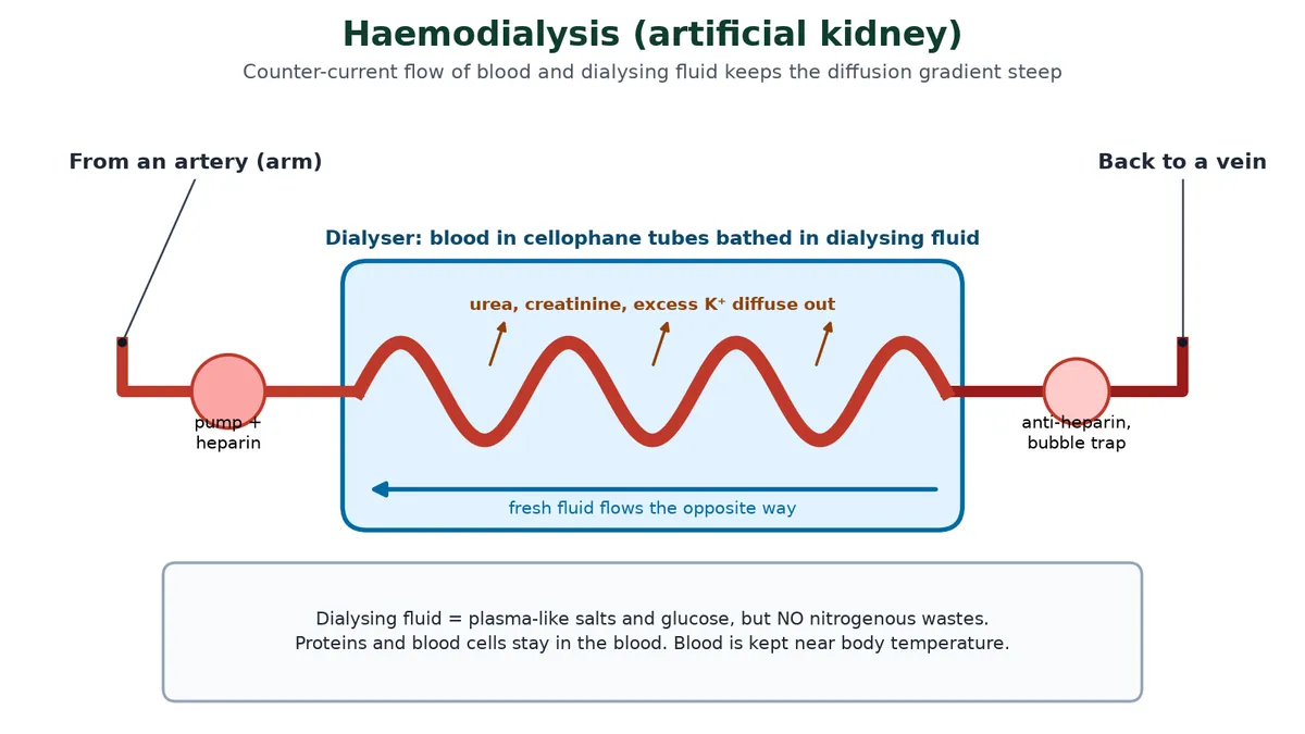
- Blood is taken from a convenient artery (often through a fistula in the arm) and an anticoagulant, heparin, is added.
- It is pumped through a dialysing unit (artificial kidney) containing coiled cellophane tubes, a partially permeable membrane.
- The tubes are bathed in dialysing fluid with the same composition as plasma except for the nitrogenous wastes. Urea, creatinine and excess K+ diffuse out down their concentration gradient; proteins and cells stay in.
- The fluid flows in the opposite direction to the blood and is constantly replaced, so the gradient stays steep.
- Anti-heparin is added and the cleared blood is pumped back into a vein. A session takes about 4 hours, usually three times a week.
Kidney transplant
Transplantation is the ultimate treatment for acute renal failure (NCERT). A functioning kidney is taken from a donor, preferably a close relative, to reduce the chance of rejection by the recipient’s immune system. Only a kidney from an identical twin is a perfect match. Other recipients take immunosuppressants such as cyclosporin A (obtained from the fungus Trichoderma polysporum) for life. A transplant gives a better quality of life than dialysis but is limited by the shortage of donors.
IB only Compare dialysis and transplant
Swipe the table sideways to see every column.
| Haemodialysis | Transplant | |
|---|---|---|
| How it works | Diffusion across an artificial membrane | A living kidney does all functions, including hormones (erythropoietin, renin) |
| Drawbacks | Hours per session, several times a week; diet and fluid limits | Surgery, risk of rejection, lifelong immunosuppressants (more infections), few donors |
| Cost over time | High, continuing | High at first, lower later |
Role of other organs in excretion
The kidneys are not the only excretory organs: the lungs, liver and skin support the human excretory system.
Swipe the table sideways to see every column.
| Organ | What it removes | Notes |
|---|---|---|
| Lungs | CO2 (about 200 mL a minute, roughly 18 L a day) and water vapour | See our chapter on Gas Exchange |
| Liver | Bile pigments (bilirubin, biliverdin), cholesterol, steroid hormones, some vitamins and drugs, via bile | Main site of urea formation; detoxifies harmful substances arriving from the gut through the hepatic portal vein |
| Skin: sweat glands | Water, NaCl, a little urea and lactic acid | Main job of sweat is cooling |
| Skin: sebaceous glands | Sterols, hydrocarbons and waxes in sebum | Sebum mainly protects the skin |
| Saliva | Small amounts of nitrogenous waste | Minor route |
For more on kidney health and disease, see the NIDDK guide to how the kidneys work.
Click to check: why is anti-heparin added before blood returns to the patient?
Heparin stops blood clotting inside the machine. If it stayed active in the patient, any cut or internal injury could bleed uncontrollably, so it is neutralised before the blood goes back.
8 Key terms
Learn these key terms for the human excretory system; most one-mark questions test a definition.
Swipe the table sideways to see every column.
| Term | Meaning |
|---|---|
| Excretion | Removal of metabolic waste (urea, CO2) from the body. |
| Osmoregulation | Control of the water and solute concentration of body fluids. |
| Ammonotelic / ureotelic / uricotelic | Excreting mainly ammonia / urea / uric acid. |
| Nephron | Functional unit of the kidney; about 1 million per kidney. |
| Malpighian body (renal corpuscle) | Glomerulus plus Bowman’s capsule. |
| Podocytes | Cells of the inner wall of Bowman’s capsule with filtration slits. |
| Ultrafiltration | Filtration under pressure that separates small molecules from cells and proteins. |
| GFR | Filtrate made by both kidneys per minute: about 125 mL. |
| Renal threshold | Blood level above which a substance (e.g. glucose, ~180 mg dL−1) appears in urine. |
| Obligatory reabsorption | Water reabsorbed in the PCT whatever the body’s needs. |
| Facultative reabsorption | Water reabsorbed in the collecting duct according to need (ADH). |
| Counter-current mechanism | Opposite flows in the loop of Henle and vasa recta that build and keep the medullary gradient. |
| Vasa recta | U-shaped capillaries beside the loop of Henle. |
| Aquaporin | Membrane channel protein for water. |
| ADH | Hormone that increases water reabsorption in the collecting duct. |
| JGA | Macula densa, JG cells and lacis cells; controls GFR and renin release. |
| Uraemia | Build-up of urea in the blood. |
| Haemodialysis | Removing wastes from blood by diffusion through an artificial membrane. |
9 Common doubts about the human excretory system
These are the questions students keep asking on Reddit, Quora and Stack Exchange. Each answer is ours; the link shows where the doubt comes from.
Why is the descending limb permeable to water and the ascending limb to salt? Wouldn’t the other way round work too?
Water can only leave by osmosis, so it needs a salty surrounding. The ascending limb makes the medulla salty by pumping NaCl out while staying watertight; the descending limb then simply lets water fall out into that salt, for free. If the roles were swapped, the salt pump would face a dilute medulla and water would have nothing to move towards, so the gradient could never build. Opposite flow plus opposite permeability is the whole trick of the counter-current multiplier. (Doubt seen on reddit.com)
What does the vasa recta actually do? Doesn’t blood just wash the salt away?
Blood flowing straight through the medulla would wash the gradient away. Because the vasa recta is a hairpin, blood going down gains salt and loses water, and on the way back up it gives the salt back and picks the water up. It leaves with only a little more water and solute than it brought, carrying off reabsorbed water without destroying the gradient: a counter-current exchanger, not a multiplier. (Doubt seen on reddit.com)
If uric acid saves water, why don’t humans excrete uric acid too?
Making uric acid costs much more energy than making urea, and its low solubility is a risk inside the body (it causes gout and stones in humans). Uricotely pays off when water is very scarce or, as in birds and reptiles, when the embryo grows inside a shelled egg: insoluble uric acid can be stored in the egg without poisoning the embryo, while urea would build up to toxic levels. Mammal embryos pass their urea to the mother through the placenta, so urea works for us. (Doubt seen on biology.stackexchange.com)
Glucose is filtered at the glomerulus, so why isn’t it in normal urine?
Because the PCT reabsorbs all of it with Na+-glucose co-transporters. These carriers have a limit (transport maximum). Normal blood glucose (about 100 mg dL−1) is well below the renal threshold (about 180 mg dL−1); in uncontrolled diabetes it goes above, the carriers are saturated and glucose spills into urine, taking water with it. (Doubt seen on quora.com)
Why does alcohol make you pass so much urine?
Alcohol suppresses ADH release from the posterior pituitary. With little ADH, aquaporins are removed from the collecting duct membranes, so less water is reabsorbed and a large volume of dilute urine is produced, even though the body may already be short of water. That is part of why people wake up dehydrated after drinking. (Doubt seen on reddit.com)
Why do sea fish drink water but freshwater fish don’t?
Both are osmoregulators with body fluids about one-third as salty as sea water. In the sea, water leaves them by osmosis, so they drink and remove the extra salt through the gills. In fresh water, water floods in, so they barely drink and make large amounts of dilute urine while taking up salt at the gills. Sharks avoid the problem by keeping urea in their blood so they are nearly isotonic with sea water. (Doubt seen on reddit.com)
Why is the efferent arteriole narrower than the afferent?
Narrow outflow raises resistance, so blood ‘backs up’ in the glomerulus and its pressure stays high (about 60 mm Hg, much higher than in most capillaries). That pressure drives ultrafiltration. The kidney can also change the diameters: angiotensin II constricts the efferent arteriole to keep GFR up when blood pressure falls. (Doubt seen on biology.stackexchange.com)
10 FAQ
Short answers to the questions we are asked most about the human excretory system.
What does the human excretory system consist of?
A pair of kidneys, a pair of ureters, a urinary bladder and a urethra. The kidneys filter blood and make urine; the others carry, store and release it.
What is the functional unit of the kidney?
The nephron. Each kidney has about one million, each made of a glomerulus and a renal tubule (Bowman’s capsule, PCT, loop of Henle and DCT).
How much filtrate and urine do the kidneys make?
About 180 L of filtrate a day (GFR about 125 mL per minute) but only about 1 to 1.5 L of urine, because about 99% of the filtrate is reabsorbed.
What is the difference between excretion and osmoregulation?
Excretion removes waste products of metabolism, such as urea. Osmoregulation keeps the water and solute concentration of body fluids steady. The kidney does both.
What does ADH do?
It makes the collecting ducts (and late DCT) permeable to water by inserting aquaporins, so more water is reabsorbed and urine becomes concentrated.
Which chapter is this for NEET?
NCERT Class XI Biology, Chapter 16 ‘Excretory Products and their Elimination’ (Chapter 19 in older editions).
Which IB topic does this page cover?
IB Biology D3.3 Homeostasis, mainly the HL parts on the kidney: excretion and osmoregulation, ultrafiltration, the PCT, the loop of Henle and ADH. IB-only and NEET-only boxes are marked.
11 Quick check
Tap each question to reveal the answer and check your grasp of the human excretory system before trying the full question set.
Test yourself on excretion and osmoregulation
1. Which animals are uricotelic?
Reptiles, birds, land snails and insects.
2. What is the excretory structure of a cockroach?
Malpighian tubules.
3. Between which vertebrae do the kidneys lie?
The last thoracic (T12) and third lumbar (L3).
4. Name the cells of the inner wall of Bowman’s capsule.
Podocytes.
5. Calculate the net filtration pressure if GHP = 60, BCOP = 30 and CHP = 20 mm Hg.
60 − (30 + 20) = 10 mm Hg.
6. What is the normal GFR?
About 125 mL per minute (180 L per day).
7. Where is most of the filtrate reabsorbed?
The PCT (70 to 80%).
8. Which limb of the loop of Henle is impermeable to water?
The ascending limb.
9. Name the capillary that runs beside the loop of Henle.
The vasa recta.
10. Which hormone causes aquaporins to be inserted in the collecting duct?
ADH (vasopressin).
11. Which enzyme converts angiotensin I to angiotensin II?
Angiotensin-converting enzyme (ACE).
12. What is uraemia?
A high level of urea in the blood.
More practice
Ready for exam-style questions? Download the IB and NEET question set below. Revise the previous chapters too: Blood, Heart and Circulation and Gas Exchange, or go back to the Human Physiology overview.
Ready to practise? Download the Excretion and Osmoregulation IB and NEET question set with markscheme (25 MCQs + 6 structured questions, 65 marks, free for signed-up members).
⬇ Download practice questions (PDF)© competitorspoint.com · Biology notes. All diagrams are original to competitorspoint.com.