
Microscopy: Light and Electron Microscopes
Microscopy is the use of microscopes to see objects too small for the naked eye. Magnification is how many times larger the image is than the real object (image size ÷ actual size); resolution is the smallest distance at which two points look separate. Electron microscopes resolve much finer detail than light microscopes because electrons have a much shorter wavelength.
Microscopy from the light microscope to cryo-EM: magnification vs resolution, the magnification formula with worked examples, graticule calibration, temporary mounts, TEM vs SEM and the student doubts that cost marks.
1 What is microscopy?
Syllabus link: IB A2.2.2 Microscopy skills, A2.2.3 Developments in microscopy; NCERT XI Chapter 8 Cell: The Unit of Life (8.1)
NCERT = statement as given in NCERT Class XI (use in NEET/CBSE answers) IB = IB Biology (first assessment 2025) link
Microscopy is the use of microscopes to see objects that are too small for the naked eye, and it is the reason we know cells exist at all. Almost every cell is far smaller than the 0.1 mm the unaided eye can separate, so every fact in cell biology, from the nucleus to the ribosome, was first a microscopy observation. This chapter covers the light microscope and the electron microscope, the magnification formula, resolution vs magnification, measuring cells with an eyepiece graticule, making a temporary mount and the newer techniques IB now names: freeze-fracture, cryogenic electron microscopy, fluorescent stains and immunofluorescence.
It is card 3 of our Cell Biology series. Cell theory and cell size are in Introduction to cells, and the organelles you will identify in micrographs are covered in cell structure. Read this chapter first if you find micrograph and magnification questions hard.
A short history of microscopy
- 1665: Robert Hooke used a compound microscope to look at thin slices of cork and named the box-like spaces cells.
- NCERT Anton von Leeuwenhoek first saw and described a live cell, using single-lens microscopes he ground himself.
- NCERT Robert Brown later discovered the nucleus (1831).
- 1931: Ernst Ruska and Max Knoll built the first electron microscope; by 1933 it beat the resolution of the light microscope.
- NCERT Improvement of the microscope leading to the electron microscope revealed all the structural details of the cell; for example the detailed structure of the cell membrane was studied only after the electron microscope arrived in the 1950s, and George Palade first saw ribosomes as dense particles with it (1953).
- 2017: the Nobel Prize in Chemistry went to Dubochet, Frank and Henderson for developing cryo-electron microscopy.
Key points
- Magnification is how many times bigger the image is than the real object; resolution is the smallest distance at which two points are still seen as separate.
- A light microscope resolves about 0.2 µm; an electron microscope resolves about 1 nm in biological specimens, roughly 200 times better.
- Magnification = image size ÷ actual size, with both sizes in the same unit.
2 The light microscope
Syllabus link: IB A2.2.2 Microscopy skills (focusing with coarse and fine adjustment); NCERT XI 8.3 observing onion peel and cheek cells
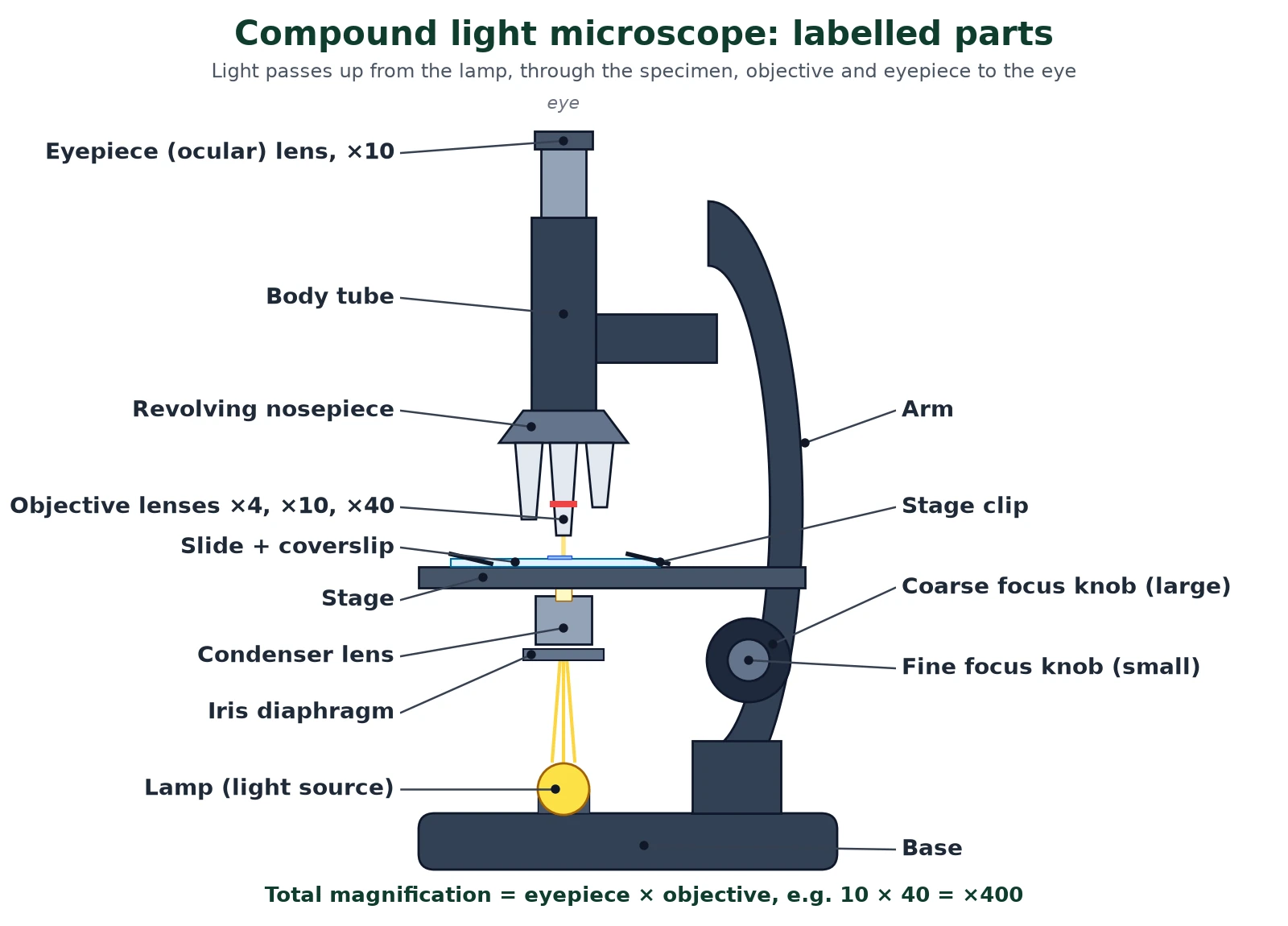
Most school microscopy is done with the light microscope, a compound microscope in which two lens systems magnify one after the other. The objective lens makes a magnified real image, and the eyepiece lens magnifies that image again. Light from a lamp is focused on the specimen by the condenser, and the iris diaphragm controls how much light passes through.
Parts and their functions
Swipe the table sideways to see every column.
| Part | Function |
|---|---|
| Eyepiece (ocular) | Magnifies the image made by the objective, usually ×10; holds the eyepiece graticule |
| Objective lenses | Main magnifying lenses, commonly ×4, ×10, ×40 (and ×100 oil immersion) |
| Revolving nosepiece | Rotates to change the objective |
| Stage and clips | Hold the slide over the hole through which light passes |
| Condenser | Focuses light onto the specimen, improving resolution and brightness |
| Iris diaphragm | Controls the amount of light; closing it raises contrast in pale, unstained cells |
| Coarse focus knob | Large movements of the stage or tube; used only with low-power objectives |
| Fine focus knob | Small movements for sharp focus, especially at high power |
How to focus a light microscope
- Start with the lowest-power objective (×4) clicked into place.
- Looking from the side, use the coarse knob to bring the objective close to the coverslip without touching it.
- Look down the eyepiece and focus by moving the objective away from the slide with the coarse knob.
- Centre the area you want, then switch to ×10 and then ×40, using only the fine focus knob at high power.
- Adjust the iris diaphragm for good contrast.
Total magnification and field of view
Total magnification is the product of the two lenses: a ×10 eyepiece with a ×40 objective gives ×400. When magnification rises, the field of view (the circle of specimen you see) shrinks in the same proportion: if the field is 1.8 mm across at ×100, it is 1.8 ÷ 4 = 0.45 mm at ×400. The image is also inverted, upside down and reversed left to right.
Exam tips
- Never use the coarse knob with the ×40 objective: it can crack the slide and the lens.
- The light microscope can show living cells, movement such as cytoplasmic streaming, and natural colour; electron microscopy cannot.
- Draw the plan view of a tissue at low power and the detail of single cells at high power.
3 Resolution vs magnification
Syllabus link: IB A2.2.2 and A2.2.3 (resolution as the advantage of electron microscopy); NCERT XI 8.1
Magnification is how many times larger the image is than the actual object. Resolution (resolving power) is the smallest distance between two points at which they can still be seen as two separate points. Resolution vs magnification is the most tested idea in microscopy: magnification makes things bigger, but only resolution adds detail.
Resolution is limited by the wavelength of the radiation used. Visible light has wavelengths of about 400 to 700 nm, so the best light microscope cannot separate points closer than about half the wavelength, roughly 0.2 µm (200 nm). Electrons accelerated at high voltage behave as waves with a wavelength thousands of times shorter, so the electron microscope resolves detail down to about 1 nm in biological specimens (and below 0.1 nm in materials science).
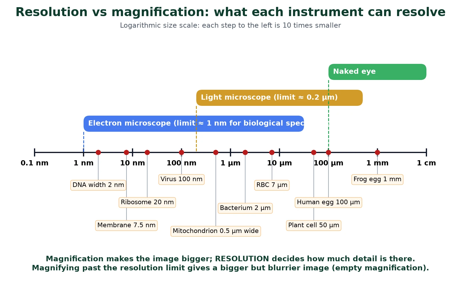
If you enlarge a light micrograph beyond about ×1500 to ×2000 you get empty magnification: the picture gets bigger, but blurred points just become bigger blurred points. In practical microscopy that is why a light microscope shows a mitochondrion as a tiny dot at best, and cristae need the electron microscope.
Swipe the table sideways to see every column.
| Feature | Magnification | Resolution |
|---|---|---|
| Meaning | How many times the image is larger than the object | Minimum distance at which two points are distinguished |
| Unit | None (×400) | Length (0.2 µm, 1 nm) |
| Limited by | Lens design; can be raised almost without limit | Wavelength of light or electrons |
| Light microscope | Up to about ×1500 useful | About 0.2 µm (200 nm) |
| Electron microscope | Up to ×500 000 or more | About 1 nm in biological specimens |
4 The magnification formula: worked examples
Syllabus link: IB A2.2.2 Calculation of actual size and magnification, use of scale bars
The magnification formula links three quantities: image size (I), actual size (A) and magnification (M). Write it as I = A × M and rearrange as needed. Almost every microscopy calculation in IB and NEET papers is one of these three rearrangements.
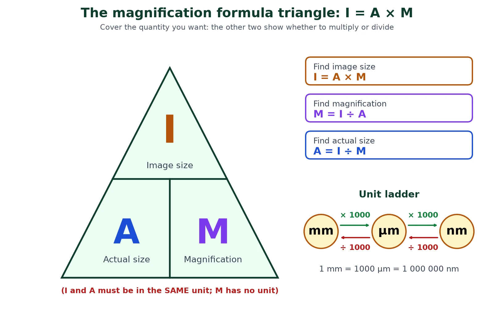
- M = I ÷ A (find magnification)
- A = I ÷ M (find actual size)
- I = A × M (find image size)
- 1 mm = 1000 µm; 1 µm = 1000 nm; so 1 mm = 1 000 000 nm
Example 1: actual size of a cheek cell
A drawing of a cheek cell is 18 mm wide and is labelled ×300. Convert: 18 mm = 18 000 µm. A = I ÷ M = 18 000 ÷ 300 = 60 µm.
Example 2: magnification of an electron micrograph
A mitochondrion known to be 2.4 µm long appears 36 mm long in a micrograph. 36 mm = 36 000 µm. M = 36 000 ÷ 2.4 = ×15 000.
Example 3: image size of a ribosome
A ribosome 25 nm wide is photographed at ×200 000. I = 25 × 200 000 = 5 000 000 nm. Convert: 5 000 000 nm = 5000 µm = 5 mm.
Example 4: using a scale bar
A scale bar labelled 5 µm measures 30 mm on the page. M = 30 000 µm ÷ 5 µm = ×6000. A bacterium on the same micrograph is 12 mm long, so A = 12 000 ÷ 6000 = 2 µm. You can skip the magnification step: the bacterium is 12/30 of the bar, so 0.4 × 5 = 2 µm.
Example 5: estimating size from the field of view
At ×100 the field of view is 1.8 mm across, and 12 cells fit end to end across it. Each cell is about 1800 µm ÷ 12 = 150 µm long.
Adding a scale bar to your own drawing
Choose a round actual length (for example 20 µm), multiply by the drawing magnification to find how long to draw it (20 µm × 500 = 10 000 µm = 10 mm), then draw a 10 mm line labelled 20 µm. A scale bar stays correct when the image is enlarged or shrunk, which a written magnification does not.
Markscheme habits
- Write the formula, substitute with units, then give the answer with a unit (actual size) or a × and no unit (magnification).
- Sense-check: cells are usually 1 to 100 µm, organelles 0.02 to 10 µm. A 600 µm mitochondrion means a slip in units.
- Give answers to sensible significant figures, normally matching the data.
5 Measuring cells: eyepiece graticule and stage micrometer
Syllabus link: IB A2.2.2 measurement of sizes using an eyepiece graticule
An eyepiece graticule is a small glass disc with a scale (often 100 divisions) that sits in the eyepiece. Its divisions have no fixed real size, because the specimen is magnified but the graticule is not. A stage micrometer is a slide with an accurately ruled scale, usually 1 mm divided into 100 parts of 10 µm each. Using the micrometer to give the graticule a value is called calibration, and it is the standard way to measure real sizes in light microscopy.
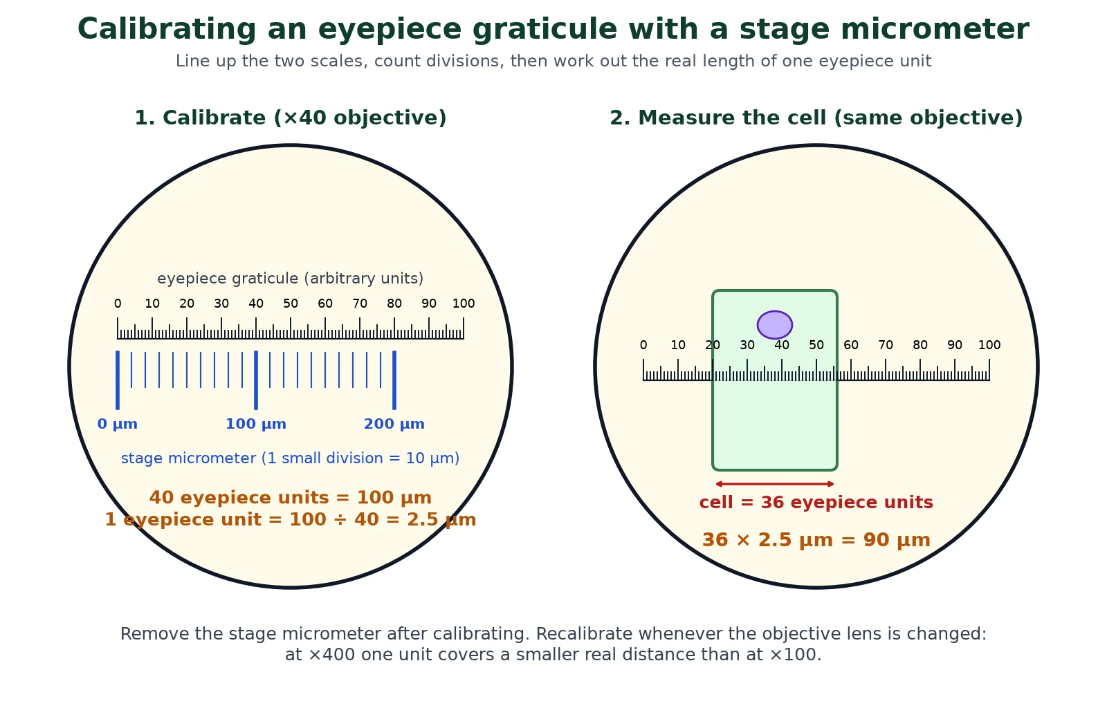
The calibration method
- Fit the graticule in the eyepiece and put the stage micrometer on the stage.
- Focus on the micrometer with the objective you will use and rotate the eyepiece until the two scales lie side by side.
- Find two points where lines on both scales coincide, as far apart as possible.
- Count the eyepiece units and read the real distance between those points on the micrometer.
- Real length of one eyepiece unit = micrometer distance ÷ number of eyepiece units.
- Replace the micrometer with the specimen and measure it in eyepiece units; multiply by the calibration value.
- Repeat for each objective lens (or scale the value exactly with the magnification).
Worked calibration
With the ×40 objective, 40 eyepiece units line up with 100 µm (10 small micrometer divisions). One eyepiece unit = 100 ÷ 40 = 2.5 µm. A cell spanning 36 eyepiece units is 36 × 2.5 = 90 µm long. Switching to the ×10 objective magnifies four times less, so each unit then covers 2.5 × 4 = 10 µm.
6 Preparing a temporary mount and drawing what you see
Syllabus link: IB A2.2.2 making temporary mounts of cells and tissues, staining; NCERT XI 8.3 onion peel and cheek cells
NCERT NCERT reminds you that you have already observed cells in an onion peel and human cheek cells under the microscope: the onion cell is a typical plant cell with a distinct cell wall and a cell membrane just inside it, while the cheek cell is bounded only by its membrane. Good microscopy starts with a good slide. A temporary mount is a quick slide made with water or stain and a coverslip, not sealed.
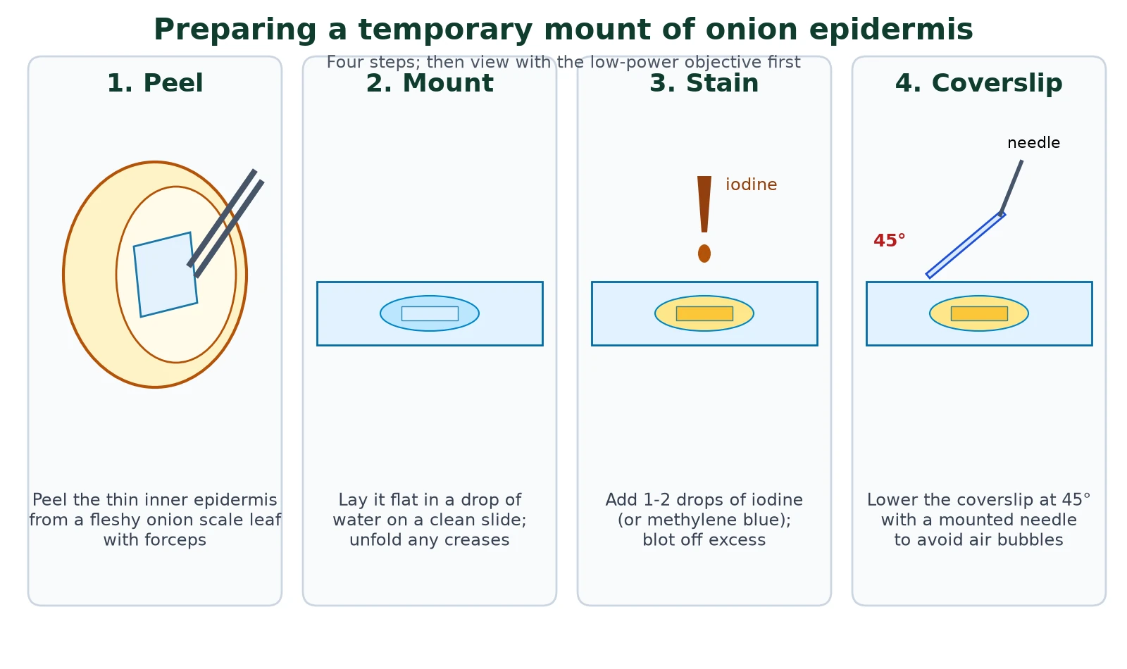
Onion epidermis method
- Cut a fleshy scale leaf from an onion and peel the thin, transparent inner epidermis with forceps.
- Place a small square flat in a drop of water on a clean slide, avoiding folds.
- Add one or two drops of iodine solution (or safranin, as in the NCERT practical); the stain makes cell walls and the nucleus stand out.
- Touch one edge of the coverslip to the liquid, hold it at about 45° and lower it slowly with a mounted needle to avoid air bubbles.
- Blot excess liquid with filter paper, then view at ×40 total, then ×100 and ×400.
Cheek cells with methylene blue
Gently scrape the inside of the cheek with a clean cotton bud, smear it on a slide, add a drop of methylene blue and cover. Methylene blue is a basic dye, so it binds acidic DNA and the nucleus stains dark blue. Put used swabs and slides in disinfectant.
Why stain?
Staining is the oldest trick in microscopy. Most cell components are colourless and almost transparent, so they show little contrast. Stains bind to particular chemicals (iodine binds starch; methylene blue binds nucleic acids) and make those structures visible. Most stains kill cells, so staining is a trade-off with observing live activity.
Rules for biological drawings
A microscopy drawing is marked on accuracy and clarity, not artistic skill.
- Use a sharp HB pencil and plain paper; draw large, using at least half the space.
- Use single, continuous, clear lines; no shading or colouring.
- Draw only what you see, keeping proportions correct; do not copy a textbook cell.
- Label lines drawn with a ruler, not crossing each other, ending exactly on the structure, with labels to the side.
- Give a title, the magnification or a scale bar, and the objective used.
7 The electron microscope: TEM and SEM
Syllabus link: IB A2.2.3 Developments in microscopy (advantages of electron microscopy)
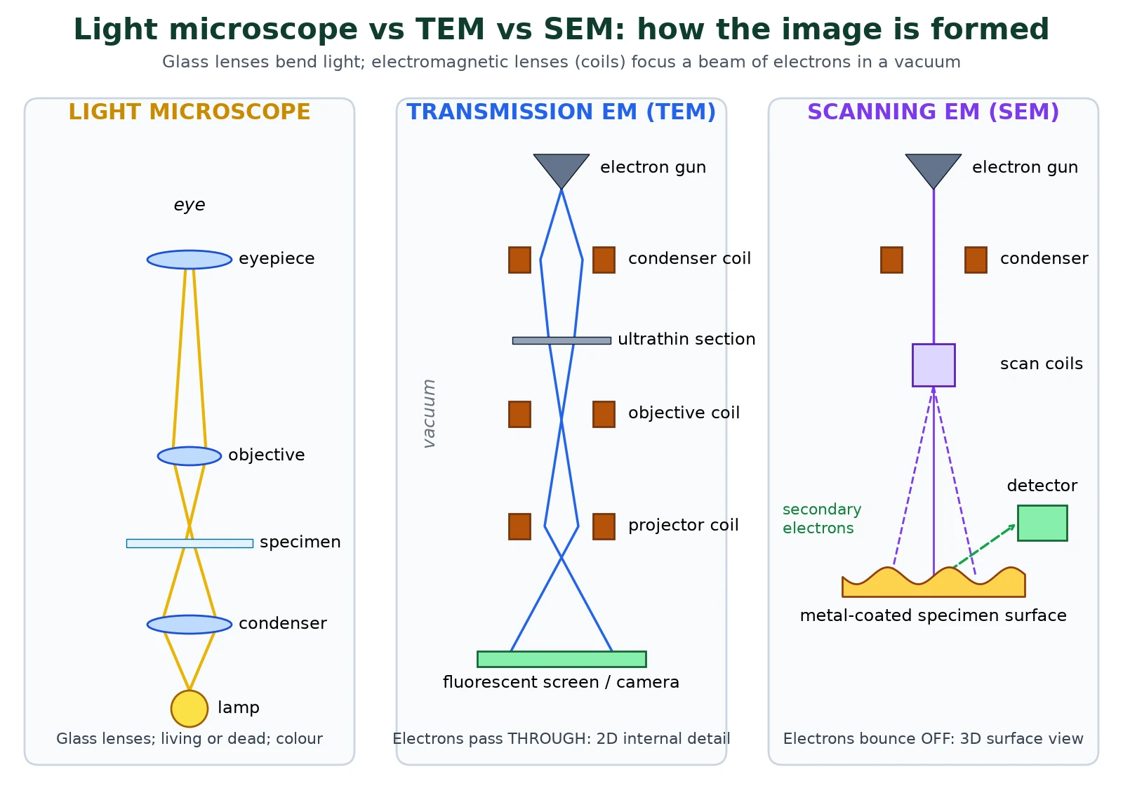
Electron microscopy changed cell biology because it showed the ultrastructure of cells. An electron microscope uses a beam of electrons instead of light, focused by electromagnets instead of glass lenses. Because electrons are scattered by air molecules, the whole column is a vacuum. The image is formed on a fluorescent screen or a camera and is black and white; any colour in a published electron micrograph is added afterwards (false colour).
Transmission electron microscope (TEM)
In transmission microscopy, electrons pass through an ultrathin section (about 50 to 100 nm thick) cut from a fixed, resin-embedded specimen and stained with heavy metals such as lead or uranium salts. Dense, stained regions scatter electrons and look dark. TEM gives the highest resolution and shows internal ultrastructure: cristae, ribosomes, the double nuclear envelope.
Scanning electron microscope (SEM)
A fine electron beam is scanned across the surface of a whole specimen coated with a thin layer of metal such as gold. Electrons knocked off the surface (secondary electrons) are collected to build a 3D-looking image of surface shape: pollen grains, insect eyes, the stomata of a leaf. SEM resolution is lower than TEM.
Light vs electron microscope compared
Swipe the table sideways to see every column.
| Feature | Light microscope | TEM | SEM |
|---|---|---|---|
| Radiation | Visible light | Electrons | Electrons |
| Lenses | Glass | Electromagnets | Electromagnets |
| Resolution | About 0.2 µm | About 1 nm (best) | A few nm (lower than TEM) |
| Useful magnification | Up to about ×1500 | Up to ×500 000 or more | Typically up to ×100 000 |
| Specimen | Living or dead; thin or whole | Dead; ultrathin section in a vacuum | Dead; whole, metal-coated, in a vacuum |
| Image | Colour, 2D | Black and white, 2D, internal detail | Black and white, 3D surface |
| Preparation | Quick and cheap; stains optional | Long: fixing, embedding, sectioning, heavy-metal staining | Long: fixing, drying, metal coating |
| Artefacts | Few | Common, from preparation | Common, from drying and coating |
| Cost and size | Cheap, portable | Very expensive, room-sized | Very expensive, large |
Exam tips
- Electron microscopes have a higher resolution because electrons have a much shorter wavelength than light: say this, not just that they magnify more.
- An artefact is a structure seen in the image that was made by the preparation, not present in the living cell. Mesosomes in bacteria are now widely thought to be artefacts of electron microscope preparation (NCERT still describes them as real structures, so use the NCERT account in NEET answers).
- Identify the type from the image: a cut section with internal membranes is TEM; a shaded 3D surface is SEM.
8 Developments in microscopy (IB A2.2.3)
Syllabus link: IB A2.2.3 freeze-fracture, cryogenic electron microscopy, fluorescent stains and immunofluorescence
IB Microscopy did not stop with the electron microscope. The 2025 IB guide asks you to describe four newer techniques and say what each can show. Each one solves a weakness of earlier microscopy.
Freeze-fracture
The specimen is frozen rapidly and then cracked with a cold blade. The fracture tends to run along the weakest plane, which is the hydrophobic centre of membranes, so the two halves of the phospholipid bilayer are split apart. A thin metal (platinum and carbon) replica of the exposed surface is made and viewed with the electron microscope. Freeze-fracture electron microscopy showed particles (proteins) spanning the middle of membranes, evidence that helped replace the Davson-Danielli model with the fluid mosaic model of the cell membrane.
Cryogenic electron microscopy (cryo-EM)
Samples such as purified proteins or viruses are frozen so fast (for example in liquid ethane) that water becomes glass-like ice without crystals. No fixing, dehydrating or staining is needed, so molecules stay close to their natural shape. Thousands of images of individual molecules lying in different orientations are combined by computer into a 3D model, often at near-atomic resolution. Cryo-EM revealed the structure of the SARS-CoV-2 spike protein within weeks in 2020.
Fluorescent stains
A fluorescent stain absorbs light of one wavelength and emits light of a longer wavelength. In fluorescence microscopy, only the stained structures glow against a dark background. Different dyes bind different targets, for example DAPI binds DNA and glows blue, so several structures can be shown in different colours in one image, sometimes in living cells.
Immunofluorescence
Immunofluorescence uses antibodies that bind one specific protein (antigen), linked to a fluorescent dye. In the direct method the dye is on the antibody that binds the target; in the indirect method a second, dye-carrying antibody binds the first, which strengthens the signal. It shows exactly where a particular protein is in a cell, such as tubulin in the cytoskeleton.
Swipe the table sideways to see every column.
| Technique | Microscope | What it shows |
|---|---|---|
| Freeze-fracture | Electron (replica) | Inside of membranes; transmembrane proteins |
| Cryo-EM | Electron | 3D shape of proteins and viruses in near-natural state |
| Fluorescent stains | Light (fluorescence) | Chosen structures such as DNA, membranes, mitochondria in colour |
| Immunofluorescence | Light (fluorescence) | Location of one specific protein using antibodies |
9 Common doubts about microscopy
These are the microscopy questions students keep asking in forums and homework sites. Each answer is ours; the source link shows where the doubt comes from.
What is the difference between magnification and resolution?
Magnification is how much bigger the image is; resolution is how close two points can be and still look separate. Raising magnification without better resolution only enlarges the blur (empty magnification). Resolution depends on wavelength, which is why electron microscopy, not a stronger lens, reveals ribosomes. (Doubt seen on quora.com)
Why can’t an electron microscope look at living cells?
The column is a vacuum, which dries out and kills cells, and the high-energy beam damages molecules. Specimens are also fixed, sectioned or coated with metal. A few experiments have imaged hardy organisms in special chambers, but for exams: electron microscopes need dead, prepared specimens. (Doubt seen on reddit.com)
To find actual size, do I divide or multiply by the magnification?
Divide: actual size = image size ÷ magnification. The microscope makes things bigger, so the real object must be smaller than the image. If your actual size comes out larger than the image, you multiplied by mistake. (Doubt seen on physicsforums.com)
How do I get the magnification from a scale bar?
Measure the bar with a ruler, convert that length to the unit written on the bar, then divide: M = measured bar length ÷ length the bar represents. A 25 mm bar labelled 50 µm gives 25 000 ÷ 50 = ×500. (Doubt seen on thestudentroom.co.uk)
Why do I need a stage micrometer if the graticule already has a scale?
The graticule sits in the eyepiece, so its divisions are not magnified with the specimen; what one division is worth depends on the objective and on the particular microscope. Only a slide with known divisions, placed where the specimen goes, tells you its real value. (Doubt seen on thestudentroom.co.uk)
Do I have to recalibrate for every objective lens?
Yes, the value of one eyepiece unit changes with each objective. In theory it scales exactly (2.5 µm per unit at ×400 becomes 10 µm at ×100), but real lenses are not exactly their stated power, so calibrate each objective you measure with. (Doubt seen on biologyeducation.co.uk)
How do I convert between mm, µm and nm without mistakes?
Each step is a factor of 1000: mm to µm multiply by 1000, µm to nm multiply by 1000. Convert everything into one unit before dividing. A common slip is jumping a unit: a quarter of 40 µm is 10 µm, not 10 nm. (Doubt seen on physicsforums.com)
Why does the image move the wrong way when I move the slide?
The objective lens forms an inverted image, upside down and back to front, and the eyepiece magnifies it without turning it round. So moving the slide left moves the image right; move the slide the opposite way to where you want the image to go. (Doubt seen on brainly.com)
Why can’t I see chloroplasts in onion epidermis cells?
The fleshy scale leaves of a bulb grow in the dark and store food; they do not photosynthesise, so their cells have no chloroplasts. Use a moss leaf or Elodea to see chloroplasts. (Doubt seen on reddit.com)
How can I tell if a micrograph was made by TEM or SEM?
TEM images are flat sections showing internal structures such as membranes and organelles. SEM images show the outside of a whole object with depth and shadows. Both are black and white unless false-coloured. (Doubt seen on thestudentroom.co.uk)
10 Quick check
Test yourself on microscopy
1. A cell is 40 mm wide in a drawing at ×800. What is its actual width?
40 000 µm ÷ 800 = 50 µm.
2. Why does an electron microscope have a higher resolution than a light microscope?
Electrons have a much shorter wavelength than visible light.
3. Which electron microscope gives a 3D surface image?
The scanning electron microscope (SEM).
4. What does freeze-fracture split apart?
The two layers of a membrane’s phospholipid bilayer, exposing proteins in it.
5. Which stain makes the nucleus of cheek cells blue?
Methylene blue.
6. 50 eyepiece units match 0.1 mm on the stage micrometer. What is one eyepiece unit worth?
100 µm ÷ 50 = 2 µm.
Ready to practise? Download the Microscopy IB and NEET question set with markscheme (25 MCQs + 6 structured questions, 65 marks, free for signed-up members).
⬇ Download practice questions (PDF)© competitorspoint.com · Biology notes. Diagrams are original to competitorspoint.com.