
IB Biology › Unity & diversity › Introduction to Cells
Introduction to Cells
Every living thing is made of cells. This introduction to cells for IB Biology covers how cells were discovered, the cell theory and its exceptions, prokaryotic and eukaryotic cells, cell size and magnification, the surface area to volume ratio, and how unicellular and multicellular organisms carry out the functions of life. It is written for IB Biology (SL and HL) and for NEET students using NCERT.
Intro What is a cell?
Syllabus link: A2.2.1 Cells as the basic structural unit of all living organisms; NCERT 8.1 What is a cell?
AHL = additional higher level (IB HL only) NCERT = extra detail for NEET / CBSE students (beyond the IB guide)
In today’s world, when everyone is obsessed with differences and fighting over them, we should not forget that we are all made of cells.
In 1809, Lamarck correctly pointed out that life cannot exist without cells and their structures. The study of cells and cell structures deepened with the discovery of different types of microscopes. The study of the cell, its structure and function, is called cytology, while the study of cell structure, function, genetics, etc., is called cell biology. Although the cell is the basic unit of all living organisms, different organisms show different cellular organization.
Some organisms are unicellular, in which a single cell performs all the functions needed to survive. For instance, Amoeba, Paramecium, Euglena and bacteria are unicellular. On the other hand, some organisms have an entire body made up of multiple cells. These are called multicellular organisms, in which different kinds of cell organization are seen. For example, sponges and jellyfish are multicellular.
A cell is the smallest unit that can carry out all the processes of life on its own. It is surrounded by a plasma membrane, it contains cytoplasm, it uses DNA as its genetic material and it makes proteins on ribosomes. Anything smaller than a cell, such as an organelle or a molecule, cannot live independently.
Introduction to cells IB Biology: what this chapter covers
- Section 1, Cell theory: who discovered cells, the principles of cell theory, and the cells that do not fit the usual picture.
- Section 2, Cell types: an overview of prokaryotic and eukaryotic cells.
- Section 3, Cell size: units, typical sizes, and how to calculate magnification and actual size.
- Section 4, Surface area to volume ratio: why cells stay small, with worked cube calculations.
- Section 5, Unicellular and multicellular organisms: the functions of life, emergent properties, differentiation and stem cells.
Key points
- The cell is the basic structural and functional unit of life.
- Cytology is the study of cells; cell biology also includes their genetics and biochemistry.
- Unicellular organisms carry out every function of life in one cell; multicellular organisms divide the work between many specialized cells.
Test yourself (click a question to reveal the answer)
Name the four structures that every cell has.
A plasma membrane, cytoplasm, ribosomes and DNA as the genetic material.
Give two unicellular and two multicellular organisms.
Unicellular: for example Amoeba, Paramecium, Euglena, yeast or any bacterium. Multicellular: for example a sponge, a jellyfish, a fern or a human.
1 Discovery of cells and the cell theory
Syllabus link: A2.2.1 Cells as the basic structural unit; A2.2.3 Developments in microscopy; A2.2.9 Atypical cell structure in eukaryotes; old 1.1.1 to 1.1.3; NCERT 8.2 Cell theory
Who discovered cells?
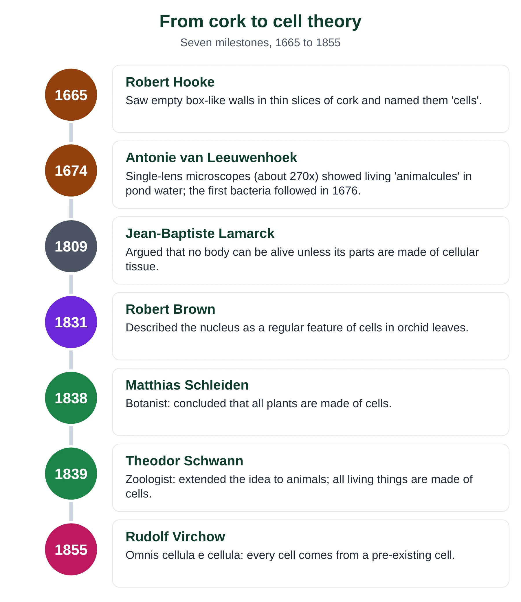
In 1665, Robert Hooke was the first scientist to observe cells under a microscope. He made thin slices of cork, a layer under the bark of a tree, and observed them under a microscope. Cork is a layer of dead cells, so when he observed it under the microscope, it looked like a honeycomb. The cells were empty, with no liquid, cytoplasm or nucleus. Robert Hooke was the first scientist to observe dead cork cells and call them cells because they looked like small chambers. He published his drawings in Micrographia (1665).
Later, Antonie van Leeuwenhoek worked with glass, made lenses with different curvatures, and built his own microscope, which could magnify objects about 270 times. This was quite something at that point of time. He observed a drop of pond water under his microscope and saw structures moving (in 1674). At that time, he had no idea he was the first to observe living cells. Most of the moving ‘animalcules’ in pond water were protists such as ciliates and algae; he went on to describe bacteria from 1676 (and drew bacteria from his own teeth in 1683). That is why Leeuwenhoek is known as the father of microbiology.
Later, in 1831, Robert Brown discovered the cell nucleus while studying orchid cells. As microscopes advanced from light microscopes to electron microscopes, scientists discovered different cell organelles. So, we now understand that observing a cell requires a magnifying apparatus called a microscope.
The cell theory
Despite Robert Hooke’s discovery of cells in 1665, scientists did not recognize the cell as the fundamental unit of life for nearly two centuries. In 1838 the German botanist Matthias Schleiden concluded that all plants are made of cells. In 1839 the German zoologist Theodor Schwann showed that animals are made of cells too, and proposed that cells are the basic units of all living things. Their work was published separately, a year apart, but together it laid the foundation of the cell theory (see the history of the cell theory on Wikipedia).
This theory states that all organisms are made up of one or more cells. A cell is the structural and functional unit of life, meaning a single cell can perform all the functions of life. In other words, anything not made of cells is not considered living. For instance, viruses are not considered living by this definition because they are not made of cells and can only reproduce inside a host cell.
Schleiden and Schwann could not explain how new cells form. In 1855 Rudolf Virchow popularised the idea that every cell arises from a pre-existing cell, summed up in the Latin phrase omnis cellula e cellula (‘all cells from cells’). Robert Remak had published evidence for this a few years earlier. In the 1860s Louis Pasteur showed that microbes do not appear by spontaneous generation in sterilised broth, which supported Virchow.
Key principles to remember about cell theory:
- Cells are the smallest units of life. All living organisms are composed of one or more cells.
- Only living organisms are made up of cells. The cell is the basic unit of structure and function.
- All cells are generally alike in structure, functions and biochemistry, with minor differences depending on their specific functions.
- All cells, except the first cell, come from pre-existing cells (omnis cellula e cellula).
Modern cell theory adds that cells contain hereditary information (DNA) that is passed to daughter cells when cells divide, and that the energy flow of life (metabolism) happens inside cells.
NCERT Cell theory in NCERT
NCERT states that Schleiden and Schwann together formulated the cell theory, and that Rudolf Virchow (1855) modified it by explaining that cells divide and new cells form from pre-existing cells. NCERT also credits Schwann with noting the thin outer layer we now call the plasma membrane, and with recognising that the cell wall is a unique feature of plant cells.
For NEET, learn the NCERT wording: (i) all living organisms are composed of cells and products of cells, and (ii) all cells arise from pre-existing cells. Historically, though, the two men published separately, Schleiden in 1838 and Schwann in 1839.
Exceptions to the cell theory
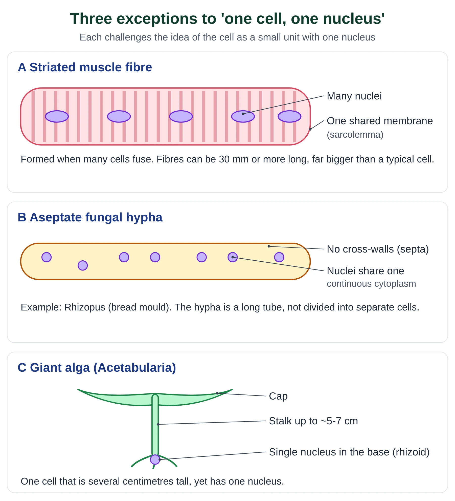
A theory in science is tested by looking for observations that do not fit it. Three structures are used in IB Biology as atypical examples because they do not consist of small units, each with one nucleus:
| Example | Why it is atypical | Does it break the cell theory? |
|---|---|---|
| Striated (skeletal) muscle | Fibres form when many cells fuse; one fibre has hundreds of nuclei inside one plasma membrane and can be several centimetres long. | It is made from cells and still has a membrane, so it is better described as an unusual cell (a syncytium). |
| Aseptate fungal hyphae | Hyphae of fungi such as Rhizopus have no cross-walls (septa); many nuclei share one long tube of cytoplasm. | The structure is not divided into cell-sized units, so it challenges the idea that organisms are made of separate cells. |
| Giant algae | Acetabularia is a single cell 5 to 7 cm tall with one nucleus in its base, much larger than expected. | It is one cell, but a very large one, so it challenges the idea that cells must be small. |
| Red blood cells (mammal) | Lose their nucleus as they mature, so they cannot divide. | They come from stem cells, so they fit the theory but lack a feature most cells have. |
| Phloem sieve tube elements | Have no nucleus and very little cytoplasm; they depend on companion cells. | Living, but only with the support of a neighbouring cell. |
Exam tip
- IB asks you to ‘discuss’ exceptions: say what is unusual, then whether the example truly contradicts the theory. A balanced answer earns more marks than ‘this disproves cell theory’.
- The old IB guide (topic 1.1) lists striated muscle, giant algae and aseptate fungal hyphae. The 2025 guide (A2.2.9) lists red blood cells, phloem sieve tube elements, skeletal muscle and aseptate hyphae. Learn all of them.
Key points
- Hooke (1665) named cells from dead cork; Leeuwenhoek (1674) first saw living cells; Brown (1831) described the nucleus.
- Schleiden (1838, plants) and Schwann (1839, animals) published separately; Virchow (1855) added omnis cellula e cellula.
- Cell theory: all organisms are made of cells; the cell is the smallest unit of life; cells come from pre-existing cells.
- Atypical examples include striated muscle fibres, aseptate hyphae and giant algae; each tests the theory rather than simply breaking it.
Common mistakes
- Writing that Schleiden and Schwann published the cell theory together in 1838. Schleiden wrote about plants in 1838, Schwann about animals in 1839.
- Crediting omnis cellula e cellula to Schwann or Hooke. It is linked with Virchow (1855).
- Saying Hooke saw living cells. Cork cells are dead; he saw only the empty walls.
Test yourself (click a question to reveal the answer)
Why did Hooke’s cork cells look empty?
Cork cells are dead. Their contents (cytoplasm and nucleus) have gone, so only the cell walls are left, giving a honeycomb of empty chambers.
Explain why a striated muscle fibre is described as atypical.
A single fibre contains many nuclei within one plasma membrane and can be centimetres long, because it forms when many cells fuse. It does not fit the usual picture of a small cell with one nucleus.
Which scientist is linked with omnis cellula e cellula, and when?
Rudolf Virchow, 1855.
2 Cell types: prokaryotic and eukaryotic cells
Syllabus link: A2.2.4 Structures common to cells in all living organisms; A2.2.5 Prokaryote cell structure; A2.2.6 Eukaryote cell structure; NCERT 8.3 to 8.5
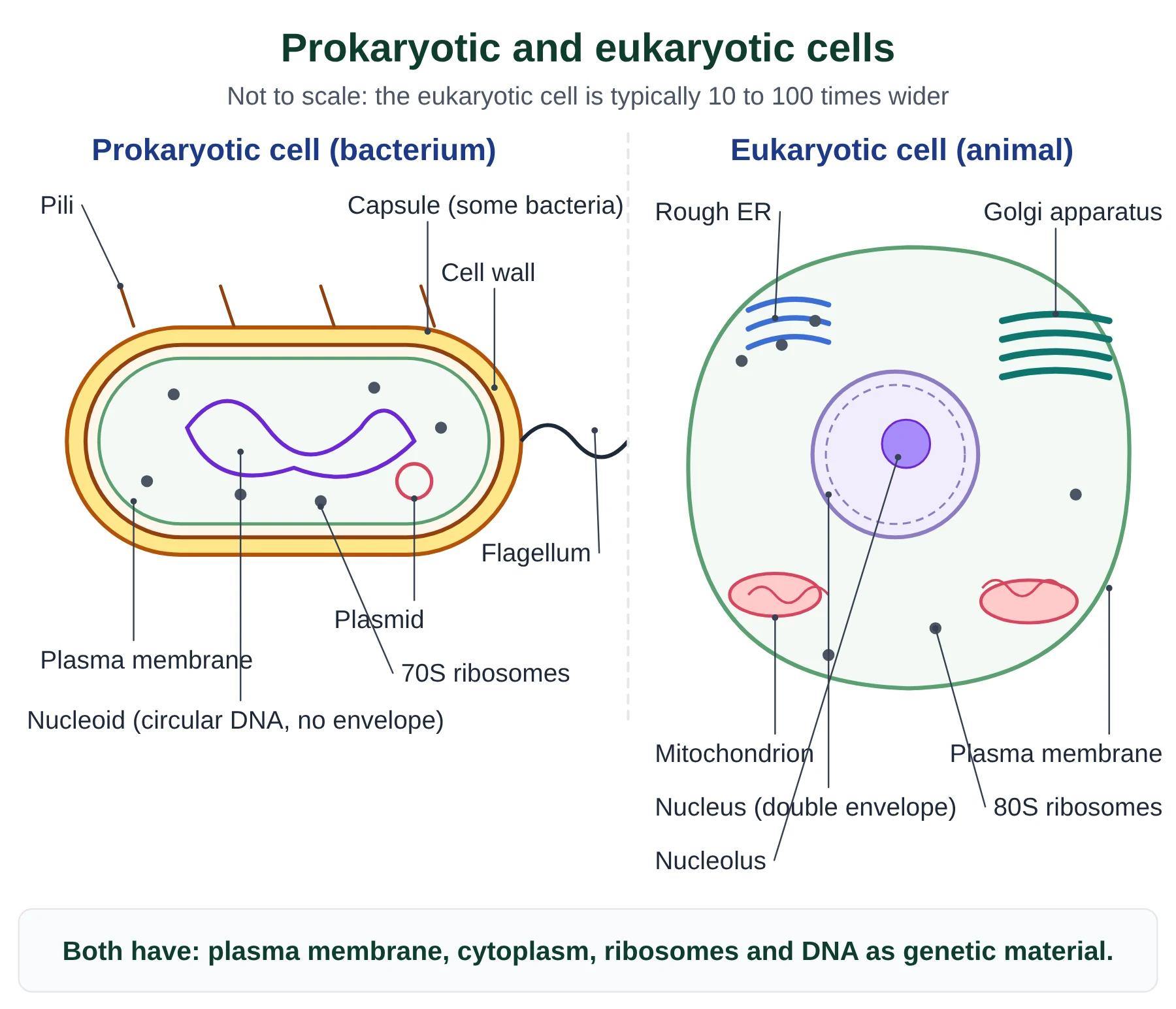
All cells fall into two groups. Prokaryotic cells (bacteria and archaea) have no nucleus: their DNA lies free in the cytoplasm in a region called the nucleoid. Eukaryotic cells (protists, fungi, plants and animals) keep their DNA inside a nucleus bounded by a double membrane, the nuclear envelope, and divide their cytoplasm into membrane-bound compartments called organelles. The words come from Greek: pro = before, eu = true, karyon = kernel (nucleus).
Structures shared by all cells
- Plasma membrane: a phospholipid bilayer that controls what enters and leaves.
- Cytoplasm: watery fluid (cytosol) where most metabolic reactions happen.
- DNA: the genetic material that carries the instructions for making proteins.
- Ribosomes: the sites of protein synthesis (70S in prokaryotes, 80S in the eukaryotic cytoplasm).
Comparing prokaryotes and eukaryotes
| Feature | Prokaryotic cell | Eukaryotic cell |
|---|---|---|
| Examples | Bacteria (e.g. E. coli), cyanobacteria, archaea | Protists, fungi, plants, animals |
| Typical size | About 1 to 5 µm | About 10 to 100 µm |
| Nucleus | None; DNA in a nucleoid | Nucleus with a double nuclear envelope |
| DNA | One circular chromosome, not bound to histones (‘naked’); often plasmids | Several linear chromosomes wound around histone proteins |
| Membrane-bound organelles | None | Mitochondria, ER, Golgi apparatus, lysosomes, vacuoles; chloroplasts in plants and algae |
| Ribosomes | 70S (smaller) | 80S in cytoplasm; 70S inside mitochondria and chloroplasts |
| Cell wall | Usually present; made of peptidoglycan in bacteria | Cellulose in plants, chitin in fungi, absent in animals |
| Cell division | Binary fission | Mitosis or meiosis, followed by cytokinesis |
Within eukaryotes there are further differences. Plant cells have a cellulose cell wall, chloroplasts and usually a large permanent vacuole; animal cells have none of these but have centrioles and often small temporary vacuoles; fungal cells have a chitin wall and no chloroplasts. These differences are covered in detail in the cell structure chapter.
NCERT Prokaryotic cell envelope and mesosome (NCERT)
NCERT describes the bacterial cell envelope as three layers: the outer glycocalyx (a loose slime layer or a thick, tough capsule), the cell wall and the plasma membrane. Bacteria that keep the Gram stain are Gram positive; those that do not are Gram negative.
NCERT also names the mesosome, an infolding of the plasma membrane said to help in wall formation, DNA replication, respiration and secretion. Other NCERT terms to know: PPLO (pleuro-pneumonia-like organisms, i.e. mycoplasma), pili and fimbriae (surface structures that do not help in movement), and chromatophores (pigment-containing membranes in cyanobacteria).
Key points
- Prokaryotes: no nucleus, no membrane-bound organelles, 70S ribosomes, circular naked DNA, binary fission.
- Eukaryotes: nucleus, membrane-bound organelles, 80S ribosomes, linear DNA with histones, mitosis.
- All cells share a plasma membrane, cytoplasm, ribosomes and DNA.
Common mistakes
- Saying prokaryotes have no DNA or no ribosomes. They have both; they lack a nucleus and membrane-bound organelles.
- Calling ribosomes membrane-bound organelles. Ribosomes have no membrane, which is why prokaryotes can have them.
Test yourself (click a question to reveal the answer)
State two structures found in a eukaryotic cell but never in a prokaryotic cell.
Any two of: a nucleus with a nuclear envelope, mitochondria, endoplasmic reticulum, Golgi apparatus, lysosomes, 80S ribosomes, linear chromosomes with histones.
A cell has 70S ribosomes, a cell wall and a single loop of DNA. Is it prokaryotic or eukaryotic? Explain.
Prokaryotic: a single circular DNA molecule and only 70S ribosomes show that there is no nucleus and no eukaryotic cytoplasmic ribosomes.
3 Cell size: units, typical sizes and magnification
Syllabus link: A2.2.2 Microscopy skills (magnification, scale bars, actual size); A2.2.3 Developments in microscopy; B2.3.5 Cell size as an aspect of specialization; NCERT 8.3
Most cells are far too small to see with the naked eye, which can only resolve objects about 0.1 mm (100 µm) apart. Biologists therefore measure cells in micrometres (µm) and their parts in nanometres (nm).
Units of length
Swipe sideways to see the whole table →
| Unit | Symbol | Value in metres | Used for |
|---|---|---|---|
| millimetre | mm | 10-3 m | Large cells, e.g. a frog egg or Acetabularia |
| micrometre | µm | 10-6 m | Cells, nuclei, chloroplasts, mitochondria |
| nanometre | nm | 10-9 m | Viruses, ribosomes, membranes, molecules |
To convert, multiply or divide by 1000: 1 mm = 1000 µm and 1 µm = 1000 nm. Going to a smaller unit, multiply; going to a larger unit, divide.
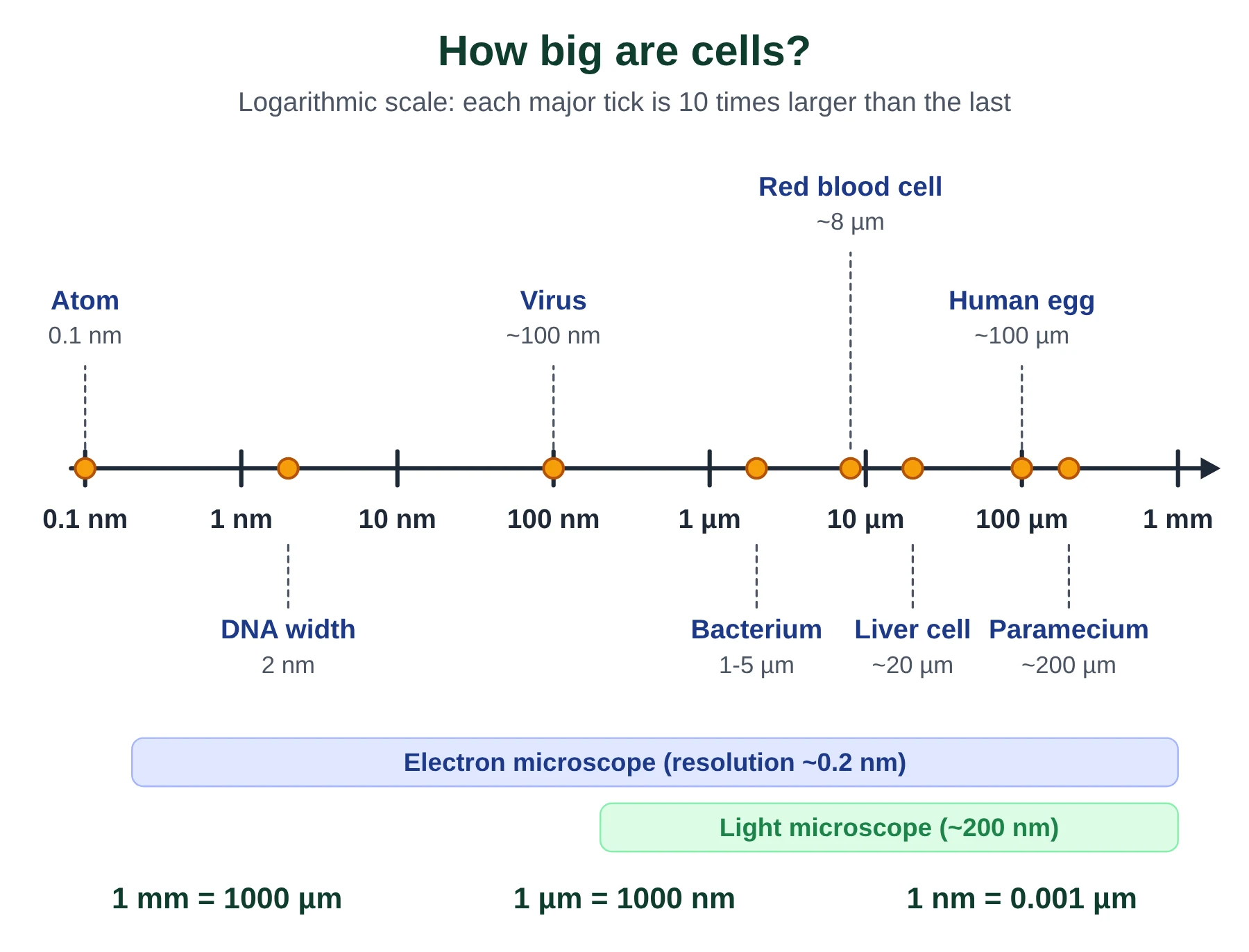
Typical sizes
- Mycoplasma, among the smallest cells: about 0.3 µm.
- Bacteria: about 1 to 5 µm (NCERT gives 3 to 5 µm).
- Human red blood cell: about 7 to 8 µm across.
- Typical plant or animal cell: about 10 to 100 µm.
- Human egg (ovum): about 100 to 120 µm, just visible to the eye.
- Largest single cell: the ostrich egg (the yolk is one cell). Longest cells: nerve cells, some over 1 m long in large animals.
Magnification and resolution
Magnification is how many times larger the image is than the real object. Resolution is the ability to tell two close points apart as separate. A light microscope can magnify about 1500 times with a resolution of about 200 nm; an electron microscope can resolve objects about 0.2 nm apart, which is why ribosomes and membranes were only seen after the 1930s to 1950s.
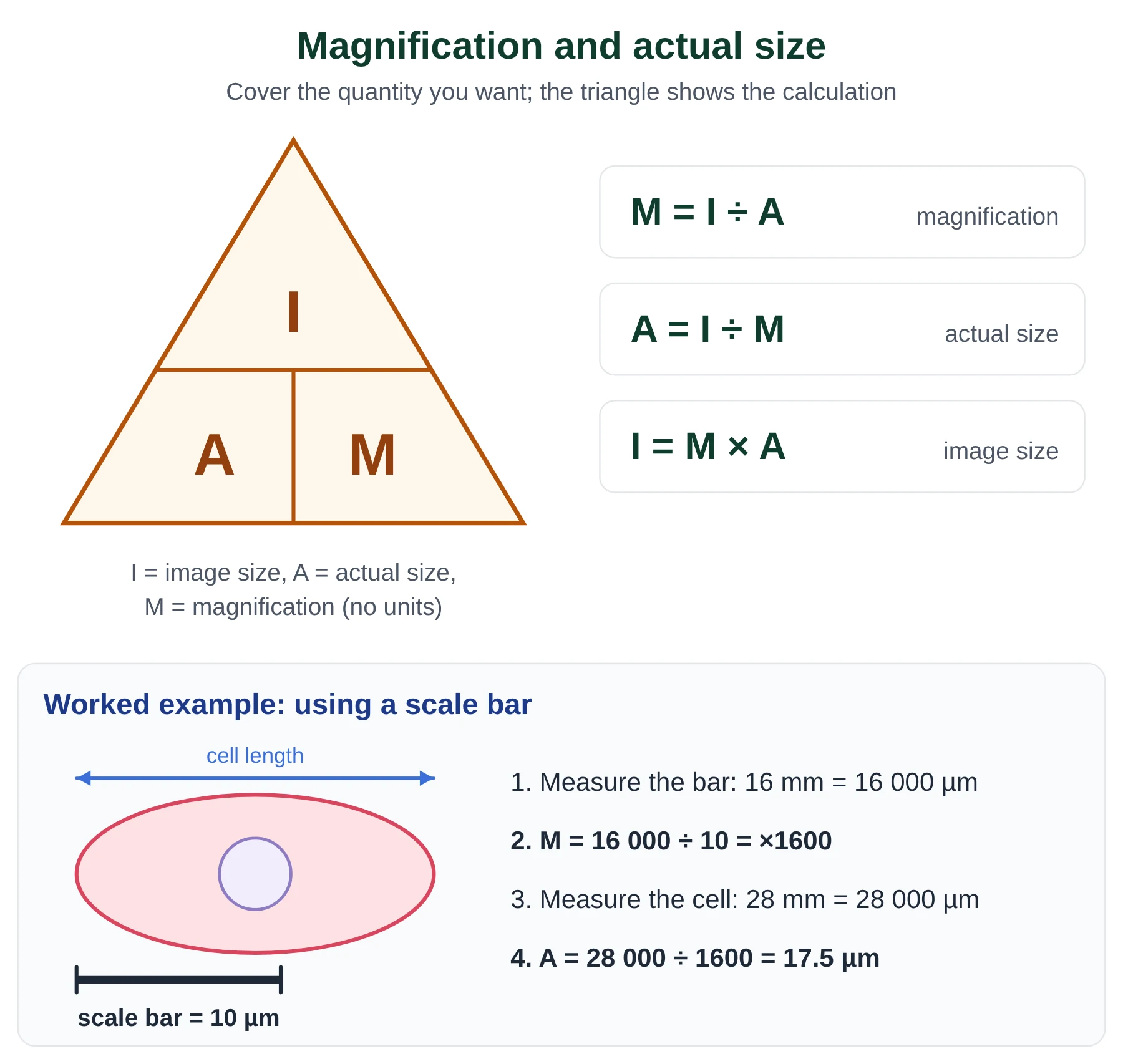
Use one formula, rearranged as needed: magnification = image size ÷ actual size. Before you divide, put both lengths in the same unit (usually µm). Magnification has no units; write it as ×400 or 400x.
Worked examples
Q1. Finding actual size. In an electron micrograph a mitochondrion is 30 mm long. The magnification is ×15 000. Find its actual length.
- Convert the image size to µm: 30 mm × 1000 = 30 000 µm.
- Actual size = image size ÷ magnification = 30 000 ÷ 15 000.
- Answer: 2 µm.
Q2. Finding magnification. A cheek cell 60 µm across is drawn 48 mm wide. What is the magnification of the drawing?
- Convert: 48 mm = 48 000 µm.
- Magnification = 48 000 ÷ 60.
- Answer: ×800.
Q3. Using a scale bar. A scale bar labelled 5 µm is 20 mm long on a micrograph. A bacterium in the same image is 36 mm long. Find the magnification and the actual length.
- Magnification = 20 000 µm ÷ 5 µm = ×4000.
- Actual length = 36 000 µm ÷ 4000.
- Answer: ×4000 and 9 µm. (Shortcut: 36 ÷ 20 × 5 µm = 9 µm.)
NCERT Shapes and sizes of cells (NCERT)
NCERT stresses that cells vary greatly in shape as well as size: disc-like (red blood cells), polygonal, columnar, cuboidal, thread-like or irregular (white blood cells, Amoeba). Shape is linked to function; long, branched nerve cells carry impulses over long distances, while flat, disc-shaped red blood cells exchange gases quickly.
Frequently asked NEET facts: smallest cell Mycoplasma (0.3 µm); bacteria 3 to 5 µm; largest isolated single cell ostrich egg; human red blood cell about 7.0 µm; longest cells nerve cells.
Exam tip
- Always show the unit conversion line; it is where most marks are lost.
- Give answers to a sensible number of significant figures and include the unit (µm) for actual size; give no unit for magnification.
- If a question gives a scale bar, measure it with a ruler in mm and treat it exactly like an object of known actual size.
Key points
- 1 mm = 1000 µm; 1 µm = 1000 nm.
- Magnification = image size ÷ actual size, both in the same unit.
- Resolution, not magnification, limits what you can see: about 200 nm for light, about 0.2 nm for electron microscopes.
- Prokaryotes are about 1 to 5 µm; most eukaryotic cells are 10 to 100 µm.
Common mistakes
- Dividing mm by µm without converting. The answer will be 1000 times wrong.
- Confusing magnification with resolution. A blurred image can be magnified more but you will not see more detail.
- Giving a unit for magnification, such as ‘×400 µm’.
Test yourself (click a question to reveal the answer)
A nucleus is 8 µm across. How big will it appear at ×500?
Image size = 8 × 500 = 4000 µm = 4 mm.
Why can an electron microscope show ribosomes when a light microscope cannot?
Ribosomes are about 20 to 30 nm across, smaller than the resolution of a light microscope (about 200 nm). Electrons have a much shorter wavelength than light, so the electron microscope resolves about 0.2 nm.
4 Surface area to volume ratio
Syllabus link: B2.3.6 Surface area-to-volume ratios and constraints on cell size; B2.3.7 Adaptations to increase surface area-to-volume ratios of cells; old 1.1.4
A cell exchanges materials with its surroundings through its surface (the plasma membrane): oxygen and nutrients come in, carbon dioxide, waste and heat go out. But the amount of material it needs, and the waste it makes, depends on its volume, because metabolism happens throughout the cytoplasm. The balance between the two is the surface area to volume ratio (SA:V) (read more about the surface-area-to-volume ratio on Wikipedia).
Worked cube calculations
Model a cell as a cube with side length L. A cube has six square faces, so surface area = 6L²; its volume = L³. Therefore SA:V = 6L² ÷ L³ = 6 ÷ L.
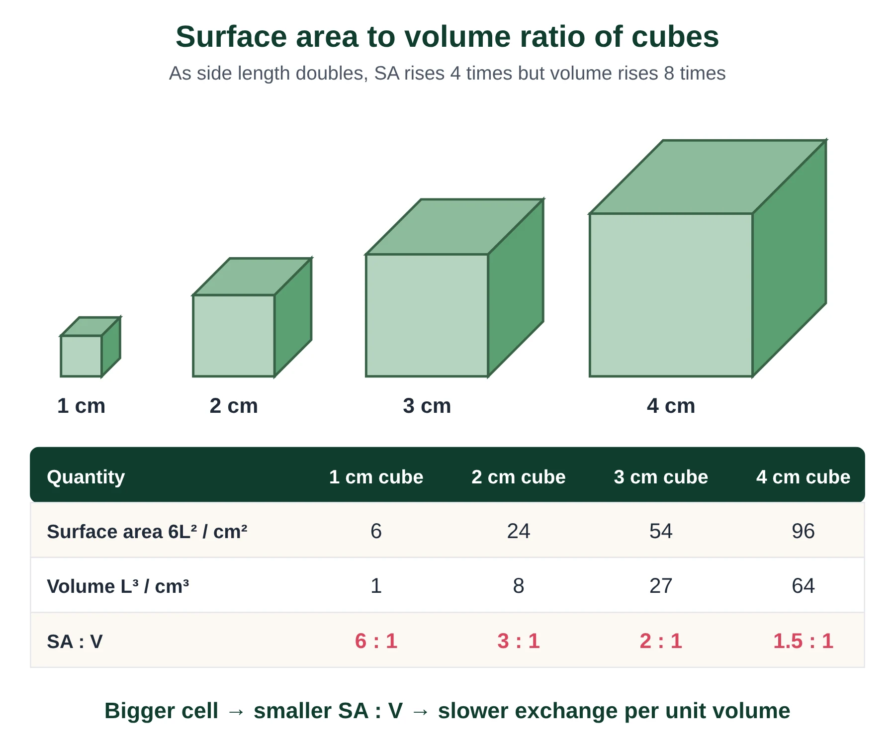
- 1 cm cube: SA = 6 × 1² = 6 cm²; V = 1³ = 1 cm³; SA:V = 6 : 1.
- 2 cm cube: SA = 6 × 4 = 24 cm²; V = 8 cm³; SA:V = 24 ÷ 8 = 3 : 1.
- 3 cm cube: SA = 6 × 9 = 54 cm²; V = 27 cm³; SA:V = 2 : 1.
- 4 cm cube: SA = 6 × 16 = 96 cm²; V = 64 cm³; SA:V = 1.5 : 1.
Doubling the side length from 1 cm to 2 cm multiplies the surface area by 4 (2²) but the volume by 8 (2³), so the ratio halves. The same rule holds for real cells: a cell 20 µm across has half the SA:V of a cell 10 µm across.
Why SA:V limits cell size
- Exchange becomes too slow. As a cell grows, its need for oxygen and nutrients rises with volume, but the membrane available to supply them rises only with area.
- Waste and heat build up. Waste products and heat are produced throughout the volume but can only leave across the surface.
- Diffusion distances get longer. Diffusion is fast over a few micrometres but very slow over millimetres, so the centre of a large cell would be starved.
- Control from one nucleus is harder. A single nucleus can only make enough mRNA to control a limited volume of cytoplasm.
This is why cells divide when they reach a certain size, rather than simply growing bigger, and why large organisms are made of many small cells instead of a few giant ones.
Adaptations that increase SA:V
| Adaptation | Example | How it helps |
|---|---|---|
| Flattened shape | Red blood cells are biconcave discs about 7 µm across and 2 µm thick | Large surface for oxygen uptake and short diffusion distance to the centre |
| Microvilli | Cells of the small intestine and the proximal convoluted tubule of the kidney | Finger-like folds of membrane multiply the area for absorption and reabsorption |
| Long thin extensions | Root hair cells | Increase area for absorbing water and mineral ions |
| Thin, flat cells | Type I pneumocytes lining the alveoli | Very short distance for gas exchange |
| Staying small or dividing | Bacteria; cells that divide by mitosis | Keeps SA:V high |
At the level of the whole organism, the same problem is solved with exchange surfaces (lungs, gills, villi, leaves) and transport systems (blood, xylem, phloem). A small, flat organism such as a flatworm can rely on diffusion alone because every cell is close to the surface.
Exam tip
- When asked to ‘explain’ why cells are small, link three ideas: rate of exchange depends on surface area; demand depends on volume; SA:V falls as size increases.
- Show the formulas (6L² and L³) in calculations, and write ratios as x : 1.
Key points
- SA:V of a cube = 6 ÷ L; it falls as the cell gets bigger.
- Doubling length: area ×4, volume ×8, SA:V halved.
- Small cells exchange materials and heat efficiently; large cells cannot supply their centre fast enough.
- Adaptations include flattening, microvilli, long extensions and cell division.
Common mistakes
- Saying large cells have a bigger surface area so they exchange more efficiently. The total area is larger, but the area per unit volume is smaller.
- Forgetting the units: surface area in cm² (or µm²), volume in cm³ (or µm³); the ratio itself has units of 1/length but is usually written x : 1.
Test yourself (click a question to reveal the answer)
Calculate the SA:V ratio of a cube with sides of 5 µm.
SA = 6 × 25 = 150 µm²; V = 125 µm³; SA:V = 150 ÷ 125 = 1.2 : 1 (or 6 ÷ 5).
Explain why a red blood cell’s biconcave shape is an advantage.
It gives a large surface area relative to the volume and keeps every part of the cytoplasm close to the membrane, so oxygen diffuses in and out quickly.
5 Unicellular and multicellular organisms
Syllabus link: A2.2.7 Processes of life in unicellular organisms; AHL A2.2.13 Cell differentiation; AHL A2.2.14 Evolution of multicellularity; B2.3.1 to B2.3.4 Differentiation and stem cells; old 1.1.2, 1.1.5 to 1.1.10
A unicellular organism must carry out every function of life inside one cell. The IB guide lists eight: homeostasis, metabolism, nutrition, movement, excretion, growth, response to stimuli and reproduction. The old IB mnemonic MR H GREN covers seven of them (metabolism, response, homeostasis, growth, reproduction, excretion, nutrition); the 2025 guide adds movement.
Functions of life in Paramecium
Paramecium is a slipper-shaped ciliate, about 200 µm long, that lives in fresh water and feeds on bacteria.
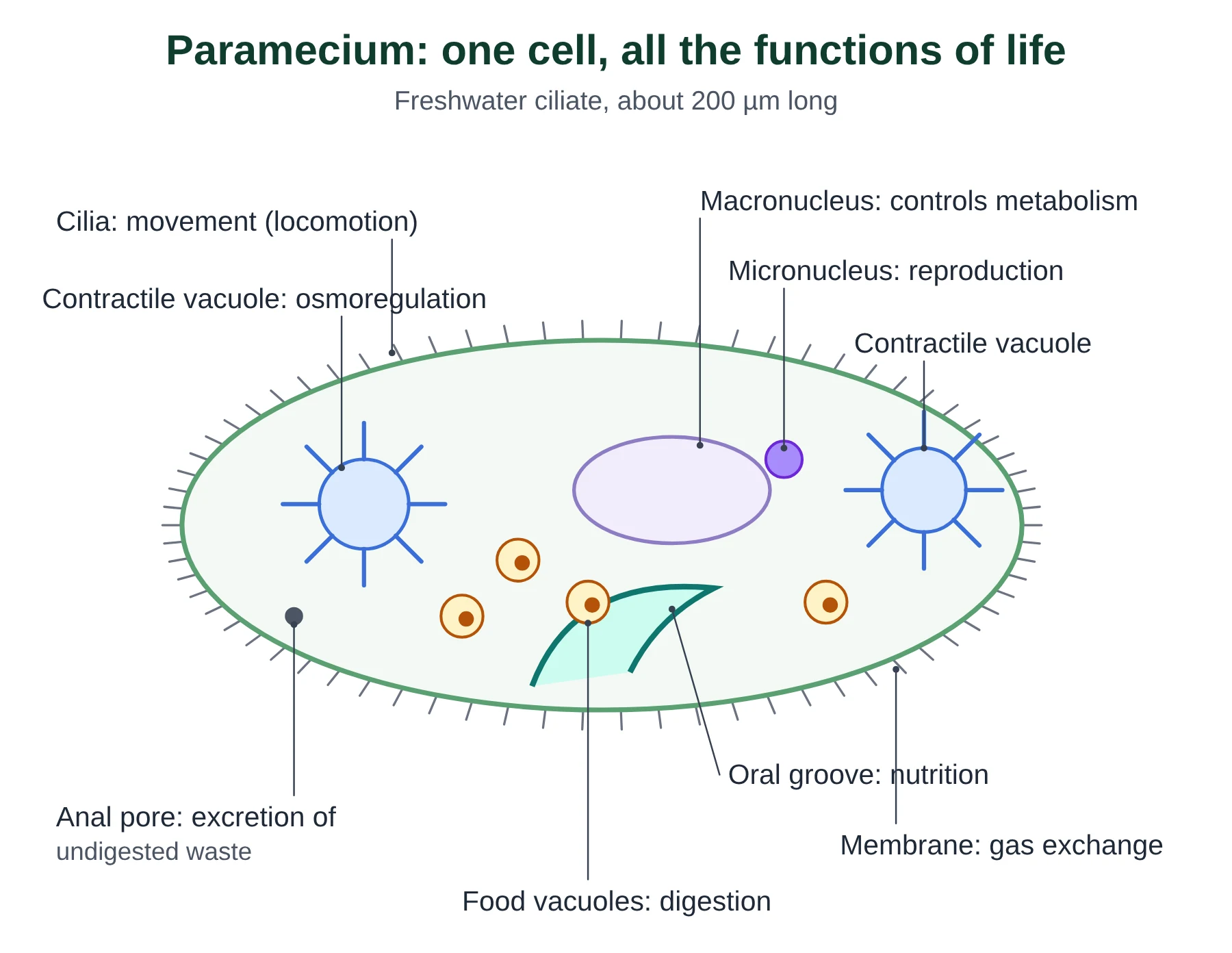
| Function | How Paramecium does it |
|---|---|
| Nutrition | Cilia sweep bacteria into the oral groove; they are taken in by endocytosis to form food vacuoles (heterotrophic). |
| Metabolism | Enzymes in food vacuoles digest food; respiration in mitochondria releases energy. |
| Movement | Thousands of cilia beat in coordinated waves to move the cell. |
| Response | Reverses its cilia and changes direction when it bumps into an obstacle or meets a harmful chemical. |
| Homeostasis | Two contractile vacuoles collect excess water that enters by osmosis and expel it, keeping the internal water content steady. |
| Excretion | Carbon dioxide and ammonia diffuse out through the membrane; undigested waste leaves through the anal pore. |
| Growth | Takes in and builds new molecules, increasing in size until it divides. |
| Reproduction | Asexually by binary fission (the micronucleus divides by mitosis); sexually by conjugation, exchanging micronuclei with another cell. |
Functions of life in Chlamydomonas
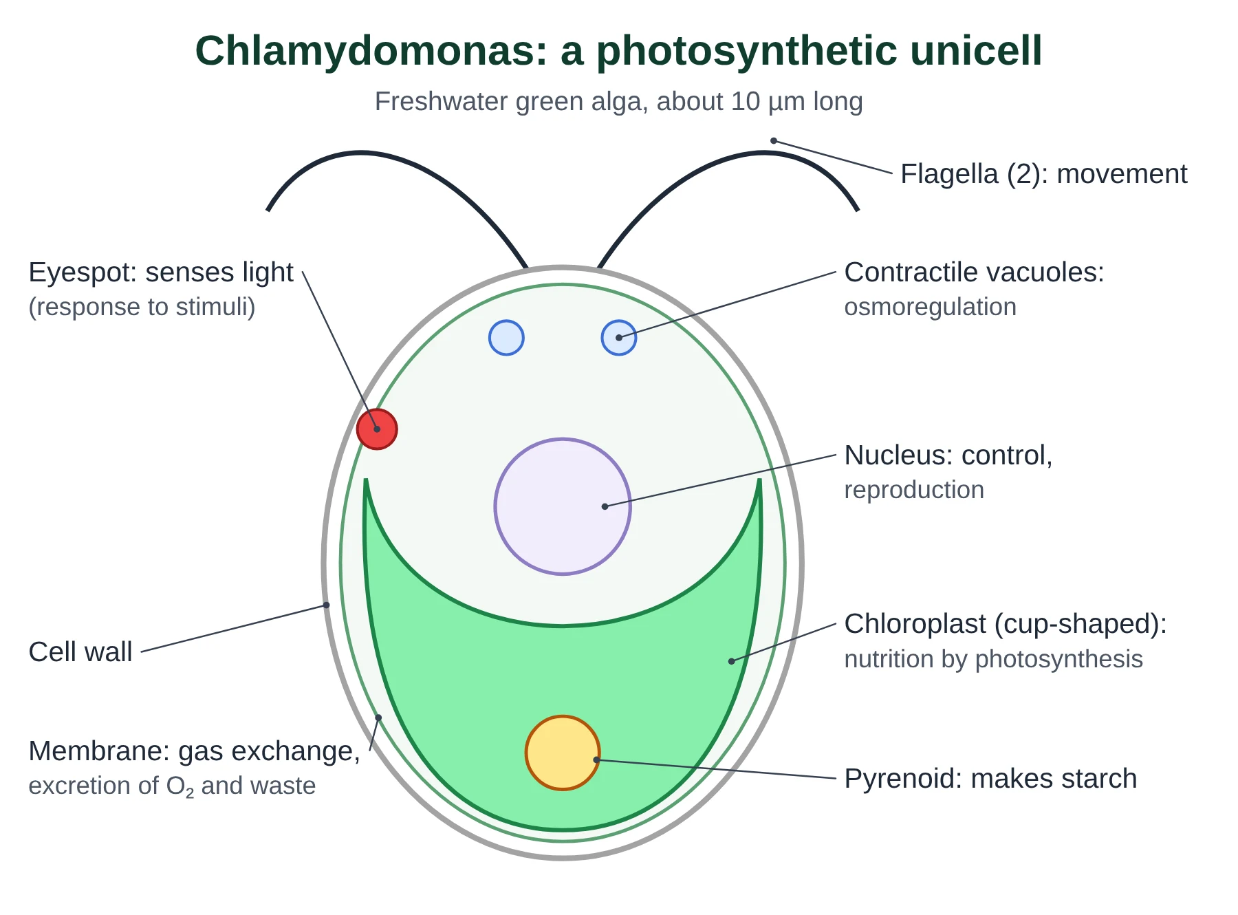
Chlamydomonas is a green alga about 10 µm long. Unlike Paramecium, it is autotrophic: it makes its own food by photosynthesis.
| Function | How Chlamydomonas does it |
|---|---|
| Nutrition | Photosynthesis in one large cup-shaped chloroplast; the pyrenoid stores starch. |
| Metabolism | Photosynthesis and respiration, catalysed by enzymes in the cytoplasm, chloroplast and mitochondria. |
| Movement | Two flagella beat to pull the cell through water. |
| Response | The light-sensitive eyespot lets the cell swim towards light of suitable intensity (phototaxis). |
| Homeostasis | Contractile vacuoles near the flagella remove excess water. |
| Excretion | Oxygen from photosynthesis and carbon dioxide from respiration diffuse out across the membrane. |
| Growth | Increases in size and dry mass using the products of photosynthesis. |
| Reproduction | Asexually by mitosis to form zoospores; sexually by fusion of gametes. |
Multicellular organisms and emergent properties
In a multicellular organism, groups of cells specialize for different tasks and depend on each other. Cells form tissues, tissues form organs, organs form organ systems. At each level new abilities appear that the parts do not have on their own. These are called emergent properties: the whole is greater than the sum of its parts. A single heart muscle cell can contract, but only a whole heart, with nerves, valves and many cells beating in time, can pump blood.
Multicellularity lets organisms grow large, live longer and specialize their cells, but it means each cell can no longer survive alone. Sponges and jellyfish show simple multicellular organization, with few cell types and no true organs.
AHL Multicellularity evolved many times (A2.2.14)
Multicellular life has evolved independently in several groups, including animals, plants, fungi, red algae and brown algae. Colonial forms such as Volvox, a hollow ball of hundreds to thousands of Chlamydomonas-like cells, show how cells can begin to cooperate and specialize.
Cell differentiation
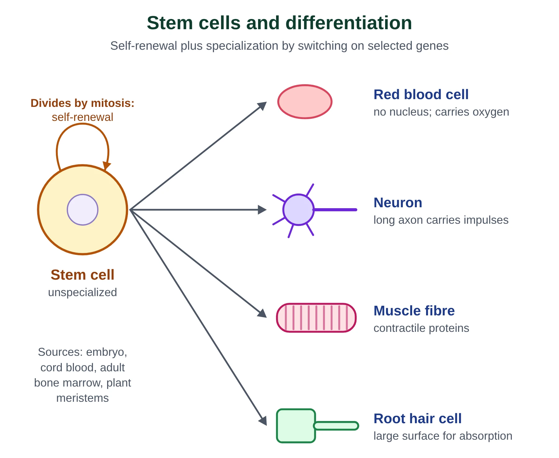
After fertilization the zygote divides by mitosis to form an embryo of unspecialized cells. These cells then differentiate: they develop a particular structure and function. Every cell of the body has the same genes, so differentiation happens because each cell type expresses (switches on) only some of its genes. A red blood cell precursor expresses the haemoglobin genes; a pancreatic cell expresses the insulin gene; neither expresses the other’s genes. Signals from neighbouring cells and the position of a cell in the embryo (for example gradients of signalling molecules called morphogens) decide which genes are switched on.
Stem cells
Stem cells are unspecialized cells with two key properties: they can divide repeatedly to make more stem cells (self-renewal), and they can differentiate along different pathways into specialized cell types.
| Type | What it can become | Example |
|---|---|---|
| Totipotent | Any cell type, including the placenta and the whole organism | Zygote and the cells of the first few divisions |
| Pluripotent | Any cell type of the body, but not the placenta | Embryonic stem cells from the inner cell mass of a blastocyst |
| Multipotent | A limited range of related cell types | Adult stem cells in bone marrow (blood cells) or skin |
Stem cells sit in stem cell niches, places that keep them dividing and release new cells as needed: for example the bone marrow, hair follicles and the base of intestinal crypts. In plants, meristems at root and shoot tips play the same role throughout life.
- Therapeutic uses: bone marrow (blood stem cell) transplants treat leukaemia after chemotherapy; research aims to replace retinal cells in Stargardt disease and damaged tissue after heart attacks or spinal injury.
- Sources: early embryos (pluripotent, but the embryo is destroyed), umbilical cord blood (multipotent, easily stored), adult tissues (multipotent, no embryo used) and induced pluripotent stem cells made by reprogramming adult cells.
- Ethical issues: whether it is right to destroy embryos, the risk of tumours from cells that keep dividing, and consent for using donated tissue.
NCERT Levels of organization (NCERT link)
NCERT Chapter 8 introduces the cell as the unit of life; the levels cell → tissue → organ → organ system → organism are developed in the chapters on animal and plant tissues. NEET questions may ask which unicellular organisms are prokaryotic (bacteria, cyanobacteria, mycoplasma) and which are eukaryotic (Amoeba, Paramecium, Euglena, Chlamydomonas, yeast).
Key points
- Unicellular organisms carry out all eight functions of life in one cell.
- Paramecium is heterotrophic and moves with cilia; Chlamydomonas is autotrophic and moves with two flagella.
- Emergent properties arise from interactions between parts: the whole is more than the sum of its parts.
- Differentiation is caused by differential gene expression; all body cells have the same genes.
- Stem cells self-renew and can differentiate; totipotent > pluripotent > multipotent.
Common mistakes
- Saying specialized cells lose genes. They keep all their genes but only express some of them (mature red blood cells are an exception, as they lose the nucleus).
- Mixing up excretion and egestion. Excretion removes waste made by metabolism; egestion removes undigested food.
- Calling all stem cells ‘embryonic’. Adult and cord blood stem cells are multipotent stem cells too.
Test yourself (click a question to reveal the answer)
Explain how Paramecium carries out homeostasis.
Water enters by osmosis because the cytoplasm is more concentrated than fresh water. Contractile vacuoles collect the excess water and expel it, so the cell does not burst and its water content stays steady.
What is the difference between a pluripotent and a multipotent stem cell?
A pluripotent stem cell can form any cell type of the body; a multipotent stem cell can form only a limited range of related cell types (e.g. blood cells).
Give an example of an emergent property.
For example, a heart pumping blood, a brain thinking, or a leaf photosynthesising as an organ; none of these can be done by a single cell of that organ.
6 Quick check
Ten questions: six multiple choice and four short answer. Try them all before opening the answers.
-
Who published the conclusion that all plants are made of cells, and in which year?
- A. Theodor Schwann, 1839
- B. Matthias Schleiden, 1838
- C. Rudolf Virchow, 1855
- D. Robert Hooke, 1665
-
Which structure is found in both prokaryotic and eukaryotic cells?
- A. Nuclear envelope
- B. Mitochondrion
- C. Golgi apparatus
- D. Ribosome
-
A cell 25 µm wide appears 50 mm wide in a drawing. What is the magnification?
- A. ×2000
- B. ×200
- C. ×20
- D. ×0.5
-
What is the SA:V ratio of a cube with sides of 3 cm?
- A. 6 : 1
- B. 3 : 1
- C. 2 : 1
- D. 1 : 2
-
Which is an example of an aseptate structure?
- A. A Rhizopus hypha
- B. A red blood cell
- C. A root hair cell
- D. A bacterium
-
Which cells are totipotent?
- A. Bone marrow stem cells
- B. Cord blood stem cells
- C. Cells of the inner cell mass
- D. The zygote and its first few daughter cells
-
Explain why Robert Hooke could not see a nucleus in the cork cells he described.
-
A mitochondrion is 3 mm long in a micrograph taken at ×1500. Calculate its actual length in µm.
-
State two ways in which Chlamydomonas differs from Paramecium in carrying out the functions of life.
-
Explain why cells divide rather than growing very large.
Show answers
- B. Matthias Schleiden, 1838. Schwann extended the idea to animals in 1839.
- D. Ribosomes (70S in prokaryotes, 80S in the eukaryotic cytoplasm).
- A. 50 000 µm ÷ 25 µm = ×2000.
- C. 54 cm² ÷ 27 cm³ = 2 : 1.
- A. A Rhizopus hypha has no cross-walls, so many nuclei share one cytoplasm.
- D. The zygote and the cells of the first few divisions.
- Cork cells are dead; their cytoplasm and nucleus have broken down, leaving only the cell walls. His simple microscope also had low resolution.
- 3 mm = 3000 µm; 3000 ÷ 1500 = 2 µm.
- Any two: Chlamydomonas photosynthesises (autotrophic) while Paramecium ingests bacteria (heterotrophic); it moves with two flagella rather than many cilia; it responds to light with an eyespot.
- As a cell grows its volume increases faster than its surface area, so SA:V falls. Exchange of oxygen, nutrients, waste and heat across the membrane can no longer keep up with the needs of the cytoplasm, so the cell divides to restore a high SA:V.
7 Glossary
© competitorspoint.com · IB Biology notes. Diagrams and text are original to competitorspoint.com.
Ready to practise? Download the Introduction to Cells practice questions with answers (free for signed-up members).
⬇ Download practice questions (PDF)