
IB Biology › Continuity & change › Cell Cycle and Cell Division
Cell Cycle and Cell Division
The cell cycle is the ordered series of events in which a cell grows, copies its DNA and divides into two. This chapter covers interphase, mitosis, cytokinesis and meiosis, how cyclins and checkpoints control the cell cycle, and what happens when that control fails in cancer. It is written for IB Biology (SL and HL) and for NEET students using NCERT.
Intro What is the cell cycle?
Syllabus link: D2.1.1 Generation of new cells by cell division; D2.1.4 Roles of mitosis and meiosis; NCERT 10.1
AHL = additional higher level (IB HL only) NCERT = extra detail for NEET / CBSE students (beyond the IB guide)
The cell cycle is the sequence of events by which a cell grows, replicates its DNA and then divides to form two daughter cells. Every cell in your body, apart from the very first one (the zygote), was made this way, so understanding the cell cycle explains how organisms grow, heal and reproduce.
Cell division is the biological process in which a parent cell (often called the mother cell) divides into two daughter cells. In 1855 Rudolf Virchow proposed that all cells arise from pre-existing cells (omnis cellula e cellula), and cell division is the process that makes this true.
The formation of daughter cells is much simpler in prokaryotic cells than in eukaryotic cells. A prokaryotic cell contains a single circular chromosome. The chromosome first replicates and the two copies of DNA separate as the cell elongates. The parent cell then splits into two daughter cells. This is called binary fission.
Eukaryotic cells are more complex: they have many linear chromosomes inside a nucleus, and many membrane-bound organelles. All these components must be duplicated and shared out during cell division. To do this, eukaryotic cells pass through a series of stages called the cell cycle.
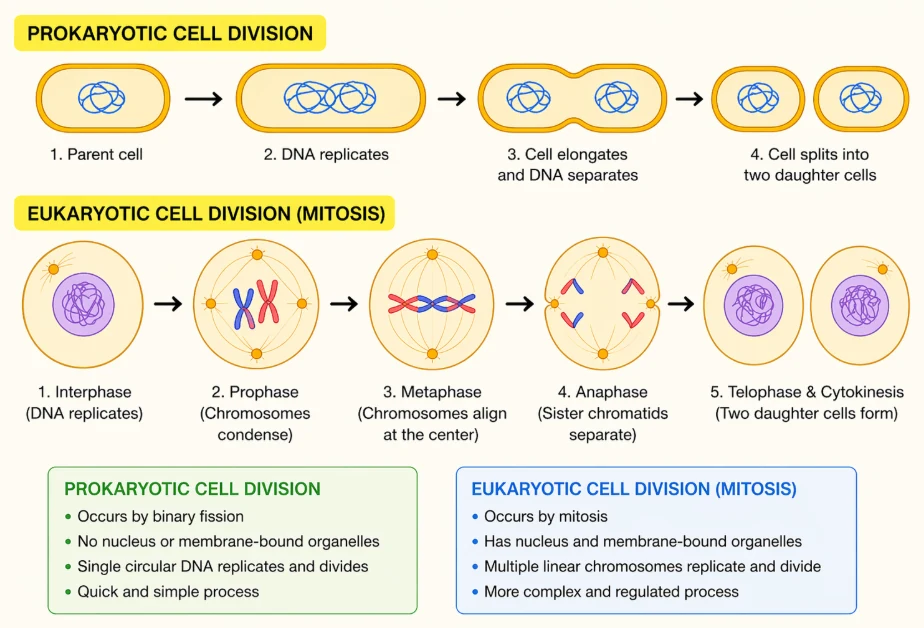
Why do cells divide?
- Growth. A human starts as one cell, the zygote, and an adult body has roughly 30 to 40 trillion cells.
- Replacement. Cells of the skin, the lining of the gut and the blood are lost all the time and must be replaced.
- Repair. Cells divide to close a wound or regrow damaged tissue.
- Asexual reproduction. Single-celled organisms such as Amoeba and yeast reproduce by dividing.
- Sexual reproduction. Meiosis makes the haploid gametes (or, in plants, spores) needed for a sexual life cycle.
Two kinds of nuclear division
In eukaryotes the nucleus must divide before the cell divides, otherwise one daughter cell would have no nucleus (an anucleate cell). There are two kinds of nuclear division:
- Mitosis keeps the chromosome number the same and gives two genetically identical nuclei. It is used for growth, repair, replacement and asexual reproduction.
- Meiosis halves the chromosome number (diploid to haploid) and gives four genetically different nuclei. It is used to make gametes or spores.
The division of the cytoplasm that follows nuclear division is called cytokinesis. Sections 2 to 4 deal with mitosis, cytokinesis and meiosis in turn; sections 5 and 7 show how the cell cycle is controlled and how cancer results when that control breaks down.
Key points
- The cell cycle is the sequence of growth, DNA replication and division that produces two daughter cells.
- All cells come from pre-existing cells (Virchow, 1855).
- Prokaryotes divide by binary fission; eukaryotes use the cell cycle, with mitosis or meiosis followed by cytokinesis.
- Nuclear division comes before cytoplasmic division, so that no daughter cell is left without a nucleus.
Common mistakes
- Saying prokaryotes divide by mitosis. They have no nucleus and no spindle; they divide by binary fission.
- Using ‘cell division’ and ‘mitosis’ as if they mean the same. Mitosis is division of the nucleus; cytokinesis divides the cell.
Test yourself (click a question to reveal the answer)
Why must nuclear division happen before cytokinesis?
So that each daughter cell receives a nucleus with a full set of chromosomes; otherwise one cell would be anucleate and could not make the proteins it needs.
Give one role of mitosis and one role of meiosis in a human.
Mitosis: growth, replacing skin or gut cells, or repairing a wound. Meiosis: making haploid sperm or egg cells for sexual reproduction.
1 Phases of the cell cycle: interphase, G0 and duration
Syllabus link: D2.1.5 DNA replication as a prerequisite for mitosis and meiosis; AHL D2.1.13 Phases of the cell cycle; AHL D2.1.14 Cell growth during interphase; NCERT 10.1
Every eukaryotic cell that divides goes through a well-defined sequence of events called the cell cycle. In one cell cycle a cell first grows, then replicates its DNA, and finally divides to form two daughter cells.
Features of the cell cycle
- Coordinated: the events of the cell cycle happen in a fixed order, and each one is completed before the next begins.
- Under genetic control: the events are controlled by genes, through proteins such as cyclins and kinases (section 5).
- DNA replication happens once in each cell cycle, during S phase.
- Growth is continuous: unlike DNA replication, cell growth carries on through interphase.
The two main phases: interphase and M phase
The cell cycle has two main phases: interphase, when the cell prepares for division, and the M phase (mitotic phase), when the cell actually divides.
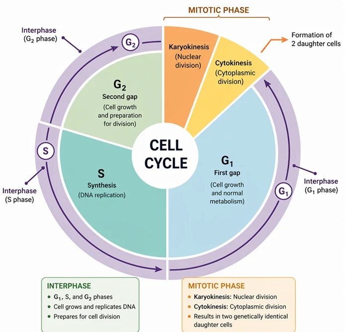
- M phase (mitosis phase) is when the cell divides. Division of the nucleus (karyokinesis) is followed by division of the cytoplasm (cytokinesis).
- M phase is short: less than 5% of the cell cycle.
- Interphase is the period between two successive M phases. The cell grows and prepares itself for division. It takes more than 95% of the cell cycle.
- Interphase is divided into three phases: G1, S and G2.
- Interphase is sometimes called the ‘resting phase’, but the cell is not resting: it is growing, making proteins and replicating its DNA. It is better to call it the preparation phase.
What happens during G1 phase?
- G1 (first gap) is the interval between the end of M phase and the start of S phase. It is also called the post-mitotic phase.
- The cell is metabolically very active and grows a lot. Growth needs proteins and enzymes, so transcription and translation are busy.
- Organelles such as mitochondria and ribosomes increase in number.
- Near the end of G1 the cell ‘decides’ what to do next: (a) continue the cycle and enter S phase, or (b) leave the cycle and enter the G0 (quiescent) phase.
- Cells that will divide pass the G1 checkpoint and move into S phase. Cells that leave the cycle stay in G0; some return to G1 later if they receive a signal to divide.
What happens during S phase?
- S stands for synthesis (DNA synthesis). It is the only phase in which DNA replication takes place.
- DNA replication is semi-conservative: the two strands of each DNA double helix separate and each acts as a template for a new strand, giving two identical double-stranded molecules.
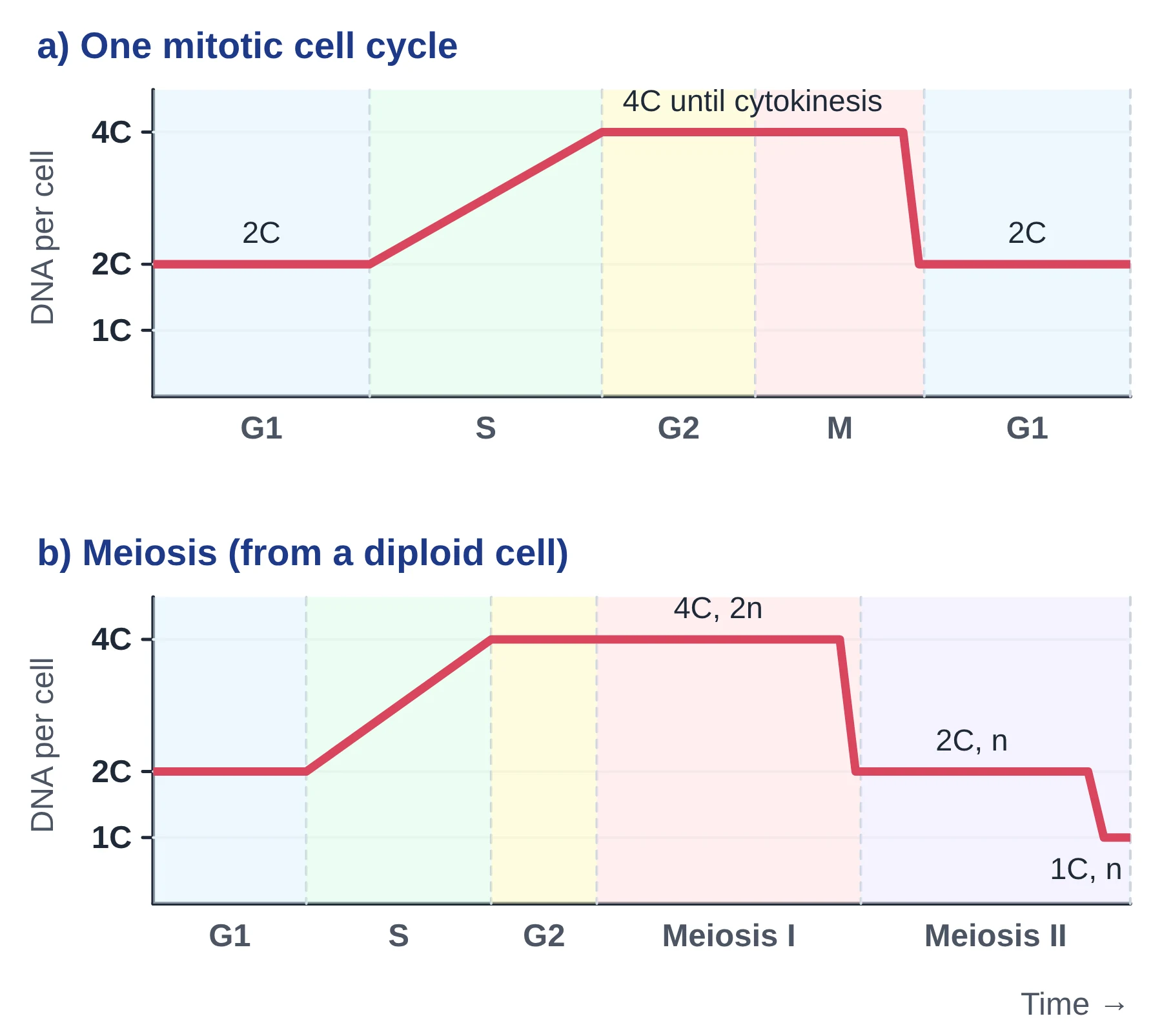
- The amount of DNA doubles. If a diploid cell has 2C DNA in G1, it has 4C at the end of S phase.
- The number of chromosomes does not change. Each chromosome now consists of two identical sister chromatids joined at the centromere.
- Histone proteins are made in large amounts, because the newly made DNA must be wrapped around histones.
- In animal cells the centriole pair duplicates in the cytoplasm. (Most plant cells have no centrioles; they still build a spindle.)
- So, DNA replication takes place in the nucleus, while histone synthesis and centriole duplication take place in the cytoplasm.
- Chromatin exists in two forms: loosely packed euchromatin and densely packed heterochromatin. Euchromatin is replicated early in S phase and heterochromatin later.
What happens during G2 phase?
- G2 (second gap) is the interval between S phase and M phase. It is also called the post-synthetic or pre-mitotic phase.
- The cell continues to grow and makes the proteins needed for mitosis.
- Tubulin is made in large amounts; it is the protein from which spindle microtubules are built.
- Mitochondria (and chloroplasts in plant cells) keep increasing in number by growth and division; this happens through interphase, not only in G2.
- The cell checks that DNA replication is complete and that any damage has been repaired before it enters mitosis (the G2 checkpoint).
AHL Cell growth during interphase (D2.1.14)
IB HL students should see interphase as a metabolically active period, not a pause. Growth involves the biosynthesis of cell components: proteins (enzymes, histones, tubulin), membranes and, in S phase, DNA. The numbers of mitochondria and chloroplasts are increased by growth and division of the existing organelles, because these organelles cannot be made from scratch.
Cell proliferation (D2.1.12) depends on the cell cycle. Examples are growth at plant meristems (root and shoot tips), the rapid divisions of an early embryo, routine replacement of skin cells, and the extra divisions that close a wound.
G0: the quiescent phase
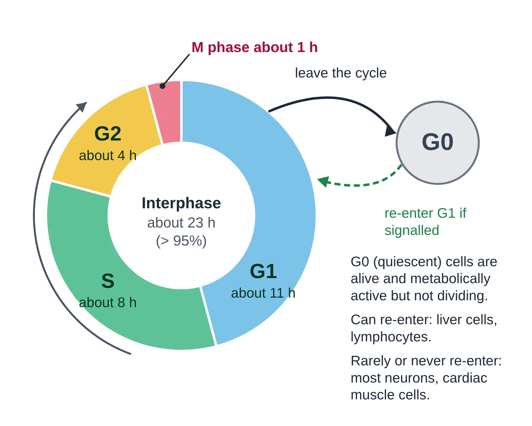
Some cells stop dividing. They leave the cycle at G1 and enter an inactive stage called G0, the quiescent stage. Cells in G0 are alive and metabolically active: they carry out their normal jobs, but they no longer proliferate unless they are signalled to do so.
- Can re-enter the cycle: liver cells (hepatocytes) return to G1 when part of the liver is damaged or removed; lymphocytes divide when they meet an antigen.
- Rarely or never re-enter: most nerve cells (neurons) and heart muscle cells are fully differentiated and stay in G0 for life. These are sometimes called permanent cells.
How long does a cell cycle take?
The length of the cell cycle varies from organism to organism and from one cell type to another. Yeast can complete a cycle in about 90 minutes. A typical human cell in culture divides about once every 24 hours, and cell division proper (M phase) lasts only about one hour of this.
For most dividing cells the phases rank G1 > S > G2 > M in duration, so G1 is usually the longest phase and M phase the shortest. G1 is also the most variable: in very fast embryonic divisions it is almost absent.
Chromatin, chromosomes and chromatids
During interphase, chromosomes cannot be seen as separate structures under the light microscope; the nucleus contains chromatin. Chromatin is the uncondensed, thread-like form of DNA with its proteins. A chromosome is the condensed, division-ready form of the same DNA, which becomes visible only during M phase.
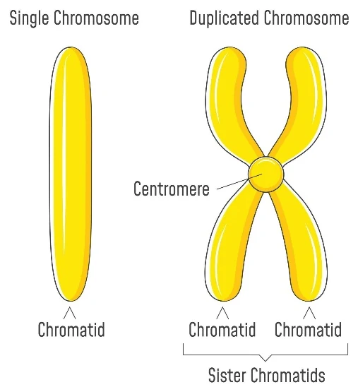
- Single chromatid = one double-stranded DNA molecule.
- Chromosomes are counted by centromeres. A duplicated chromosome (two sister chromatids, one centromere) still counts as one chromosome.
- n = one complete set of chromosomes (one copy of each type). 2n = two sets. In humans 2n = 46 and n = 23.
- C = the amount of DNA in one haploid set. It tracks DNA, not chromosomes. A diploid cell is 2C in G1 and 4C in G2.
DNA amount and chromosome number through the cycle
Take an example of a cell with 2n = 6: 6 chromosomes, made of 3 homologous pairs (n = 3, so one set has 3 chromosomes). In humans 2n = 46 and n = 23: there are two sets, each containing 23 different types of chromosome.
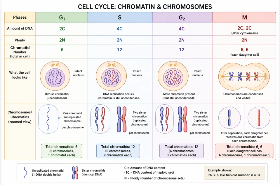
Swipe sideways to see the whole table →
| G1 | After S / G2 | Metaphase | Anaphase | Each daughter cell | |
|---|---|---|---|---|---|
| DNA amount | 2C | 4C | 4C | 4C | 2C |
| Chromosome number | 6 | 6 | 6 | 12 (sister chromatids now separate) | 6 |
| Chromatids per cell | 6 | 12 | 12 | 12 | 6 |
| Chromatids per chromosome | 1 | 2 | 2 | 1 | 1 |
| Ploidy | 2n | 2n | 2n | 2n (two sets moving to each pole) | 2n |
Notice two things in the table. First, S phase doubles the DNA (2C to 4C) but not the chromosome number. Second, in anaphase each chromatid becomes an independent chromosome, so for a short time the cell contains twice the usual number of chromosomes (12 here, 92 in a human cell) until cytokinesis shares them out.
Worked examples
Q1. The number of chromosomes in a gamete is 10. Find the amount of DNA in G2 and the number of chromosomes in a body cell.
Answer. A gamete is haploid, so n = 10 and body cells have 2n = 20. If the body cell has 2C DNA in G1, the DNA amount in G2 is 4C, but the number of chromosomes stays 20.
Q2. You have studied mitosis in onion root tip cells, where each cell contains 16 chromosomes. How many chromosomes are present in a cell during G1, after S phase and after M phase? If the DNA content of a cell after M phase is 2C, what is the DNA content during G1, after S phase and during G2?
- Chromosome number (counted by centromeres): G1 = 16; after S phase = 16 (each now has two sister chromatids); after M phase = 16 in each daughter cell.
- DNA content changes because DNA is replicated in S phase: G1 = 2C (the same as after M phase); after S phase = 4C; G2 = 4C (still duplicated, no division yet).
Key points
- Interphase (G1, S, G2) takes over 95% of the cell cycle; M phase takes less than 5%.
- G1: growth and normal metabolism; S: DNA replication, histone synthesis, centriole duplication; G2: growth, tubulin synthesis, preparation for mitosis.
- G0 cells have left the cycle but are metabolically active; some (liver cells) can re-enter G1.
- S phase doubles the DNA (2C to 4C) but not the chromosome number.
- A human cell in culture divides about every 24 hours; M phase lasts about 1 hour.
Common mistakes
- Saying the chromosome number doubles in S phase. Only the DNA (and the number of chromatids) doubles; each chromosome now has two chromatids.
- Calling interphase a resting phase and stopping there. The cell is very active: growing, making proteins and copying DNA.
- Writing ‘chromatin number’ when you mean chromatid number. Chromatin is the material; chromatids are the copies you count.
- Mixing up n and C. n counts chromosome sets; C measures DNA. A G2 cell is 2n but 4C.
Exam tip
- IB: the sequence G1, S, G2, mitosis, cytokinesis and the events of each phase are AHL (D2.1.13). All students need D2.1.5: DNA is replicated before mitosis and meiosis, so each chromosome has two chromatids.
- NEET often tests C-value and chromosome number together: write both for each stage in a small table before choosing an option.
Test yourself (click a question to reveal the answer)
A cell has 2n = 24. How many chromatids does it have in G2?
48. It still has 24 chromosomes, but each has two sister chromatids after S phase.
Why is G1 the most variable phase in length?
It is when the cell grows and responds to signals; the cell may wait in G1 until it is big enough and has the right signals, or leave for G0. Embryonic cells that divide very fast have almost no G1.
Name a cell type that stays permanently in G0 and one that can return to the cell cycle.
Permanent: most neurons (or cardiac muscle cells). Can return: liver cells (hepatocytes) or lymphocytes.
2 Mitosis: prophase, metaphase, anaphase and telophase
Syllabus link: D2.1.6 Condensation and movement of chromosomes; D2.1.7 Phases of mitosis; D2.1.8 Identification of phases of mitosis; AHL D2.1.12 Cell proliferation; NCERT 10.2, 10.3
Mitosis is the division of the nucleus that produces two nuclei with the same number and type of chromosomes as the parent nucleus. Because the chromosome number stays the same, mitosis is called an equational division.
Features of M phase
- Major reorganisation of the nucleus and the cytoplasm: the nuclear envelope breaks down and a spindle is built.
- Equational division: daughter nuclei are genetically identical to the parent nucleus.
- Can occur in both haploid and diploid cells.
- M phase has two parts: karyokinesis (nuclear division) and cytokinesis (division of the cytoplasm).
- Karyokinesis is divided into four stages: prophase, metaphase, anaphase and telophase. Prophase usually takes the longest and anaphase the shortest time.
Condensing and moving chromosomes
Two problems have to be solved in every mitosis and meiosis (D2.1.6). First, very long DNA molecules must be packed into short, compact chromosomes that can be moved without tangling. DNA is wound around histone proteins to form nucleosomes, and the chain of nucleosomes is then coiled and looped again and again (supercoiling). A human chromosome can become thousands of times shorter than its DNA.
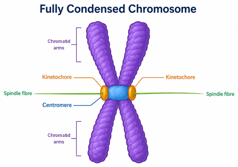
Second, the chromosomes must be moved. This is done by the spindle, made of microtubules. Microtubules attach to the kinetochore, a protein structure on each centromere. Motor proteins at the kinetochore and along the spindle use ATP to move chromosomes, while microtubules shorten by losing tubulin subunits, pulling chromatids towards the poles.
Prophase
Prophase is the first stage of karyokinesis. It follows the S and G2 phases of interphase, so every chromosome already consists of two sister chromatids. By the end of prophase:
- Chromatin condenses to form compact chromosomes, each with two visible sister chromatids.
- The two centrosomes, which duplicated during interphase, move towards opposite poles of the cell, and the spindle starts to form between them.
- The nucleolus disappears, and the Golgi apparatus and endoplasmic reticulum break up into small vesicles, so they are no longer seen under the microscope.
- The nuclear envelope breaks down into fragments.
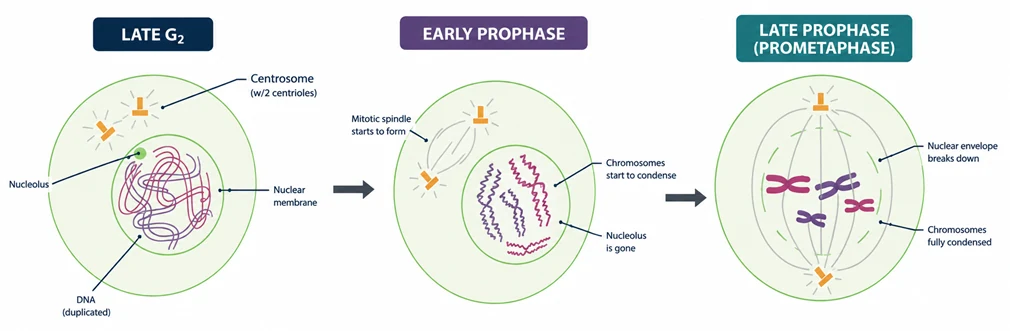
A centrosome contains two centrioles at right angles, surrounded by protein material. Microtubules radiating from each centrosome form a star shape called an aster. Animal cells have two asters, so their spindle is called amphiastral. Most plant cells have no centrioles and no asters; their spindle is anastral. The two asters together with the spindle fibres form the mitotic (spindle) apparatus. In prophase the spindle fibres have not yet attached to the chromosomes.
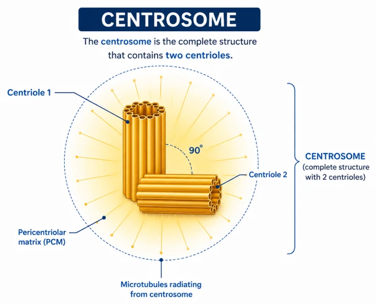
NCERT Prometaphase in NCERT and IB
Some textbooks add a stage called prometaphase between prophase and metaphase, when the nuclear envelope has broken down and spindle fibres start to attach to kinetochores. Neither NCERT nor the IB guide asks for it as a separate stage: both use the four stages prophase, metaphase, anaphase and telophase. NCERT describes these events as the end of prophase and says that metaphase begins with the complete disintegration of the nuclear envelope. In exams, include prometaphase events in prophase unless a question names prometaphase.
Metaphase
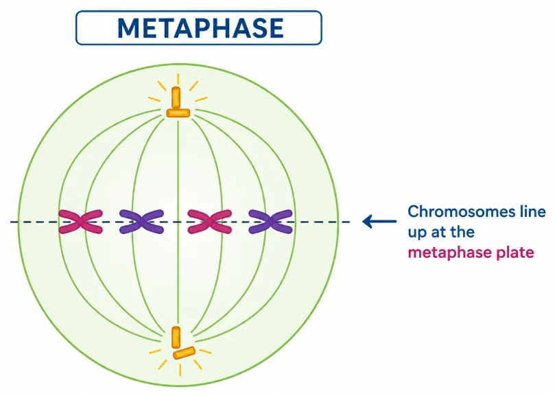
- The nuclear envelope has completely disappeared, and the chromosomes are spread through the cytoplasm.
- Chromosomes are fully condensed. Metaphase is the best stage to study chromosome number and shape (morphology), which is why karyotypes are made from metaphase cells.
- Spindle fibres attach to the kinetochores, the disc-shaped protein structures on the surface of each centromere. The kinetochore of one sister chromatid is attached to fibres from one pole, and the kinetochore of its sister to fibres from the opposite pole.
- Pulling from both sides brings all the chromosomes to the equator of the spindle. The plane where they line up is called the metaphase plate.
Anaphase
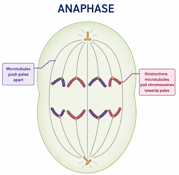
- The centromere of each chromosome splits and the sister chromatids separate from each other, all at the same moment.
- Each separated chromatid is now called a daughter chromosome of the future nuclei.
- Kinetochore microtubules shorten and pull the daughter chromosomes to opposite poles. The centromere leads, and the arms trail behind.
- Other spindle microtubules lengthen and push the poles apart, so the cell elongates.
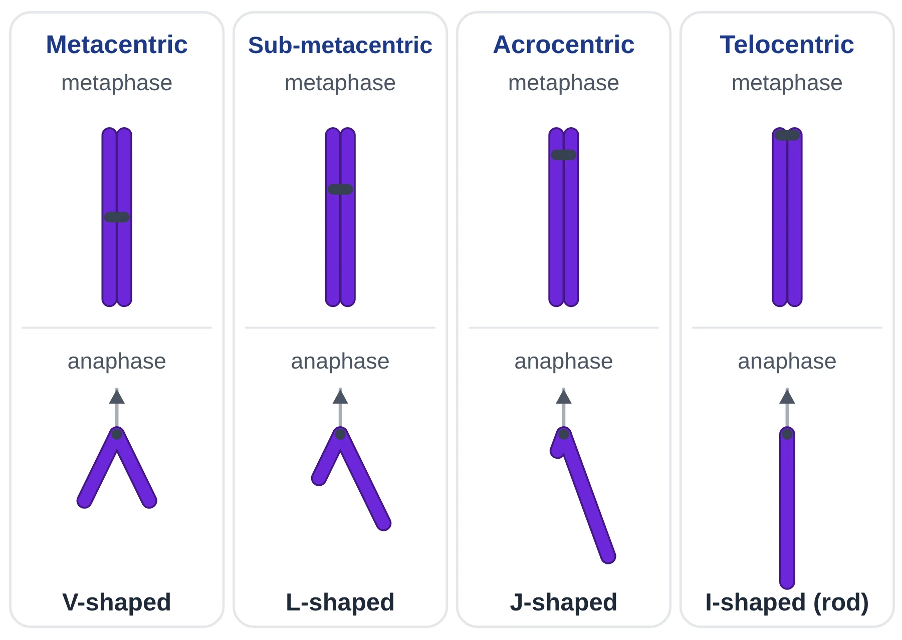
Because the arms trail behind the centromere, chromosomes take different shapes in anaphase depending on where the centromere is:
- Metacentric (centromere in the middle): V-shaped.
- Sub-metacentric (centromere slightly off-centre): L-shaped.
- Acrocentric (centromere very close to one end): J-shaped.
- Telocentric (centromere at the end): I-shaped, a straight rod.
How sister chromatids are held together and released
Sister chromatids are held together along their length, and most strongly at the centromere, by rings of a protein called cohesin. When every kinetochore is attached to the spindle, a protein complex called the anaphase-promoting complex (APC/C) becomes active. APC/C is not itself the enzyme that dissolves cohesin. It tags an inhibitor protein, securin, for destruction. With securin gone, the enzyme separase becomes active and cuts the cohesin, so the sister chromatids can be pulled apart. APC/C also tags mitotic cyclin for destruction, which lets the cell finish mitosis.
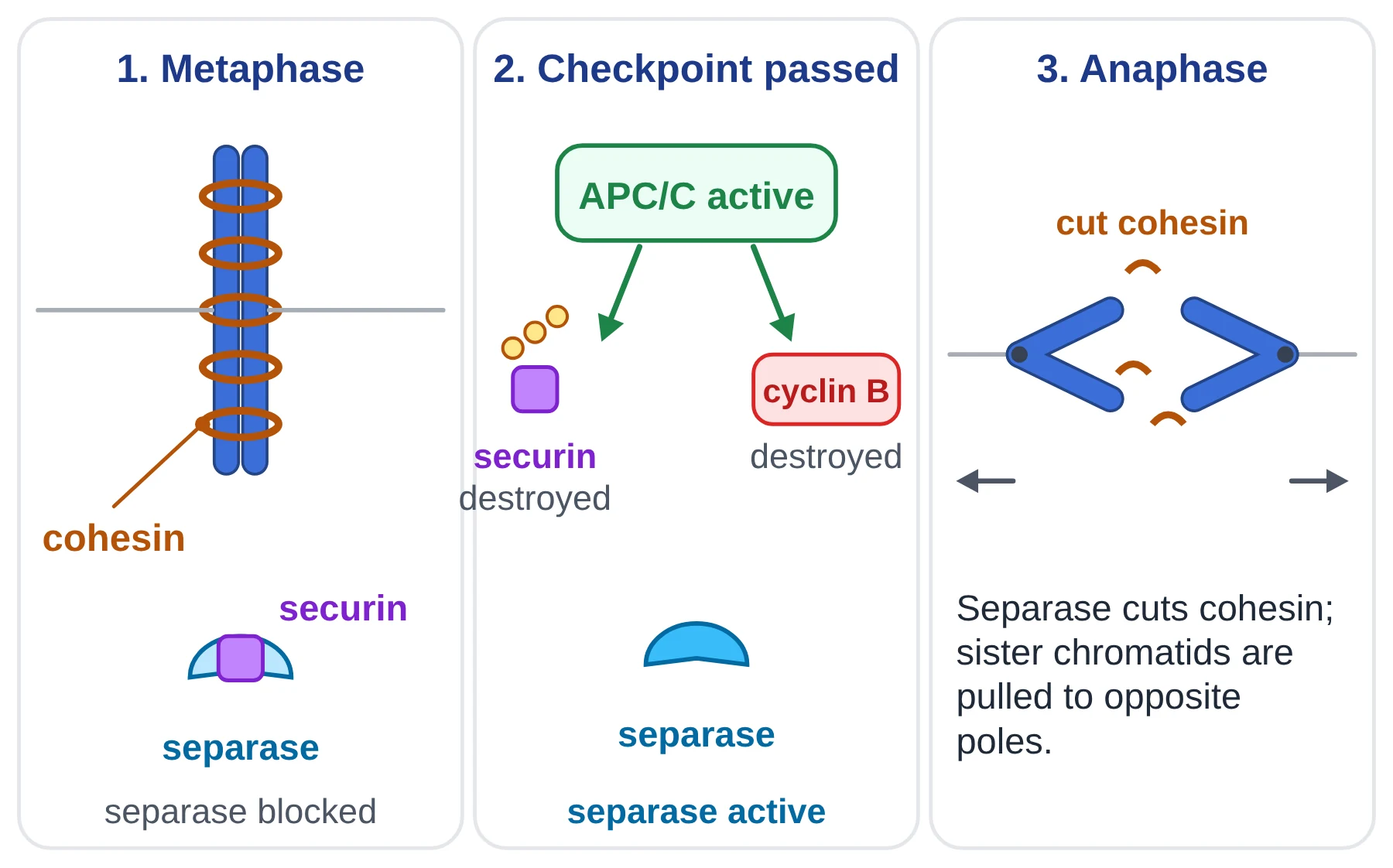
If APC/C does not work, securin is not destroyed, cohesin is not cut and the cell stays stuck in metaphase. (This is different from polyteny: polytene chromosomes, as in the salivary glands of Drosophila larvae, form when DNA is replicated many times without any mitosis, so hundreds of chromatids stay side by side.)
Colchicine and polyploidy
The alkaloid colchicine, from the autumn crocus, binds to tubulin and stops microtubules forming, so no spindle can be built. The chromosomes condense and the centromeres still split, but the chromatids are not pulled to the poles. When the nuclear envelope reforms, the cell has a single nucleus with double the chromosome number. Repeating this gives polyploid cells, and plant breeders use colchicine to make polyploid crop varieties.
Telophase
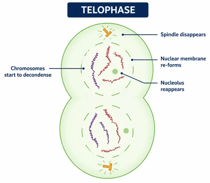
- Chromosomes reach the poles and decondense, losing their identity as separate chromosomes and becoming chromatin again.
- A nuclear envelope re-forms around each group of chromosomes.
- The nucleolus, endoplasmic reticulum and Golgi apparatus re-form.
- The spindle disappears.
- Two genetically identical nuclei are formed. Cytokinesis usually begins during telophase.
Identifying the phases of mitosis
IB students must be able to identify the phases in diagrams, micrographs and slides (D2.1.8). Root tip squashes of onion or garlic are the usual practical. Look for these clues:
| Phase | What you see under the microscope |
|---|---|
| Interphase | Intact nucleus with a clear boundary; chromatin evenly spread or granular; one or more nucleoli visible. |
| Prophase | Chromosomes visible as threads inside the nucleus area; nuclear boundary fading; nucleolus disappearing. |
| Metaphase | Chromosomes lined up in a band across the middle of the cell; no nuclear envelope. |
| Anaphase | Two groups of V-shaped chromosomes moving apart towards opposite ends of the cell. |
| Telophase | Two clusters of chromosomes at opposite ends, nuclear envelopes re-forming; in plant cells a cell plate between them. |
Where does mitosis occur?
- Gut lining: cells lining the alimentary canal are replaced every few days.
- Skin: cells in the deepest layer of the epidermis divide continuously to replace cells lost from the surface.
- Red bone marrow: stem cells divide to produce blood cells. Mature red blood cells have no nucleus and cannot divide themselves.
- Plant meristems: apical meristems at root and shoot tips cause growth in length (primary growth); lateral meristems (vascular cambium and cork cambium) increase girth (secondary growth).
Mitosis in haploid and diploid cells
In animals such as humans (2n = 46), body cells are called somatic cells. They are diploid, and new somatic cells are formed by mitosis. Diploid germ cells in the ovaries and testes form haploid gametes (n = 23) by meiosis. In most animals the gametes never divide by mitosis, so mitosis happens only in diploid cells.
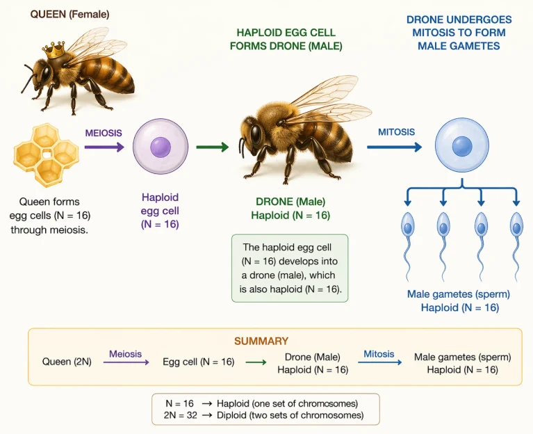
Exception: the honey bee. A queen honey bee (2n = 32) forms egg cells (n = 16) by meiosis. An unfertilised egg develops into a drone (male), which is therefore haploid (n = 16). The drone makes its male gametes by mitosis, so in honey bees mitosis happens in haploid cells too. Fertilised eggs (2n = 32) develop into females: workers or queens.
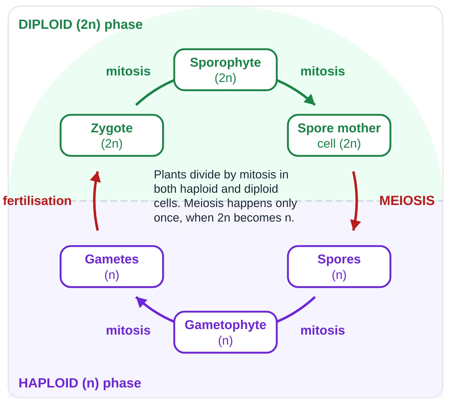
In plants, mitosis occurs in both haploid and diploid cells. The diploid sporophyte grows by mitosis; meiosis makes haploid spores; the spores grow into a haploid gametophyte by mitosis; and the gametophyte makes gametes by mitosis. Fertilisation restores the diploid number in the zygote.
Significance of mitosis
- Produces daughter cells with the same chromosome number and identical genetic information as the parent (genetic stability).
- Growth of multicellular organisms, from the zygote onwards.
- Restores the nucleo-cytoplasmic ratio: as a cell grows, its cytoplasm increases but its nucleus does not, and division restores the balance.
- Cell repair and replacement, for example in the epidermis, the lining of the gut and the production of blood cells.
- Continuous growth of plants from meristems (apical and lateral).
- Asexual reproduction and vegetative propagation, which produce genetically identical offspring.
- It occurs in both haploid and diploid cells.
Key points
- Mitosis is an equational division: two genetically identical nuclei with the parent’s chromosome number.
- Prophase: condensation, centrosomes move apart, nucleolus and nuclear envelope disappear.
- Metaphase: chromosomes at the metaphase plate, sister kinetochores attached to opposite poles.
- Anaphase: cohesin is cut, centromeres split and sister chromatids move to opposite poles.
- Telophase: nuclear envelopes and nucleoli re-form, chromosomes decondense, spindle disappears.
- Histones supercoil DNA; microtubules and motor proteins move chromosomes.
Common mistakes
- Saying chromosomes replicate in prophase. DNA replication happened in S phase of interphase.
- Writing that spindle fibres attach to the centromere ‘in prophase’. Attachment to kinetochores is completed by metaphase.
- Saying APC dissolves cohesin. APC/C triggers destruction of securin; separase cuts the cohesin.
- Saying colchicine stops chromatids separating. It stops the spindle forming, so separated chromatids stay in one nucleus.
- Listing ‘blood cells’ as cells that undergo mitosis. Blood cells are made by mitosis of stem cells in the bone marrow.
Exam tip
- When asked how mitosis produces genetically identical cells, link three ideas: DNA replication in S phase makes identical sister chromatids; each chromosome attaches to both poles at metaphase; one chromatid of every chromosome goes to each pole at anaphase.
- In ‘identify the phase’ questions, give the evidence you used (for example, ‘chromosomes aligned at the equator, so metaphase’).
Test yourself (click a question to reveal the answer)
Why is metaphase the best stage to count chromosomes?
The chromosomes are fully condensed and lined up separately at the metaphase plate, so each one can be seen and counted.
A cell with 2n = 8 is in anaphase of mitosis. How many chromosomes are there in the cell?
16: the sister chromatids of all 8 chromosomes have separated, and each separated chromatid counts as a chromosome until cytokinesis.
Explain why colchicine can make a diploid plant cell tetraploid.
It stops spindle formation, so after the centromeres split the chromatids are not separated into two cells; one nucleus re-forms containing twice the chromosome number (4n).
3 Cytokinesis: cleavage furrow and cell plate
Syllabus link: D2.1.2 Cytokinesis as splitting of cytoplasm; D2.1.3 Equal and unequal cytokinesis; NCERT 10.2.5
Cell division is complete only after cytokinesis, the division of the cytoplasm and its organelles between the two daughter cells. It usually starts in late anaphase or telophase.
In some organisms karyokinesis is not followed by cytokinesis. The nuclei keep dividing inside one mass of cytoplasm, giving a multinucleate cell called a syncytium. A well-known example is the liquid endosperm of coconut (coconut water), which contains many free nuclei; endosperm nuclei are triploid (3n).
Cytokinesis in animal cells: the cleavage furrow
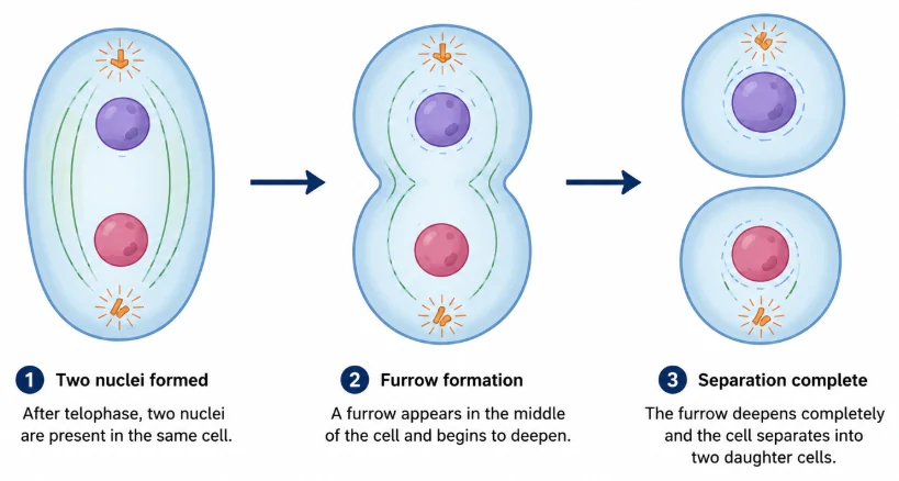
Animal cells have no cell wall; they are bounded only by the plasma membrane. Just under the membrane, around the equator of the cell, a contractile ring of actin filaments and myosin forms. Myosin pulls the actin filaments past each other, so the ring tightens like a drawstring. The membrane is pulled inwards, forming a cleavage furrow that deepens until the cell is pinched into two daughter cells.
Cytokinesis in plant cells: the cell plate
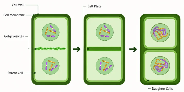
Plant cells have a rigid cell wall that cannot be pinched inwards. Instead, vesicles from the Golgi apparatus, carrying wall materials such as pectins, are guided by microtubules to the equator. They fuse to form a flat sac, the cell plate. The cell plate starts in the centre and grows outwards (centrifugally) until it joins the existing side walls. The vesicle membranes become the new plasma membranes of the two cells, and their contents form the middle lamella, made mainly of calcium pectate. Each daughter cell then lays down cellulose on its side to complete its new primary wall. The two daughter cells stay joined by the middle lamella; they do not separate.
| Feature | Animal cell | Plant cell |
|---|---|---|
| Method | Cleavage furrow (furrowing) | Cell plate formation |
| Direction | From the outside inwards (centripetal) | From the centre outwards (centrifugal) |
| What does the work | Contractile ring of actin and myosin | Golgi vesicles carried by microtubules |
| Product | Two separate cells | Two cells joined by a middle lamella and new walls |
Equal and unequal cytokinesis
Cytokinesis is usually equal, giving two daughter cells of similar size. It does not have to be equal, but each daughter cell must receive at least one mitochondrion (and, in plants, a plastid), because these organelles can only be made by division of an existing one.
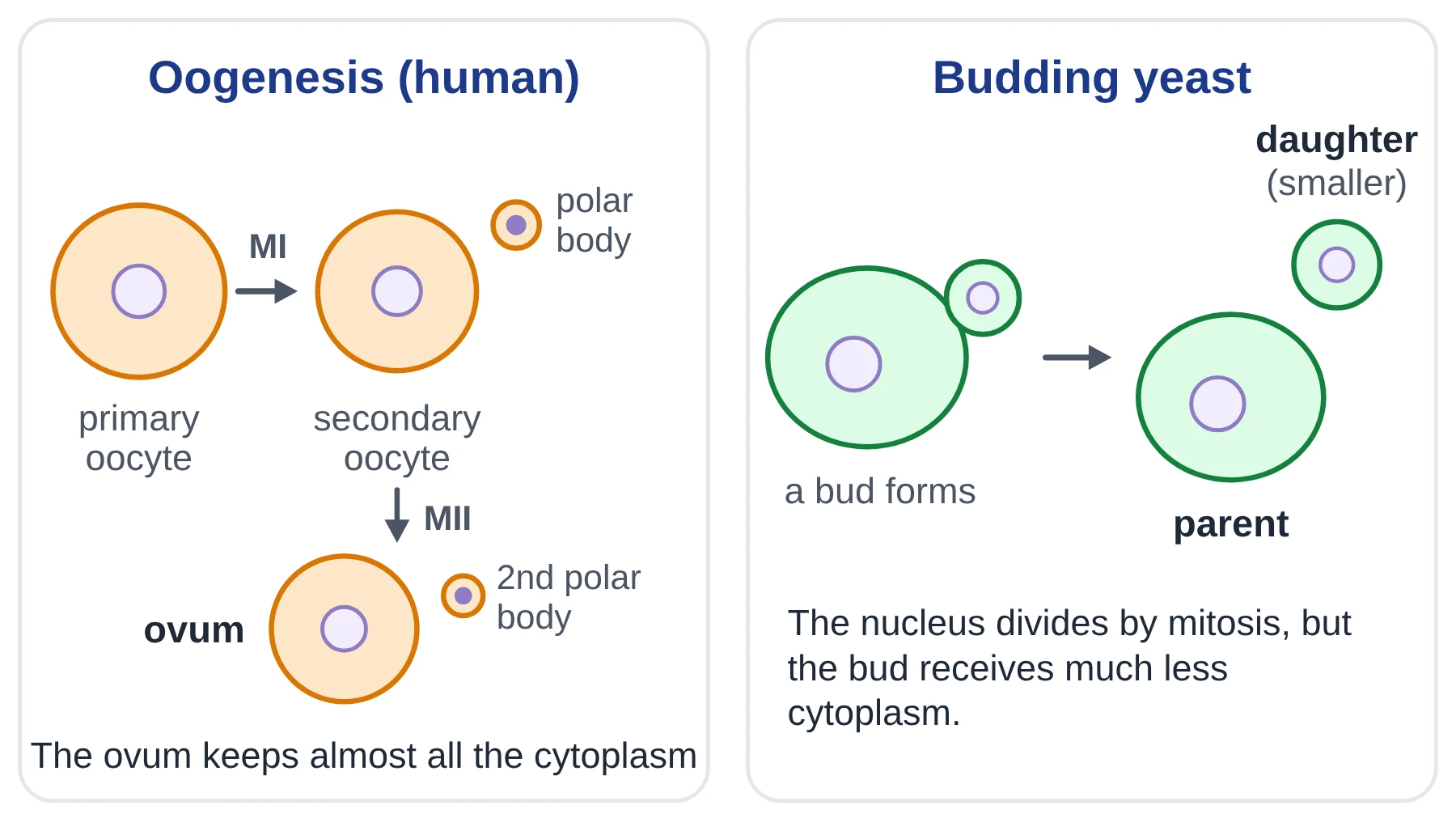
- Oogenesis in humans: both divisions of meiosis are unequal. Almost all the cytoplasm goes to the secondary oocyte and then to the ovum, while the tiny polar bodies get very little and later die. The ovum keeps a large store of nutrients and organelles for the early embryo.
- Budding in yeast: the nucleus divides by mitosis, but one nucleus moves into a small outgrowth (the bud), which pinches off as a smaller daughter cell.
Key points
- Cytokinesis divides the cytoplasm after nuclear division; without it a syncytium forms (e.g. coconut liquid endosperm).
- Animal cells: a contractile ring of actin and myosin forms a cleavage furrow (centripetal).
- Plant cells: Golgi vesicles fuse into a cell plate that grows centrifugally and becomes the middle lamella.
- Cytokinesis can be unequal (oogenesis, yeast budding), but each cell must get mitochondria.
Common mistakes
- Saying plant daughter cells separate after cytokinesis. They stay joined by the middle lamella.
- Saying the cell plate grows from the edges inwards. It starts in the centre and grows outwards.
- Calling the cell plate a cell wall straight away. It first becomes the middle lamella; cellulose walls are laid down on each side.
Test yourself (click a question to reveal the answer)
Why can plant cells not divide by furrowing?
They have a rigid cellulose cell wall around the plasma membrane, which cannot be pulled inwards by a contractile ring.
Suggest why the polar bodies in oogenesis receive so little cytoplasm.
So that the ovum keeps as much cytoplasm, nutrient store and as many organelles as possible to support the embryo after fertilisation; the polar bodies only remove extra sets of chromosomes.
4 Meiosis: the reduction division
Syllabus link: D2.1.9 Meiosis as a reduction division; D2.1.10 Down syndrome and non-disjunction; D2.1.11 Meiosis as a source of variation; NCERT 10.4, 10.5
Meiosis is the nuclear division that halves the chromosome number, producing four haploid (n) nuclei from one diploid (2n) nucleus. Because the chromosome number is reduced, meiosis is called a reduction division.
Meiosis is needed in every sexual life cycle. Fertilisation fuses two gametes and doubles the chromosome number; without a halving step somewhere in the life cycle, the chromosome number would double in every generation. In animals meiosis makes gametes; in plants it makes spores.
Key features of meiosis
- Two divisions, meiosis I and meiosis II, but only one round of DNA replication (in the S phase before meiosis I).
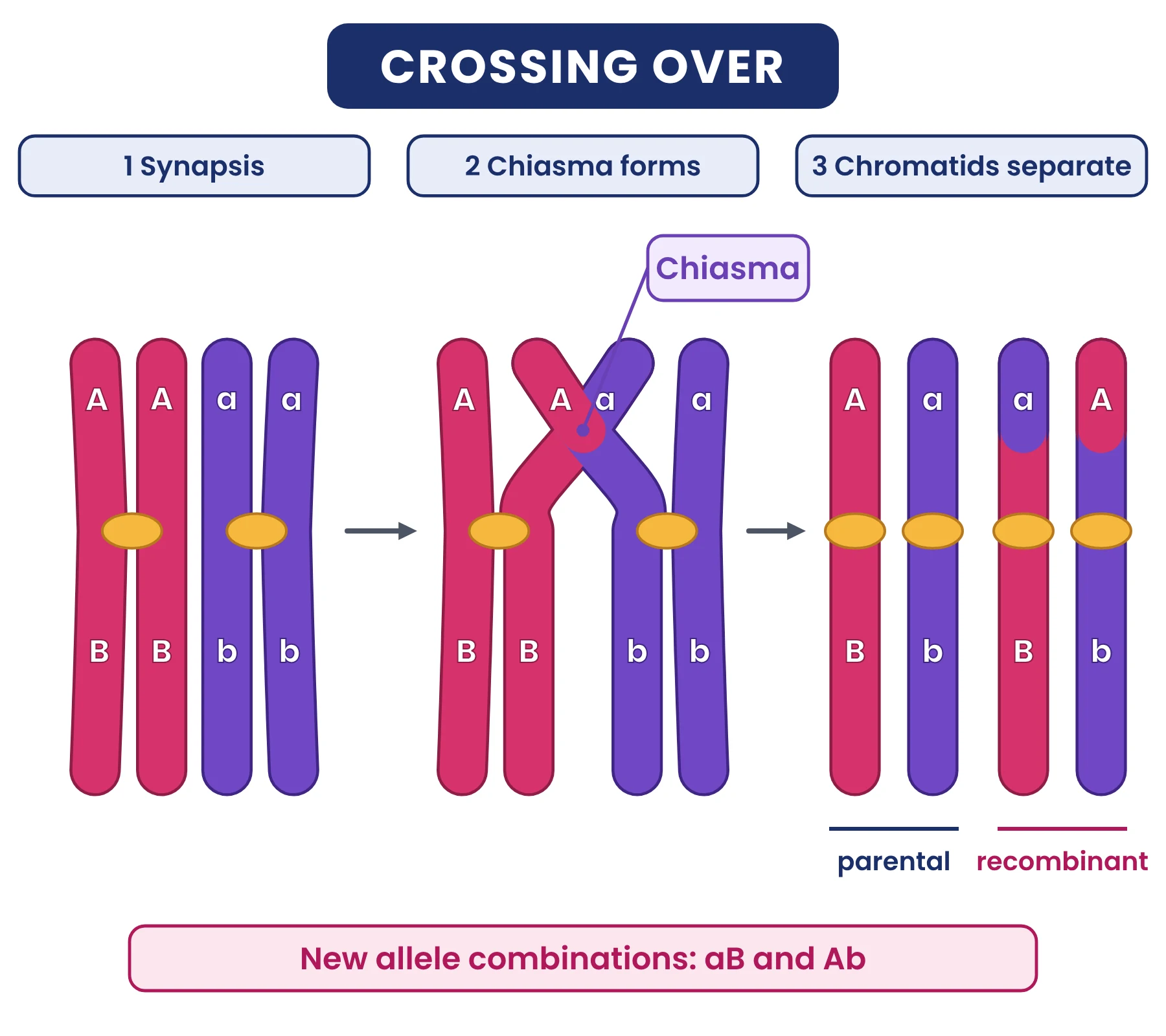
- Homologous chromosomes pair up in prophase I to form bivalents, and crossing over occurs between non-sister chromatids.
- Two rounds of segregation: homologous chromosomes separate in meiosis I; sister chromatids separate in meiosis II.
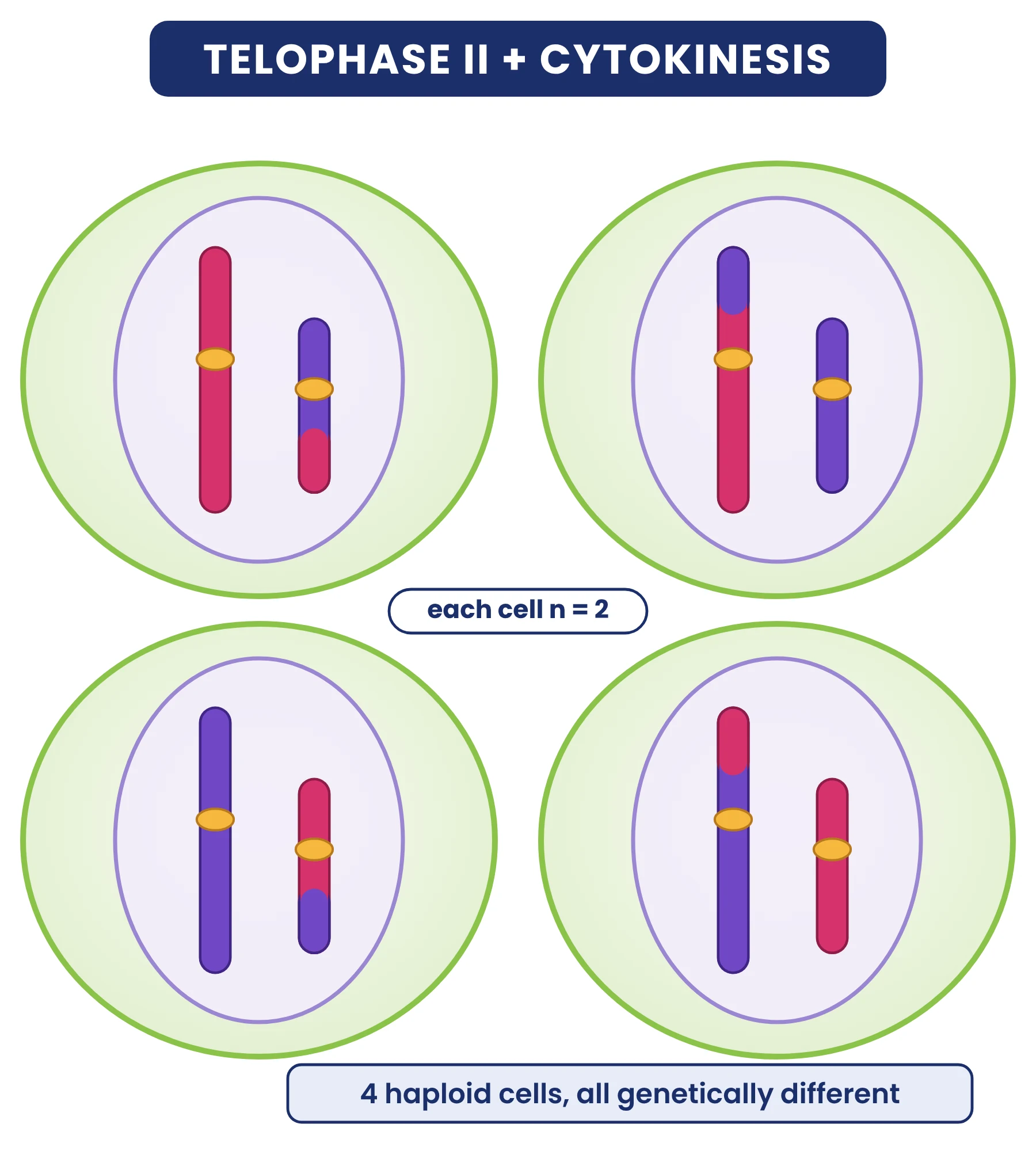
- The result is four haploid cells that are genetically different from each other and from the parent cell.
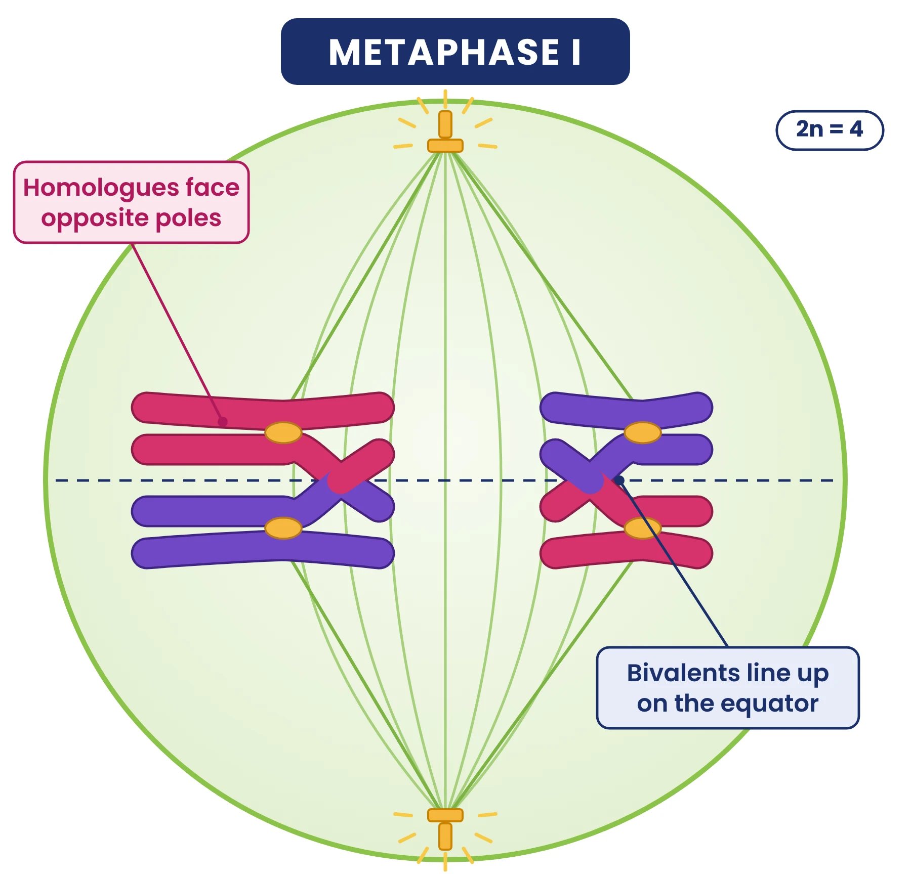
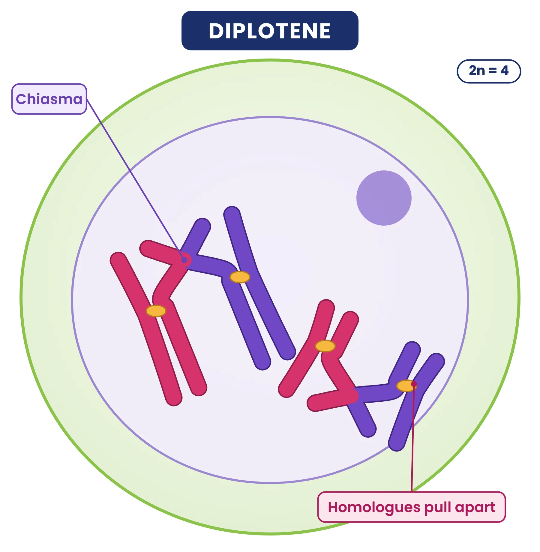
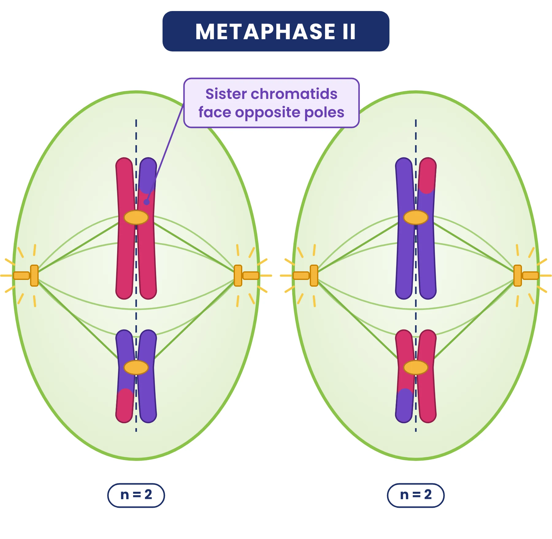
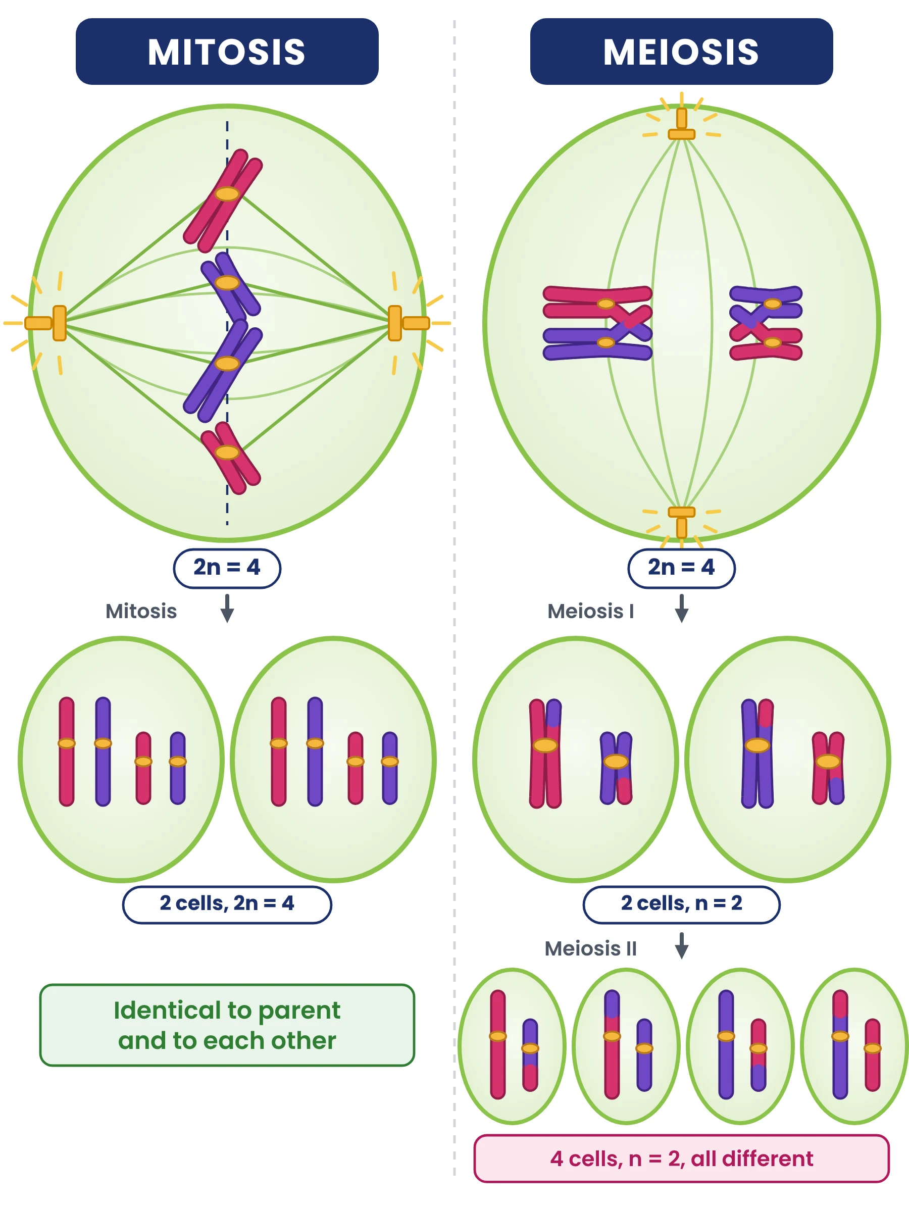
Homologous chromosomes are a matching pair, one from each parent (shown magenta for the maternal and purple for the paternal homologue in the diagrams below, in a cell with 2n = 4: one long pair and one short pair). They have the same genes in the same order, although the alleles of those genes may differ. A pair of homologous chromosomes held together in prophase I is a bivalent; because it has four chromatids it is also called a tetrad.
Meiosis I
Follow the two long and two short chromosomes through every diagram: each bivalent has one crossover, so after meiosis I one chromatid of each chromosome carries a tip of the other colour.
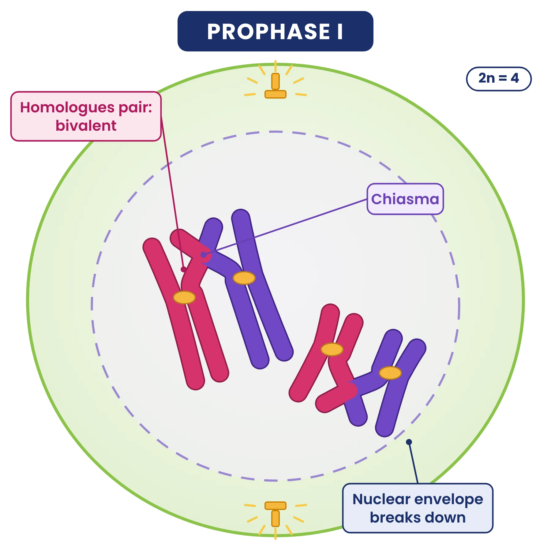
- Prophase I: chromosomes condense; homologues pair (synapsis) to form bivalents; crossing over occurs; the nuclear envelope and nucleolus disappear; the spindle forms. This is the longest stage of meiosis.
- Metaphase I: bivalents line up at the equator. Spindle fibres from one pole attach to one homologue and fibres from the other pole to its partner.
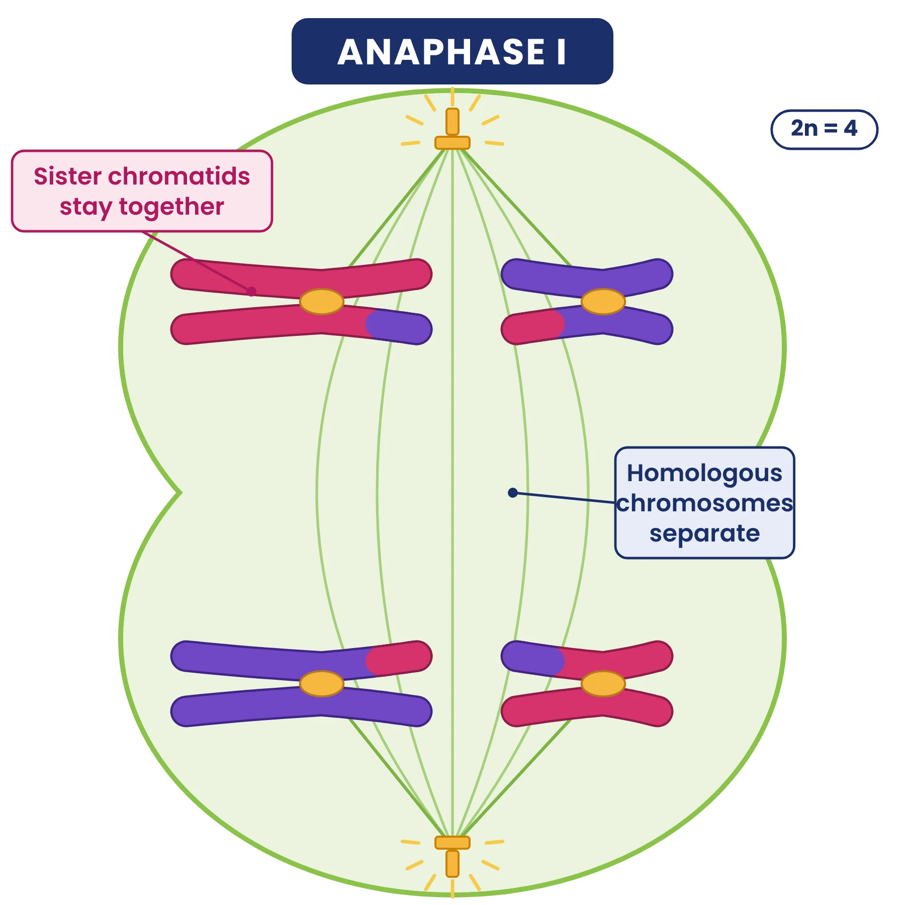
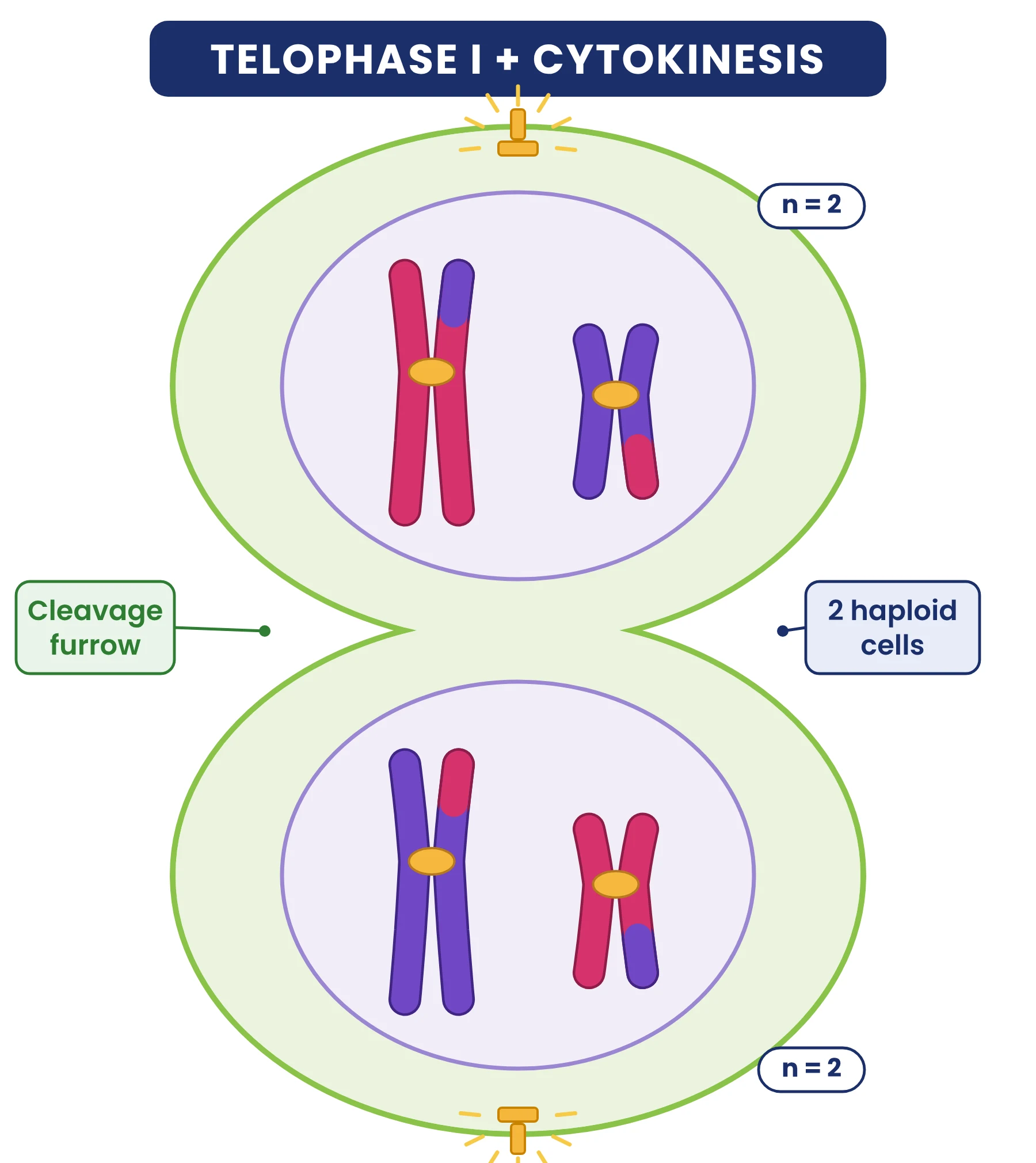
- Anaphase I: homologous chromosomes separate and move to opposite poles. The centromeres do not divide, so each chromosome still has two chromatids.
- Telophase I: the nuclear envelope and nucleolus may re-form; cytokinesis produces two cells (a dyad), each haploid.
NCERT The five substages of prophase I (NCERT)
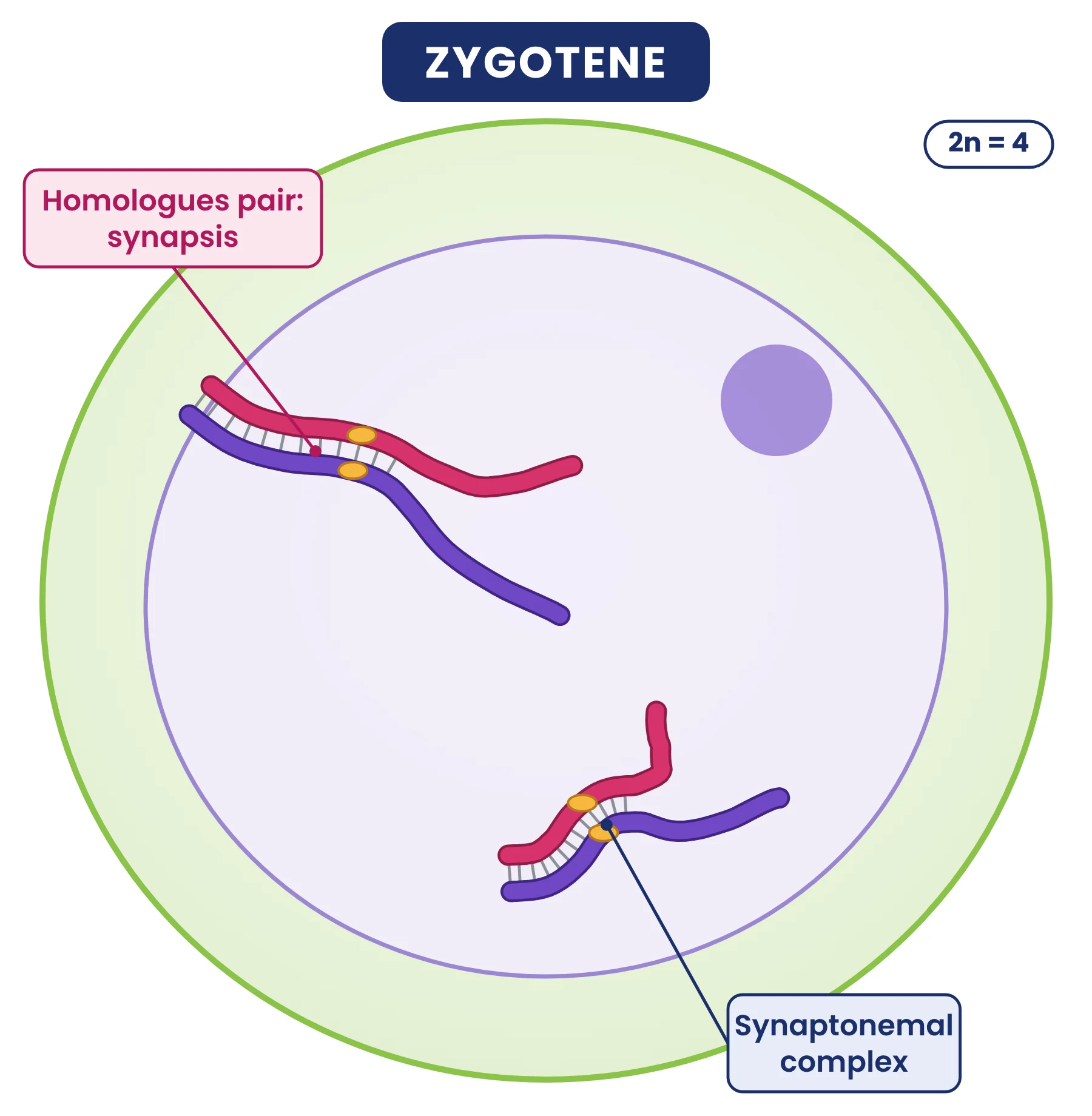
NCERT divides the long prophase I into five substages. These are not required by the IB guide but are tested often in NEET.
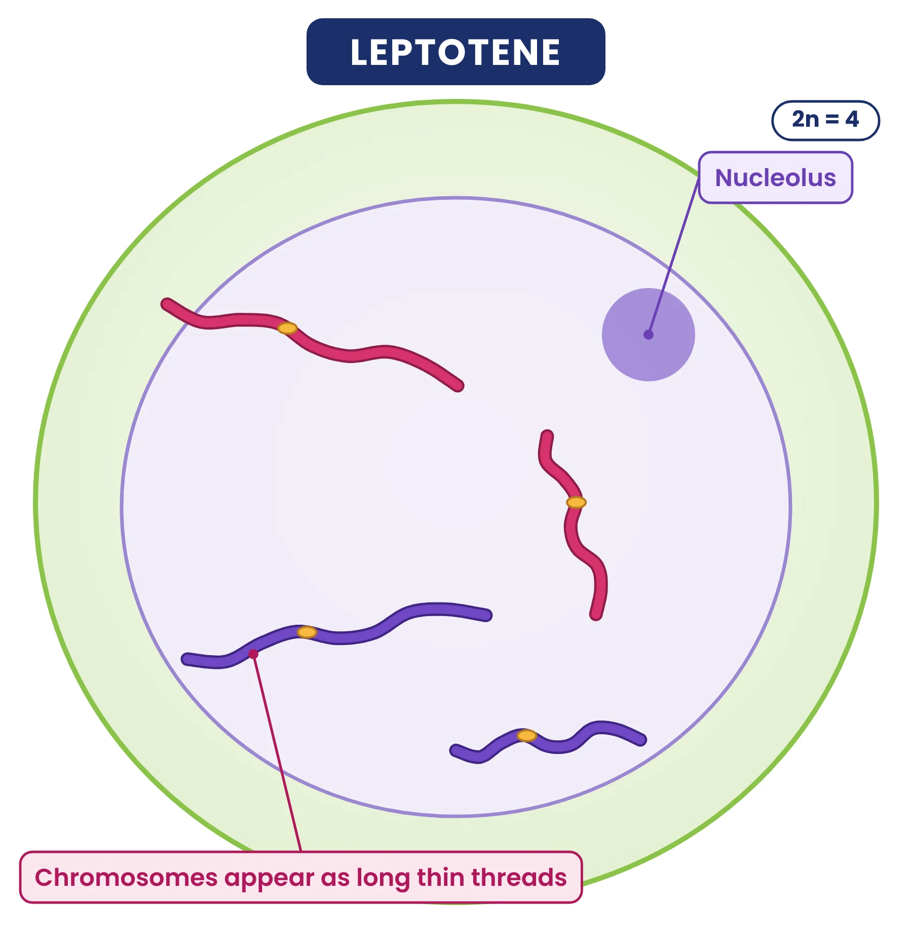
- Leptotene: chromosomes become gradually visible under the light microscope as long, thin threads; condensation continues.
- Zygotene: homologous chromosomes start pairing, a process called synapsis, helped by a protein framework called the synaptonemal complex. The paired homologues form a bivalent (tetrad).
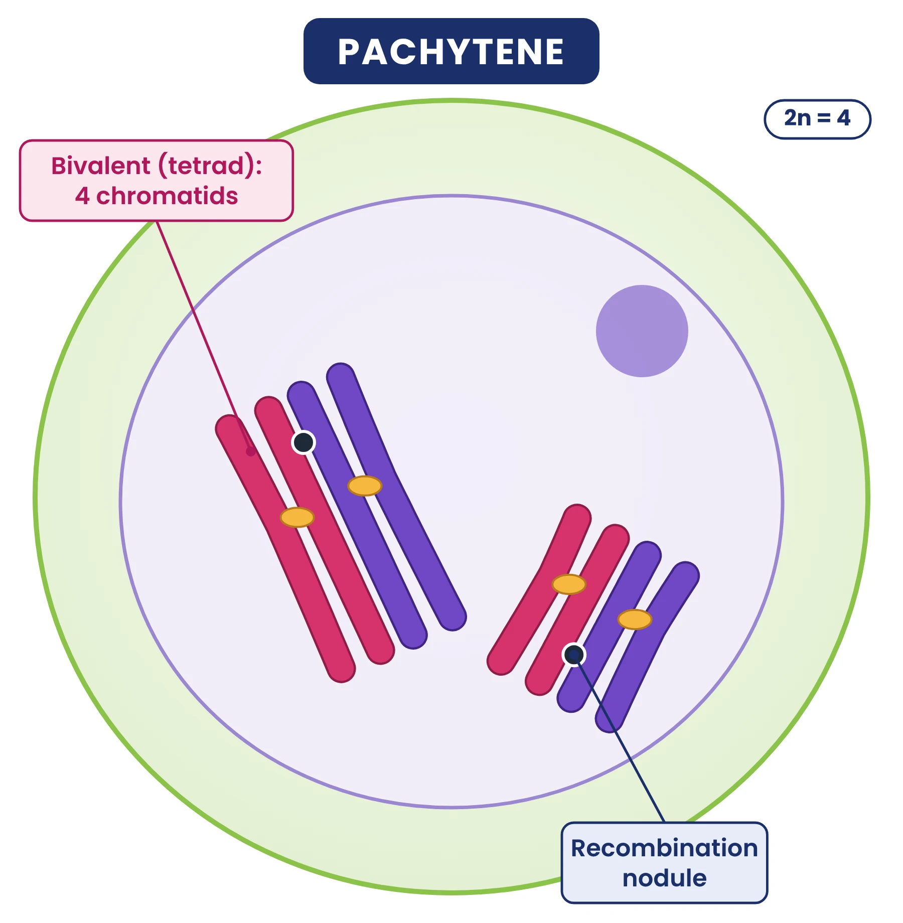
- Pachytene: the four chromatids of each bivalent become clear. Recombination nodules appear, the sites where crossing over takes place between non-sister chromatids, helped by the enzyme recombinase. Pachytene is longer than leptotene and zygotene.
- Diplotene: the synaptonemal complex dissolves and the homologues begin to separate, except at the sites of crossing over. These X-shaped points are the chiasmata. In the oocytes of some vertebrates diplotene can last for months or years.
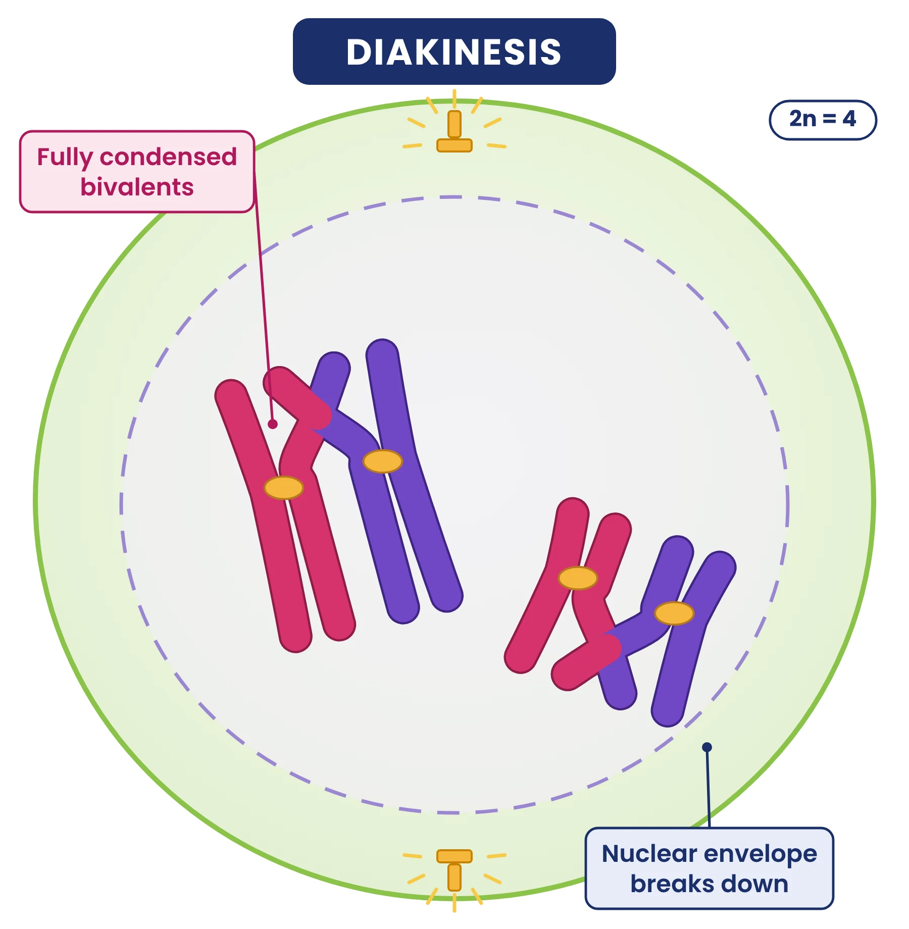
- Diakinesis: chiasmata move towards the ends of the chromosomes (terminalisation); chromosomes are fully condensed; the spindle is assembled; the nucleolus disappears and the nuclear envelope breaks down. This marks the move into metaphase I.
Interkinesis
Between the two divisions there may be a short pause called interkinesis. It is like a brief interphase, but there is no DNA replication, so each chromosome still has the two chromatids it had at the end of meiosis I.
Meiosis II
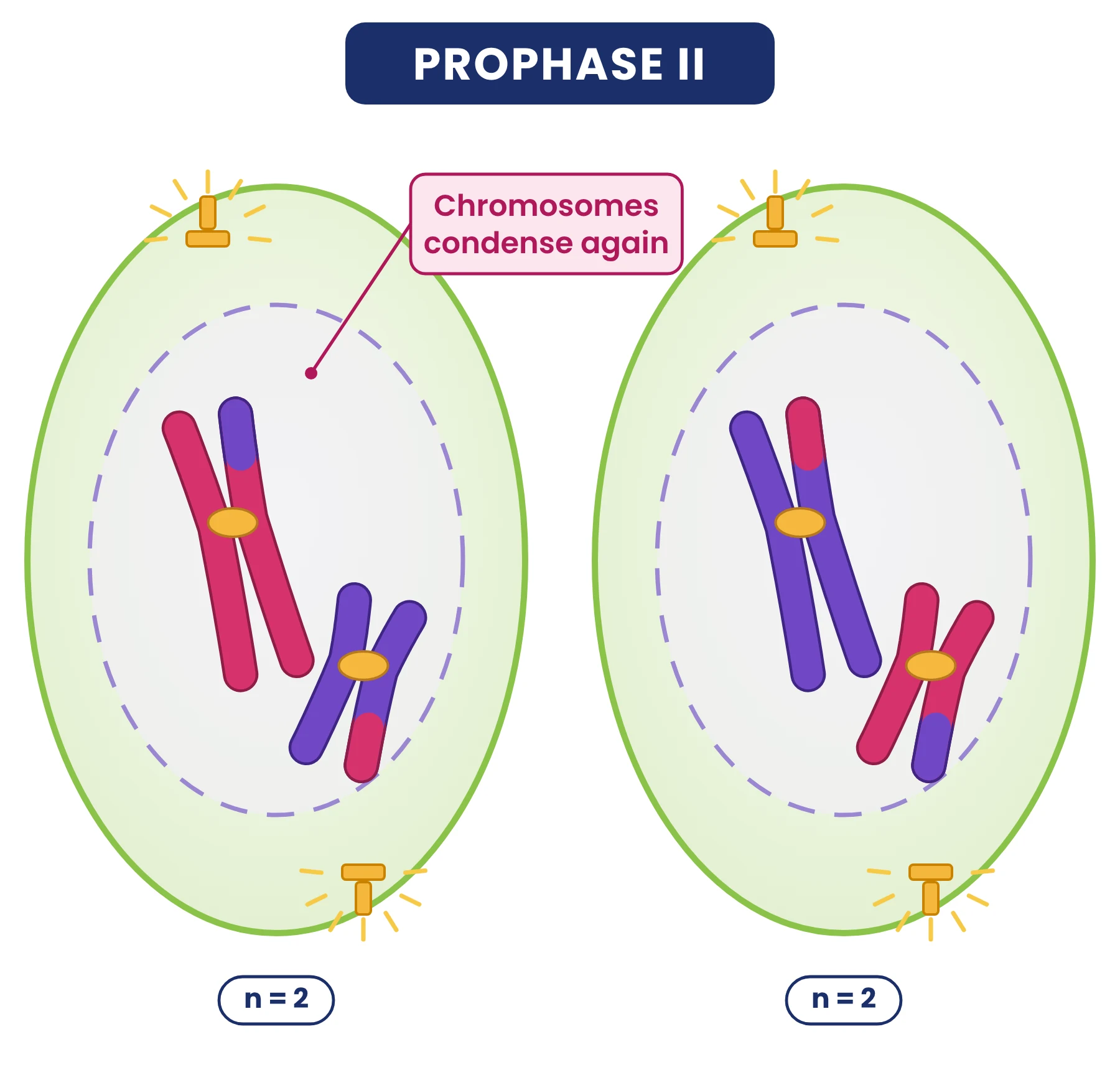
- Prophase II: starts soon after cytokinesis, often before the chromosomes have fully uncoiled. The nuclear envelope disappears and the chromosomes condense again.
- Metaphase II: chromosomes line up singly at the equator. Fibres from opposite poles attach to the kinetochores of the two sister chromatids.
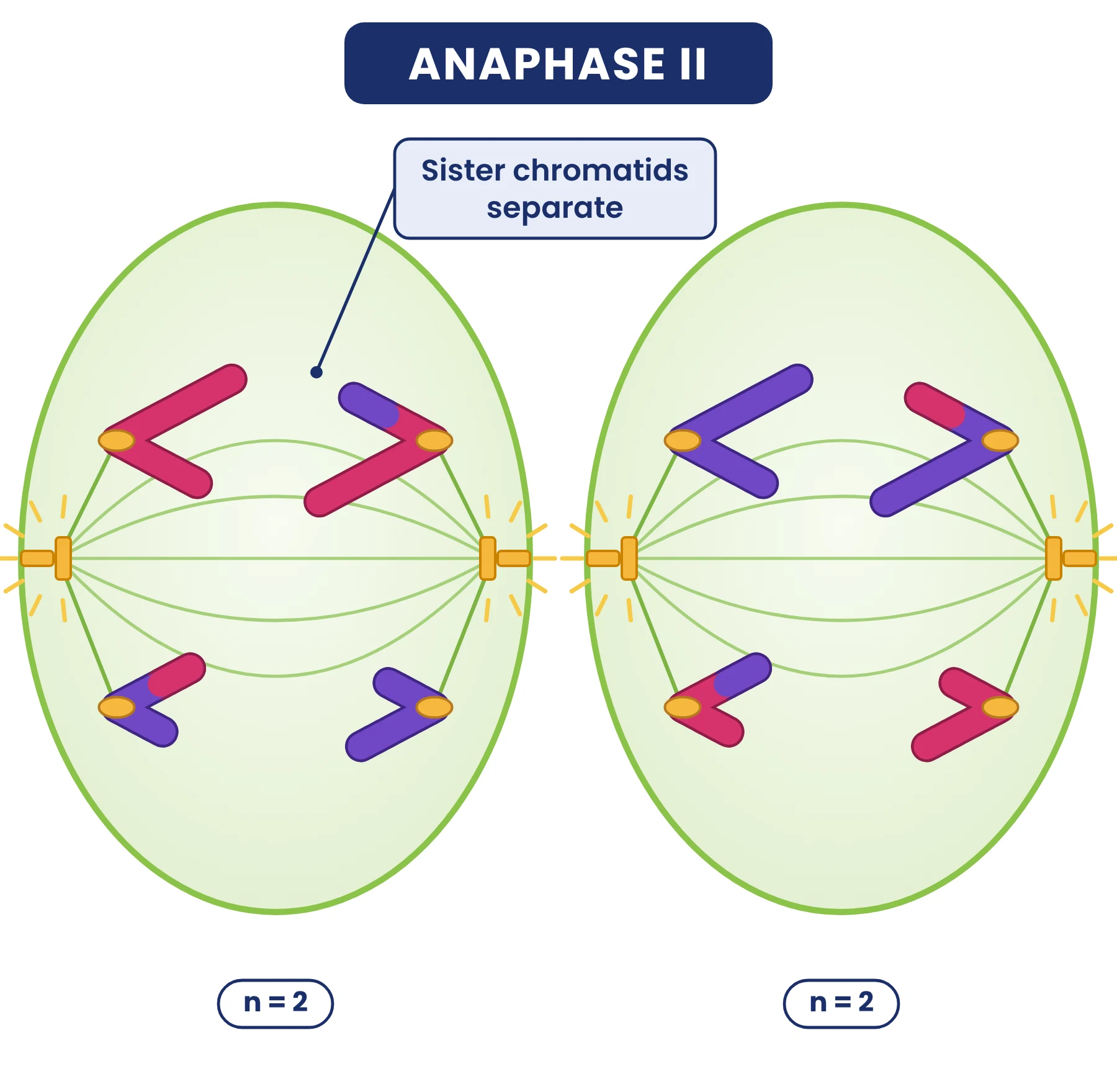
- Anaphase II: the centromere of each chromosome splits (cohesin is cut) and sister chromatids move to opposite poles.
- Telophase II: nuclear envelopes re-form around the four groups of chromosomes; cytokinesis produces four haploid cells (a tetrad of cells).
Meiosis II is very similar to mitosis, with one difference: it starts with a haploid cell. That is why meiosis II is sometimes called the equational division of meiosis.
Meiosis as a source of variation
The four cells produced by meiosis are genetically different from one another. IB requires two sources of this variation (D2.1.11), and random fertilisation adds a third.
1. Crossing over. In prophase I, non-sister chromatids of a bivalent break at the same points and exchange equivalent sections. Where they are joined after the exchange, a chiasma is visible. Each chromosome that has crossed over carries a new combination of alleles: a recombinant chromatid. Crossing over separates alleles of linked genes (genes on the same chromosome).
2. Random orientation of bivalents (independent assortment). In metaphase I, each bivalent lines up at the equator independently of every other bivalent. Whether the maternal or the paternal homologue faces a given pole is a matter of chance. With n pairs of chromosomes there are 2n possible combinations of chromosomes in the gametes.
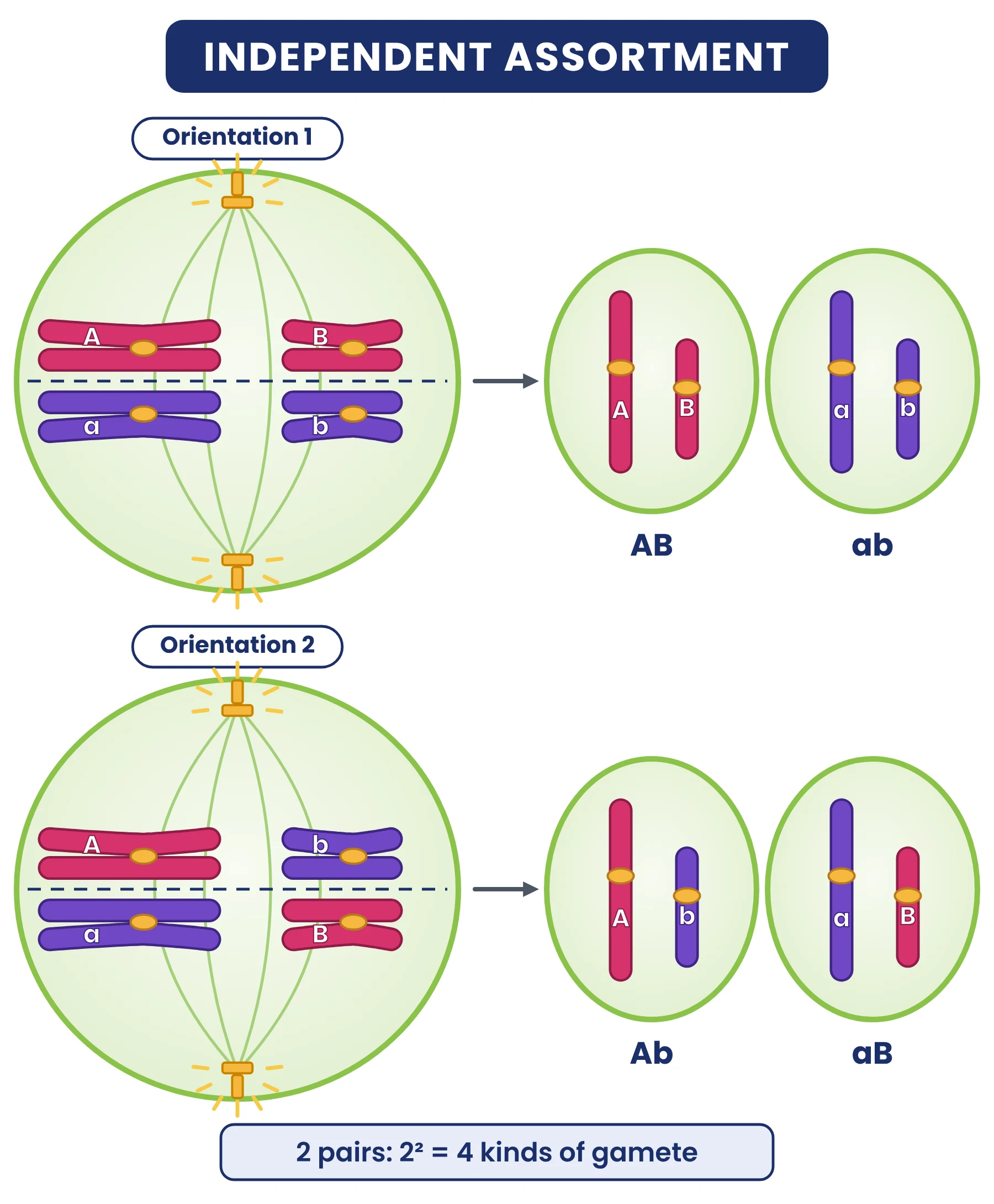
In humans n = 23, so random orientation alone gives 223 = 8 388 608 combinations in each person’s gametes. 3. Random fertilisation then combines any one of these with any one from the other parent: 223 × 223, about 70 trillion combinations, before crossing over is even counted.
Non-disjunction and Down syndrome
Non-disjunction is the failure of chromosomes to separate correctly in meiosis. It can happen in either division:
- In meiosis I, both homologues of a pair go to the same pole. All four gametes are abnormal: two have an extra copy (n + 1) and two lack that chromosome (n − 1).
- In meiosis II, the sister chromatids of one chromosome go to the same pole in one of the two cells. Two gametes are abnormal (n + 1 and n − 1) and two are normal (n).
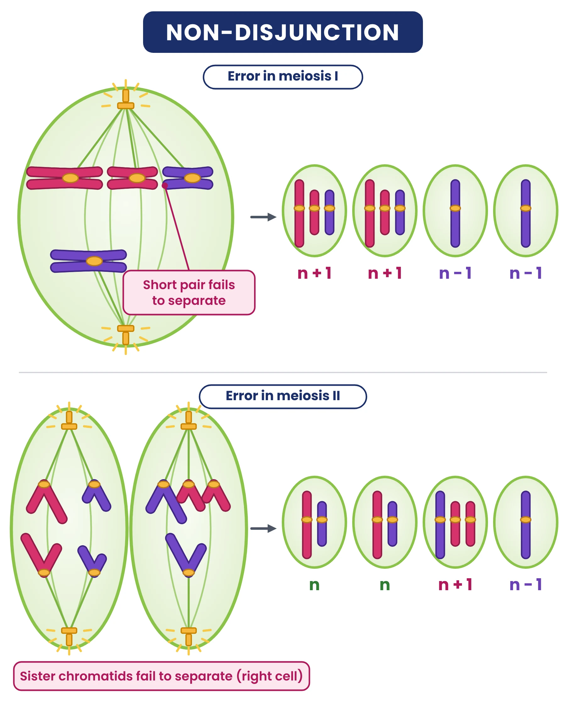
If a gamete with an extra chromosome 21 (24 chromosomes) is fertilised by a normal gamete (23), the zygote has 47 chromosomes, with three copies of chromosome 21. This trisomy 21 causes Down syndrome. Features can include learning difficulties, characteristic facial features, short stature and a higher risk of heart defects. The risk of non-disjunction rises with the age of the mother, especially after about 35, which is linked to the long time oocytes spend arrested in prophase I.
A gamete missing a chromosome gives a monosomy (45 chromosomes) after fertilisation. Monosomy of an autosome is not viable; the only viable human monosomy is 45,X (Turner syndrome).
Significance of meiosis
- Keeps the chromosome number of a species constant from generation to generation in sexually reproducing organisms, by halving it before fertilisation doubles it.
- Produces genetic variation through crossing over, random orientation of bivalents and (with fertilisation) random combination of gametes.
- Variation is the raw material for natural selection and evolution.
- Produces gametes in animals and spores in plants.
Key points
- Meiosis: one DNA replication, two divisions, four haploid, genetically different cells.
- Meiosis I separates homologous chromosomes (reduction); meiosis II separates sister chromatids.
- Prophase I (NCERT): leptotene, zygotene, pachytene, diplotene, diakinesis.
- Variation: crossing over at chiasmata and random orientation of bivalents (2n combinations), plus random fertilisation.
- Non-disjunction gives n + 1 and n − 1 gametes; trisomy 21 causes Down syndrome.
Common mistakes
- Saying sister chromatids separate in anaphase I. In anaphase I homologous chromosomes separate; sister chromatids separate in anaphase II.
- Saying DNA replicates again before meiosis II. Interkinesis has no S phase.
- Saying crossing over happens between sister chromatids. It happens between non-sister chromatids of homologous chromosomes.
- Calling meiosis II a reduction division. The reduction in chromosome number happens in meiosis I.
Exam tip
- IB: ‘outline the two rounds of segregation’ means: homologues separate in anaphase I, sister chromatids in anaphase II.
- Diagram questions: draw chromosomes with two chromatids until anaphase II, and keep the number of chromosomes consistent (2n = 4 gives n = 2 after meiosis I).
- NEET: learn the event that marks each prophase I substage (synapsis in zygotene, crossing over in pachytene, chiasmata in diplotene, terminalisation in diakinesis).
Test yourself (click a question to reveal the answer)
A cell with 2n = 12 undergoes meiosis. How many chromosomes and chromatids are in each cell at the end of telophase I?
6 chromosomes and 12 chromatids, because homologues have separated but each chromosome still has two sister chromatids.
How many different combinations of chromosomes can a pea plant (2n = 14) produce in its gametes by random orientation alone?
n = 7, so 27 = 128 combinations.
In which substage of prophase I does the synaptonemal complex dissolve?
Diplotene.
Why does non-disjunction in meiosis I produce more abnormal gametes than non-disjunction in meiosis II?
In meiosis I the error affects both daughter cells, so all four gametes are abnormal; in meiosis II only one of the two cells is affected, so two of the four gametes are normal.
5 Cell cycle regulation: cyclins, CDKs and checkpoints
Syllabus link: AHL D2.1.15 Control of the cell cycle using cyclins; links to D2.1.16 (cancer). NCERT does not cover cyclins in detail.
AHL The cell cycle must be controlled. A cell should divide only when it is large enough, when its DNA has been copied correctly and when the body needs more cells. This control is carried out by proteins whose amounts rise and fall during each cycle, and by checkpoints where the cell checks that it is ready to move on.
Cyclins and cyclin-dependent kinases
- Cyclins are a family of regulatory proteins whose concentration rises and falls during the cell cycle. Different cyclins peak at different stages.
- Cyclin-dependent kinases (CDKs) are enzymes that add phosphate groups to target proteins (phosphorylation), switching them on or off. Their concentration stays roughly constant.
- A CDK is inactive on its own. When the right cyclin reaches a threshold concentration, it binds to the CDK and activates it.
- The active cyclin–CDK complex phosphorylates proteins that carry the cell past a checkpoint, for example proteins needed for DNA replication or for nuclear envelope breakdown and spindle formation.
- The cyclin is then broken down, so the CDK becomes inactive again and the next cyclin takes over. This keeps the cycle moving in one direction.
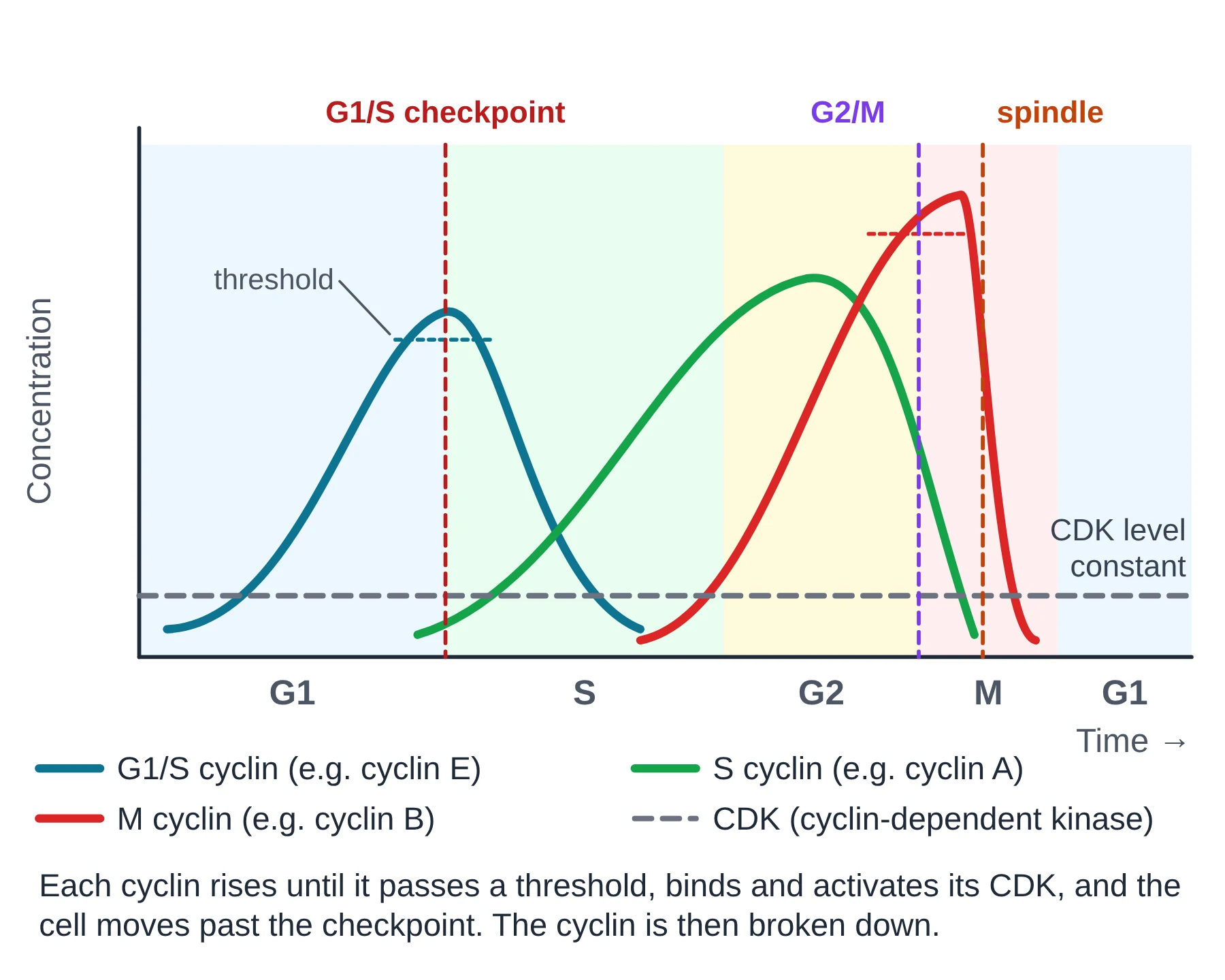
For example, the M cyclin (cyclin B) builds up through S and G2. When it passes its threshold, cyclin B–CDK1 (historically called MPF, maturation- or mitosis-promoting factor) triggers condensation of chromosomes, breakdown of the nuclear envelope and assembly of the spindle. At the metaphase to anaphase transition APC/C tags cyclin B for destruction, the CDK switches off and the cell can complete mitosis and cytokinesis.
Checkpoints
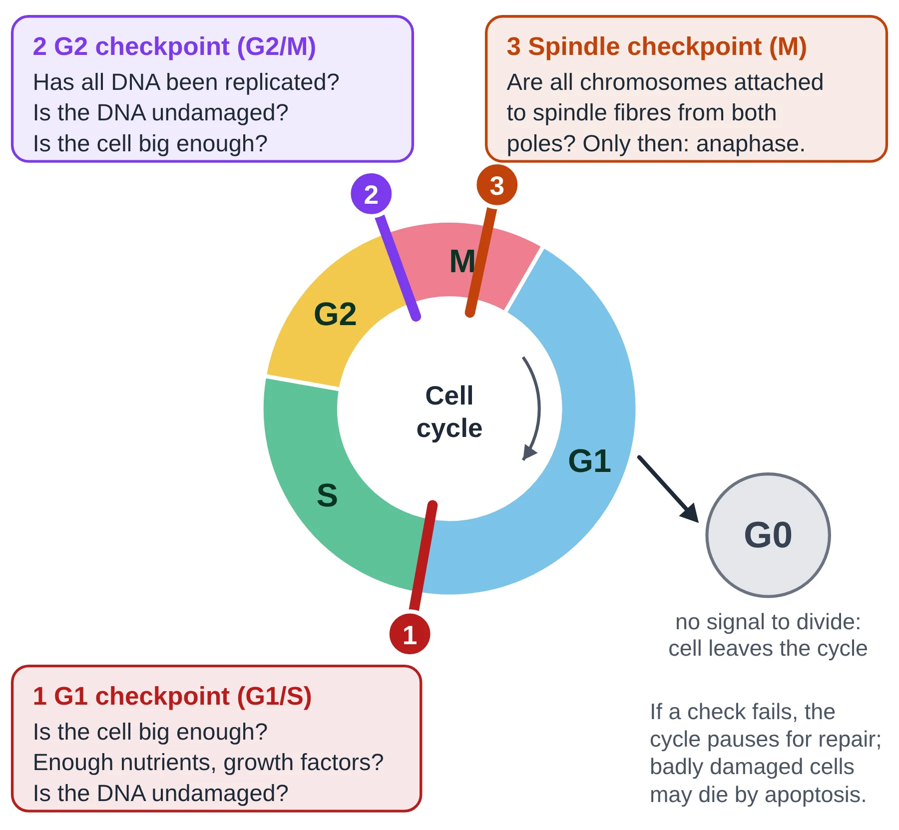
- G1 checkpoint (G1/S, also called the restriction point): is the cell large enough, are nutrients and growth factors present, and is the DNA undamaged? If not, the cell pauses or enters G0.
- G2 checkpoint (G2/M): has all the DNA been replicated, and has any damage been repaired? Only then can the cell enter mitosis.
- Spindle checkpoint (metaphase or M checkpoint): are all chromosomes attached to spindle fibres from both poles? Anaphase is delayed until they are, which protects against non-disjunction.
If damage cannot be repaired, the cell may be destroyed by apoptosis (programmed cell death). The protein p53, sometimes called the ‘guardian of the genome’, detects DNA damage and stops the cycle at G1 so that the DNA can be repaired, or triggers apoptosis if it cannot.
Signals from outside the cell
Cells also respond to their surroundings. Growth factors released by other cells (for example after a wound) stimulate cells to make cyclins and pass the G1 checkpoint. Normal cells also show contact inhibition: when they touch neighbouring cells on all sides, they stop dividing. Cancer cells have lost this property (section 7).
Key points
- Cyclins rise and fall during the cycle; CDK levels stay constant.
- A specific cyclin must reach a threshold to activate its CDK and pass each checkpoint; the cyclin is then broken down.
- Checkpoints: G1/S (size, nutrients, growth factors, DNA damage), G2/M (DNA fully replicated and undamaged), spindle (all chromosomes attached).
- p53 stops the cycle for DNA repair or triggers apoptosis.
Common mistakes
- Saying cyclins are enzymes. Cyclins are regulatory proteins; the kinases (CDKs) are the enzymes.
- Saying CDK concentration rises and falls. It is the cyclin concentration that changes; CDK activity changes because cyclin binds to it.
- Saying a checkpoint is a phase of the cycle. It is a control point at the boundary between phases (or within M phase).
Exam tip
- IB says students are not required to know the roles of specific cyclins. Learn the principle: different cyclins, rising and falling concentration, a threshold of a specific cyclin to pass each checkpoint.
- Graph questions: identify which cyclin peaks just before which phase, and link the drop in cyclin to the cell moving into the next phase.
Test yourself (click a question to reveal the answer)
Explain why CDKs are active only at certain times in the cell cycle, even though their concentration is constant.
A CDK is only active when bound to its cyclin. Cyclin concentration rises and falls, so CDK activity rises only when the cyclin passes its threshold, and falls when the cyclin is broken down.
What would happen to a cell if M cyclin could not be broken down?
The CDK would stay active, so the cell could not exit mitosis; it would stay arrested in M phase (around metaphase) and not complete division.
6 Mitosis vs meiosis: comparing the two kinds of cell division
Syllabus link: D2.1.4 Roles of mitosis and meiosis in eukaryotes; D2.1.9 Meiosis as a reduction division; NCERT 10.3, 10.5
Mitosis and meiosis both divide a nucleus, both use a spindle of microtubules and both follow one round of DNA replication. They differ in what happens to homologous chromosomes, in the number of divisions, and in the number and kind of cells produced.
| Feature | Mitosis | Meiosis |
|---|---|---|
| Number of divisions | One | Two (meiosis I and meiosis II) |
| DNA replication | Once, before division | Once, before meiosis I only |
| Daughter cells | Two | Four |
| Chromosome number | Kept the same (2n → 2n, or n → n) | Halved (2n → n) |
| Genetic make-up of daughter cells | Identical to each other and to the parent | Different from each other and from the parent |
| Pairing of homologues (synapsis) | Does not occur | Occurs in prophase I, forming bivalents |
| Crossing over and chiasmata | Do not occur | Occur in prophase I |
| Metaphase alignment | Individual chromosomes line up at the plate | Bivalents line up in metaphase I; single chromosomes in metaphase II |
| What separates at anaphase | Sister chromatids | Homologous chromosomes (anaphase I), then sister chromatids (anaphase II) |
| Duration of prophase | Relatively short | Prophase I is long, with five substages (NCERT) |
| Where it happens | Somatic cells: growth, repair, replacement; asexual reproduction | Germ cells in gonads (animals); spore mother cells (plants) |
| Function | Growth, repair, asexual reproduction, genetic stability | Gametes or spores for sexual reproduction; genetic variation |
Key points
- Mitosis: one division, two identical cells, chromosome number unchanged.
- Meiosis: two divisions, four different haploid cells, chromosome number halved.
- Only meiosis has synapsis, bivalents, crossing over and separation of homologues.
- Both start after a single S phase; DNA content falls from 4C to 2C in mitosis and from 4C to 1C over meiosis.
Common mistakes
- Saying mitosis only happens in diploid cells. It also happens in haploid cells (plant gametophytes, male honey bees).
- Saying meiosis makes ‘two cells with half the DNA’. Meiosis I gives two haploid cells, but each chromosome still has two chromatids; meiosis II gives four cells.
Test yourself (click a question to reveal the answer)
State two ways in which meiosis I differs from mitosis.
Any two: homologous chromosomes pair to form bivalents; crossing over occurs; bivalents (not single chromosomes) line up at the equator; homologues (not sister chromatids) separate at anaphase; the chromosome number is halved.
A cell has 10 pg of DNA in G1. How much DNA is in each cell after meiosis II?
5 pg. DNA doubles to 20 pg in S phase, falls to 10 pg in each cell after meiosis I and to 5 pg after meiosis II, which is half the G1 value.
7 Cancer: when the cell cycle goes out of control
Syllabus link: AHL D2.1.16 Consequences of mutations in genes that control the cell cycle; AHL D2.1.17 Differences between tumours (including the mitotic index); NCERT 10.3 (link)
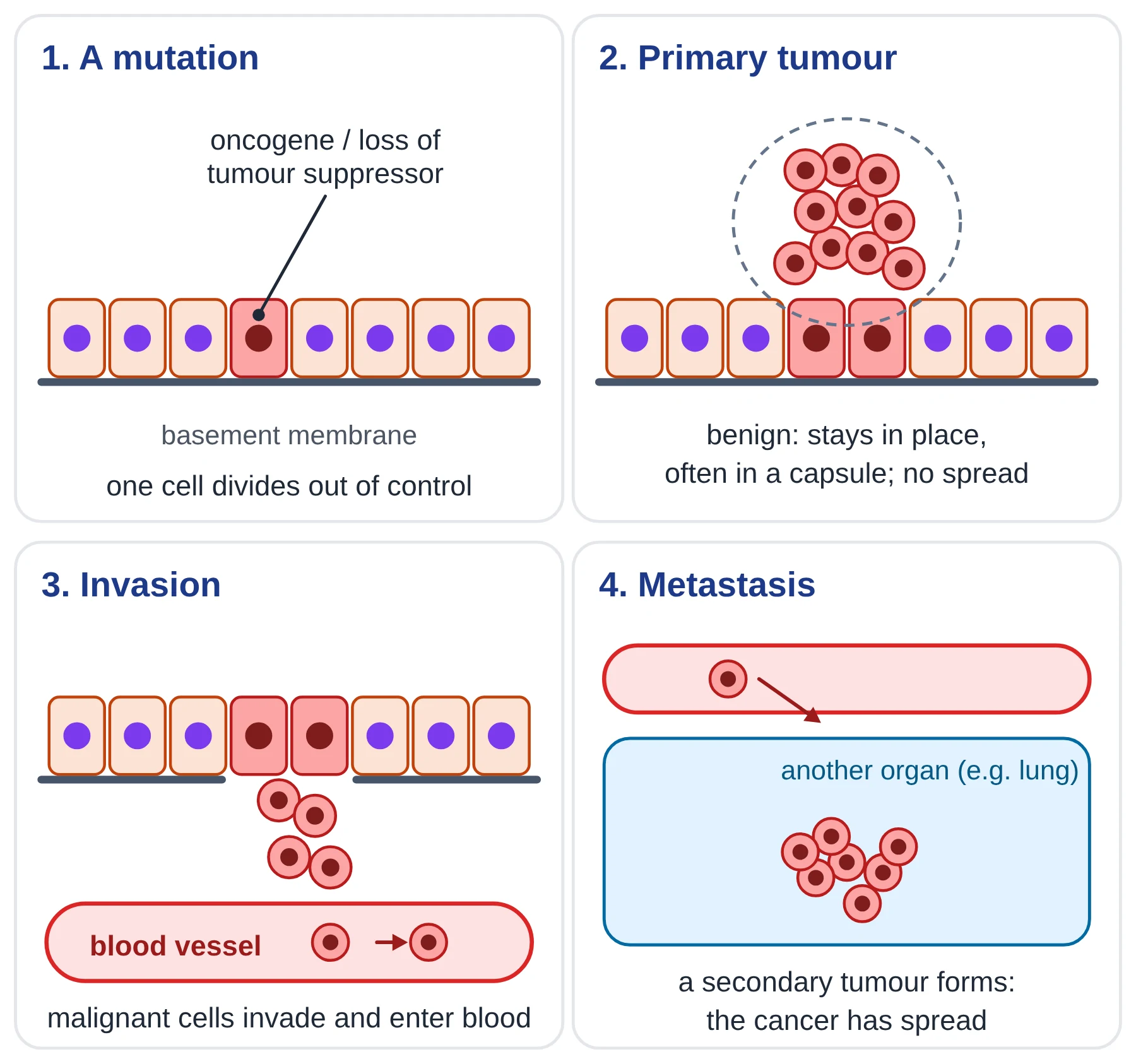
In a healthy body, cells divide only when they need to. Cancer is a group of diseases in which cells divide uncontrollably because the genes that control the cell cycle have been damaged. The mass of cells formed by this uncontrolled cell division is a tumour.
Oncogenes and tumour suppressor genes
AHL Two groups of genes control the cell cycle, and mutations in either group can lead to cancer:
- Proto-oncogenes code for proteins that stimulate cell division, such as growth factors, their receptors and cyclins. A mutation can turn a proto-oncogene into an oncogene, which is permanently ‘switched on’, so the cell divides even without a signal. Only one mutated copy is needed (the mutation acts like an accelerator stuck down).
- Tumour suppressor genes code for proteins that stop cell division or trigger apoptosis when something is wrong, such as p53 and the retinoblastoma protein (Rb). If both copies are inactivated by mutation, the brake is lost and damaged cells keep dividing.
Cancer usually needs several mutations in the same cell, in both kinds of gene. Because mutations build up over time, the risk of most cancers rises with age.
Mutagens and carcinogens
A mutagen is an agent that increases the rate of mutation. A mutagen that causes cancer is a carcinogen. Examples:
- Chemical: tar and other chemicals in tobacco smoke, asbestos fibres, benzene, some food contaminants such as aflatoxins.
- Radiation: ultraviolet (UV) light from the Sun (skin cancer), X-rays and gamma rays, radiation from radioactive materials such as radon gas.
- Biological: some viruses, such as human papillomavirus (HPV, cervical cancer) and hepatitis B virus (liver cancer).
Mutations can also arise spontaneously, through errors in DNA replication, and some mutations are inherited (for example in the BRCA1 and BRCA2 tumour suppressor genes).
Benign and malignant tumours
| Feature | Benign tumour | Malignant tumour (cancer) |
|---|---|---|
| Rate of division and growth | Usually slow | Usually fast |
| Boundary | Often enclosed in a capsule; clear boundary | No capsule; irregular boundary |
| Invasion of neighbouring tissue | No | Yes |
| Metastasis (spread) | No | Yes, through blood or lymph |
| Cell appearance | Similar to normal, differentiated cells | Abnormal, poorly differentiated cells with large nuclei |
| Danger | Usually not life-threatening, unless it presses on a vital organ (e.g. in the brain) | Life-threatening if not treated |
- The tumour where the cancer starts is the primary tumour.
- Metastasis is the spread of cancer cells from the primary tumour to other parts of the body, through the blood or lymphatic system.
- A tumour formed at a new site by cells that have spread is a secondary tumour. It is made of the same type of cell as the primary tumour: breast cancer that spreads to the lung is still breast cancer.
Normal cells show contact inhibition: they stop dividing when they touch their neighbours. Cancer cells have lost contact inhibition, so they pile up into a mass, and many also lose the ability to stick to each other, which lets them break away and spread.
The mitotic index
The mitotic index is the proportion of cells in a tissue that are in mitosis at a given moment. It is a measure of how fast cells in the tissue are dividing:
Mitotic index = number of cells in mitosis ÷ total number of cells observed
Cells in prophase, metaphase, anaphase and telophase are counted as ‘in mitosis’; cells in interphase are not. The result can be given as a decimal or as a percentage. A high mitotic index shows rapid cell division. Doctors use it to grade tumours: a malignant tumour usually has a much higher mitotic index than the normal tissue around it, and a falling mitotic index during treatment suggests the treatment is working. Many anti-cancer drugs and radiotherapy target dividing cells for this reason.
Worked example: calculating a mitotic index
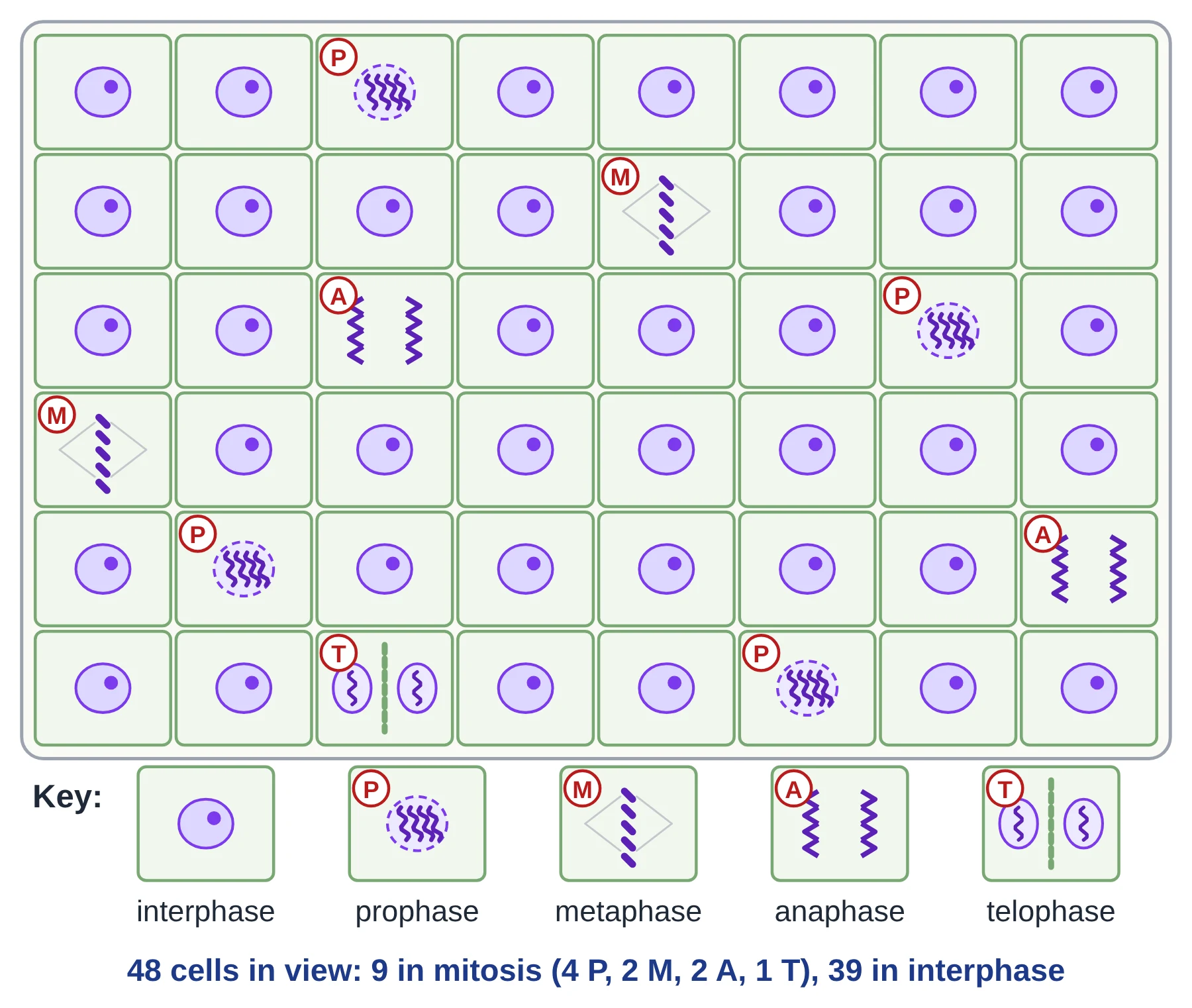
- Count the cells in mitosis. In the field of view above: prophase 4 + metaphase 2 + anaphase 2 + telophase 1 = 9 cells.
- Count all the cells. 9 in mitosis + 39 in interphase = 48 cells.
- Divide. Mitotic index = 9 ÷ 48 = 0.1875.
- Round sensibly. Mitotic index = 0.19 (2 significant figures), or 18.75% ≈ 19%.
- Interpret. About one cell in five is dividing: this is a region of rapid growth (the meristem near the root tip).
Estimating time in each phase. If the cells are dividing at random, the proportion of cells in a phase is the same as the proportion of the cell cycle spent in that phase. If this cell cycle takes 24 hours, time in mitosis ≈ 0.1875 × 24 h = 4.5 hours, and time in prophase ≈ (4 ÷ 48) × 24 h = 2 hours.
Comparing tissues. In a biopsy, 1000 cells from a tumour included 120 in mitosis, while 1000 cells from nearby normal tissue included 15. Mitotic index of tumour = 120 ÷ 1000 = 0.12; normal tissue = 15 ÷ 1000 = 0.015. The tumour cells are dividing about 8 times as often.
NCERT Practical note (NCERT and IB)
The standard practical is a squash of onion (Allium cepa) or garlic root tips. Root tips are softened in warm dilute hydrochloric acid (this hydrolyses the middle lamella so the cells separate), stained with a DNA stain such as toluidine blue, acetocarmine or acetic orcein, then squashed gently under a cover slip so the cells form a single layer. Count at least several hundred cells over a few fields of view, and count each cell only once, for a reliable mitotic index.
Key points
- Cancer is uncontrolled cell division caused by mutations in genes that control the cell cycle.
- Oncogenes (from mutated proto-oncogenes) stimulate division; tumour suppressor genes such as p53 normally stop it.
- Mutagens that cause cancer are carcinogens: chemicals, radiation and some viruses.
- Benign tumours stay in place; malignant tumours invade tissue and metastasise to form secondary tumours.
- Mitotic index = cells in mitosis ÷ total cells; tumours have a high mitotic index.
Common mistakes
- Saying any tumour is cancer. Only malignant tumours are cancers.
- Leaving out telophase or prophase cells when counting cells in mitosis. All four stages count.
- Dividing cells in mitosis by cells in interphase. The denominator is the total number of cells.
- Saying that a secondary tumour in the lung is lung cancer. It is the original cancer type that has spread.
Exam tip
- ‘Explain’ questions on cancer: link mutation → loss of cell cycle control (oncogene or tumour suppressor) → uncontrolled division → tumour → metastasis.
- Show working in mitotic index calculations and give the answer to the number of significant figures used in the data.
Test yourself (click a question to reveal the answer)
In a sample of 250 cells, 30 were in mitosis. Calculate the mitotic index.
30 ÷ 250 = 0.12 (or 12%).
Why is a single mutation in a tumour suppressor gene usually not enough to cause loss of control?
Cells have two copies of each gene; the remaining normal copy can still make enough functional protein (such as p53) to stop the cycle. Both copies usually have to be inactivated.
Explain why radiotherapy has side effects in tissues such as hair follicles and the gut lining.
Radiotherapy damages rapidly dividing cells most. Hair follicles and the gut lining also have a high mitotic index, so they are damaged as well as the tumour.
9 Quick check
Ten questions: six multiple choice and four short answer. Try them all before opening the answers.
-
During which phase of the cell cycle is DNA replicated?
- A. G1
- B. S
- C. G2
- D. Prophase
-
A cell in G1 has 2C DNA and 2n = 14. What are the DNA content and chromosome number of the same cell in G2?
- A. 2C, 14
- B. 4C, 14
- C. 4C, 28
- D. 2C, 28
-
Which event occurs in anaphase I of meiosis but not in anaphase of mitosis?
- A. Sister chromatids separate
- B. Homologous chromosomes separate
- C. Centromeres split
- D. The nuclear envelope re-forms
-
In which substage of prophase I does crossing over take place?
- A. Leptotene
- B. Zygotene
- C. Pachytene
- D. Diakinesis
-
Which statement about cyclins is correct?
- A. Cyclins are enzymes that phosphorylate proteins
- B. Cyclin concentration stays constant through the cycle
- C. Cyclins activate cyclin-dependent kinases
- D. Cyclins are only found in cancer cells
-
Cytokinesis in a plant cell involves:
- A. a contractile ring of actin and myosin
- B. a cleavage furrow growing inwards
- C. Golgi vesicles fusing to form a cell plate
- D. the formation of asters
-
State two differences between benign and malignant tumours.
-
Explain how non-disjunction can lead to Down syndrome.
-
A root tip field of view has 400 cells, of which 36 are in mitosis. Calculate the mitotic index and state what a higher value in a tumour would show.
-
Outline two ways in which meiosis generates genetic variation.
Show answers
- B. S (synthesis) phase.
- B. 4C, 14: DNA doubles in S phase, but each chromosome now has two chromatids, so the chromosome number is unchanged.
- B. Homologous chromosomes separate.
- C. Pachytene (recombination nodules).
- C. Cyclins bind to and activate CDKs; their concentration rises and falls.
- C. Golgi vesicles fusing to form a cell plate.
- Any two: malignant tumours invade neighbouring tissue; they metastasise to form secondary tumours; they usually grow faster; they have no capsule; benign tumours stay in one place.
- Chromosome 21 homologues (meiosis I) or chromatids (meiosis II) fail to separate, so a gamete has two copies of chromosome 21 (n + 1 = 24). Fertilisation by a normal gamete gives a zygote with three copies (trisomy 21, 47 chromosomes).
- 36 ÷ 400 = 0.09 (9%). A higher mitotic index shows that cells are dividing more rapidly than in normal tissue.
- Crossing over between non-sister chromatids in prophase I produces recombinant chromatids; random orientation of bivalents in metaphase I gives 2n combinations of maternal and paternal chromosomes.
10 Glossary
© competitorspoint.com · IB Biology notes. Diagrams and text are original to competitorspoint.com.
Ready to practise? Download the Cell Cycle and Cell Division practice questions with answers (free for signed-up members).
⬇ Download practice questions (PDF)