
The Basic Concept of Biotechnology
The principles of biotechnology rest on two core techniques, genetic engineering and bioprocess engineering, and on a small set of tools: restriction enzymes, cloning vectors and a competent host. This page explains the meaning of biotechnology, the first recombinant DNA made by Cohen and Boyer, how restriction enzymes are named and how they cut DNA, the features of a good vector such as pBR322, and how a host cell is made competent to take up DNA.
1 Meaning of biotechnology
NCERT Class XII Biology, Chapter 9 Biotechnology: Principles and Processes, introduction, section 9.1 Principles of Biotechnology and section 9.2 Tools of Recombinant DNA Technology. NEET syllabus unit: Biotechnology and its applications.
In this section, I’m going to discuss Chapter 9 of the current NCERT Class XII Biology textbook, i.e., Biotechnology: Principles and Processes. This is a very important chapter from the examination point of view. I will make this chapter very simple and easy to understand. When you read the NCERT book, the content is written in brief and can be hard to understand.
Considering all these problems, I have made this chapter very easy, and I guarantee that after you go through the content, you’ll be able to solve all kinds of questions related to this chapter asked in various competitive examinations, including NEET, ICAR, BIOTECH, UPSC, MPSC and LIFE SCIENCE entrance exams. So, let’s start with the chapter: Biotechnology: Principles and Processes.
I would like to break the word BIOTECHNOLOGY into three pieces: BIO, TECHNO and LOGY. BIO means life, TECHNO means technique or technology, and LOGY means study. If we want to define biotechnology from these three words, we can say that biotechnology is the study of the technique or techniques in which living organisms, cells, parts of them or molecules (DNA, RNA, proteins) are used to manufacture desired products useful to mankind.
NCERT puts it in a similar way: biotechnology deals with techniques that use live organisms or enzymes from organisms to produce products and processes useful to humans.
Let’s see the definition given by the EFB (European Federation of Biotechnology). NCERT notes that this definition covers both the traditional view and modern molecular biotechnology:
EFB definition of biotechnology
“The integration of natural science and organisms, cells, parts thereof, and molecular analogues for products and services.”
Terms used in the EFB definition
- Natural science: the branch of science concerned with the description, prediction and understanding of natural phenomena based on empirical evidence (observation and documentation of patterns and behaviour through experimentation). Life science comes under the natural science domain.
- Molecular analogues: DNA, RNA, proteins.
- Parts thereof: it means any part of a substance or a thing (for example, part of an organism or a cell).
Classical and modern biotechnology
It is important to mention that the definition given above refers to modern biotechnology, but classical biotechnology practices were used in the past as well. For example, people used to make curd, bread and wine using microorganisms such as bacteria and fungi; these microbe-mediated processes are also a form of biotechnology. However, in modern biotechnology, advanced techniques are used even at the molecular level.
Today the word is used in a narrower sense, for processes that use genetically modified organisms to do the same work on a much larger scale. Many other techniques also count as biotechnology, for example:
- in vitro fertilisation leading to a “test-tube” baby;
- synthesising a gene and using it;
- developing a DNA vaccine;
- correcting a defective gene.
2 Traditional hybridisation vs genetic engineering
Sexual reproduction brings variation and new combinations of genes, some of which may be useful to the organism and the population, whereas asexual reproduction preserves the genetic information as it is. Breeders have long used this variation.
Traditional hybridisation procedures are used in plant and animal breeding. However, this method very often leads to the inclusion and multiplication of undesirable genes along with the desirable genes. On the other hand, genetic engineering, which includes the creation of recombinant DNA, gene cloning and gene transfer, overcomes this limitation of the traditional breeding technique. In the genetic engineering technique, only the desired gene (or set of genes) is isolated and introduced into the target organism, without the undesirable genes.
| Feature | Traditional hybridisation | Genetic engineering |
|---|---|---|
| Genes transferred | Whole sets of genes; undesirable genes come along with the desired ones | Only one or a few chosen genes |
| Techniques | Crossing and selection | Recombinant DNA, gene cloning, gene transfer |
Swipe the table sideways on a phone.
In this chapter, we’ll mostly talk about modern biotechnology. It is important to mention the names of two scientists who laid the foundation of modern biotechnology:
- Herbert Boyer: he studied a couple of restriction enzymes of E. coli and their useful properties. He observed that these enzymes cut DNA strands in a particular fashion, which leaves “sticky ends” on the strands.
- Stanley Cohen: he worked on bacterial plasmids and developed a method of isolating plasmids from bacterial cells and then reinserting them into other cells.
Together, the two scientists recombined desired DNA segments and inserted them into bacterial cells. This experiment allowed them to manufacture specific proteins from the desired DNA inside the bacterial cell. This breakthrough became the basis upon which the discipline of modern biotechnology was established.
3 Principles of biotechnology
We’ll discuss the biotechnology chapter under the following headings:
- Principles of biotechnology (this article)
- Tools of genetic engineering: enzymes, cloning vectors and a competent host (this article)
- Process of genetic engineering (covered in the recombinant DNA technology article)
The two core techniques
Among many, two core techniques made modern biotechnology possible:
- Genetic engineering: techniques used to alter the chemistry of the genetic material (DNA, RNA) and then introduce the altered genetic material into host organisms, so as to change the phenotype of the host organism.
- Bioprocess engineering: the maintenance of sterile (microbial contamination-free) conditions in chemical engineering processes, so that only the desired microbes or eukaryotic cells grow, in large quantities, for the manufacture of biotechnological products like antibiotics, vaccines and enzymes.
Why a foreign gene needs an origin of replication
Suppose a piece of DNA is somehow transferred into an alien organism. On its own it usually cannot multiply in the progeny cells. It can be copied and inherited only if it becomes part of a chromosome, because a chromosome carries a special sequence, the origin of replication (ori), where replication starts. So the alien DNA is linked to an origin of replication; it then replicates and multiplies along with the host. Making many identical copies of a template DNA in this way is called cloning.
The first recombinant DNA (1972)
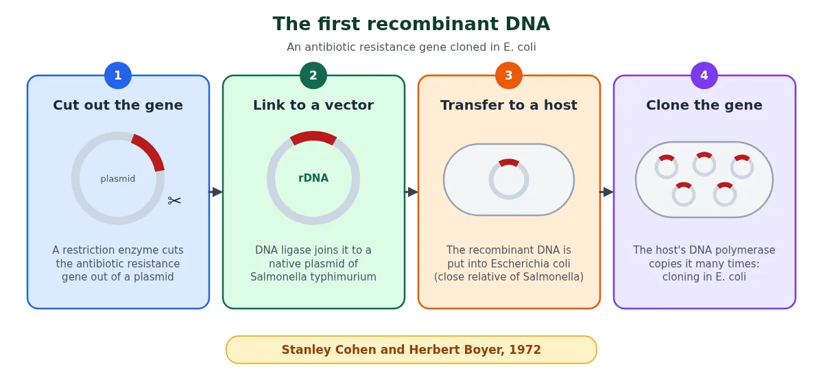
The first artificial recombinant DNA was made in 1972 by Stanley Cohen and Herbert Boyer. They cut out a piece of DNA carrying an antibiotic resistance gene from a plasmid and linked it to a native plasmid of Salmonella typhimurium. (A plasmid is an autonomously replicating, circular, extra-chromosomal DNA.)
- Cutting DNA at specific places became possible with restriction enzymes, the “molecular scissors”.
- The cut gene was joined to the plasmid by DNA ligase. The new circular, self-replicating molecule made in vitro is recombinant DNA.
- The plasmid acts as a vector: just as a mosquito carries the malarial parasite into the human body, the plasmid carries the attached DNA into the host.
- The recombinant DNA was put into Escherichia coli, a bacterium closely related to Salmonella. Using the host’s DNA polymerase it was copied many times. This was called cloning of the antibiotic resistance gene in E. coli.
Three basic steps in genetically modifying an organism
- Identify the DNA with the desirable genes.
- Introduce the identified DNA into the host.
- Maintain the introduced DNA in the host and transfer it to the host’s progeny.
Process of genetic engineering (overview)
These steps are explained in detail, with diagrams, in the recombinant DNA technology article:
- Isolation of DNA
- Fragmentation of DNA by a specific RE
- Isolation of the desired DNA fragment through agarose gel electrophoresis
- Ligation of the DNA fragment into a vector treated with the same RE
- Transferring the recombinant DNA into the host
- Culturing the host cells in a medium at a large scale
- Extraction of the desired product
4 Tools of recombinant DNA technology
Genetic engineering (recombinant DNA technology) can be done only if we have the key tools: restriction enzymes, polymerase enzymes, ligases, vectors and the host organism. We’ll group them as:
- Enzymes
- Cloning vectors
- Competent host
Enzymes used in genetic engineering
The following enzymes are involved in the genetic engineering process:
- Restriction endonucleases (molecular scissors).
- Alkaline phosphatase.
- DNA ligase: it is used for making the phosphodiester bond. It acts as a molecular glue. Generally, T4 DNA ligase is used for this purpose (T4 is a bacteriophage).
- Reverse transcriptase: it is used for converting RNA into DNA (making cDNA).
- DNA polymerases: for synthesising DNA, for example copying a short stretch of DNA (Taq polymerase is the one used in PCR).
- Cellulase, chitinase (and lysozyme for bacteria): required to break the cell wall.
Why alkaline phosphatase is used
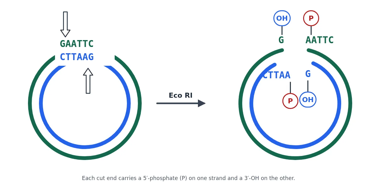
When EcoRI is used as the RE, it cuts DNA at a specific site. Once the DNA is cut, each cut end has a 5′ phosphate group on one strand and a 3′ OH group on the other. Chances are that these P and OH groups of the same cut vector join again. If they rejoin, there is no benefit in cutting the DNA, because the GAATTC base pairs are simply re-established and the vector closes without an insert. In order to prevent this self-ligation, the phosphate needs to be removed from the cut vector, and that is done by the enzyme alkaline phosphatase. This enzyme is obtained either from bacteria (BAP = bacterial alkaline phosphatase) or from the calf intestine (CIP = calf intestinal alkaline phosphatase).
5 Restriction enzymes (restriction endonucleases)
Out of the enzymes mentioned above, we’ll discuss the restriction endonuclease enzyme in detail.
Bacteria are attacked by viruses (bacteriophages). After infection, the viral DNA takes over the bacterial cell and makes it produce new viral particles. To protect itself, the bacterium produces restriction endonuclease (RE) enzymes that cut the viral DNA at specific points.
In other words, the enzyme restricts the multiplication of the viral DNA inside the bacterium. Along with restriction endonucleases, another enzyme, methylase, works in tandem: wherever the same recognition sequence occurs in the bacterium’s own DNA, methylase adds methyl groups to it, so that the bacterium’s own DNA is protected from being cut. This defence mechanism of the bacterial cell is called the RESTRICTION–MODIFICATION SYSTEM.
History note (NCERT 9.2.1)
- In 1963, two enzymes that restrict the growth of bacteriophage in Escherichia coli were isolated. One added methyl groups to DNA; the other cut DNA and was called a restriction endonuclease.
- Five years later, Hind II was isolated and characterised. It was the first restriction endonuclease whose action depended on a specific DNA nucleotide sequence: it always cut DNA at a particular point by recognising a specific sequence of six base pairs, its recognition sequence.
- Today, more than 900 restriction enzymes are known, isolated from over 230 strains of bacteria, each recognising different recognition sequences.
Restriction enzymes are nucleases
Restriction enzymes belong to a larger class of enzymes called nucleases, which are of two kinds:
- Exonucleases remove nucleotides from the ends of DNA.
- Endonucleases make cuts at specific positions within the DNA.
Each restriction endonuclease “inspects” the length of a DNA sequence. Once it finds its specific recognition sequence, it binds to the DNA and cuts each of the two strands of the double helix at specific points in their sugar-phosphate backbones.
Palindromic sequences
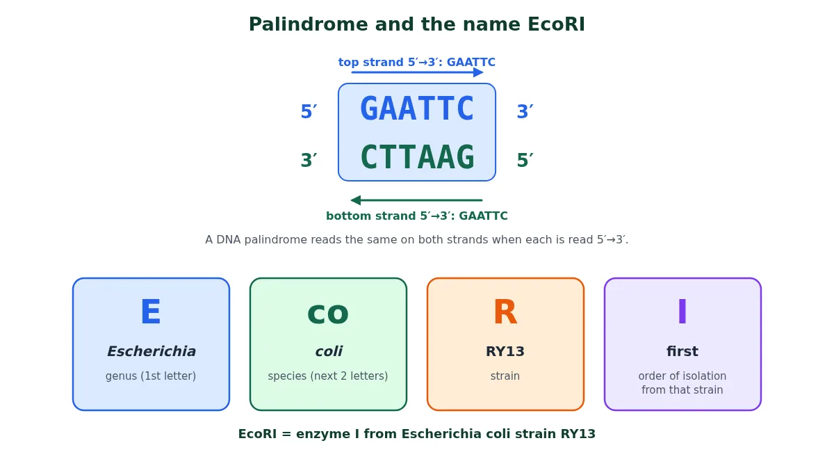
Restriction endonuclease (RE) works on a PALINDROMIC SEQUENCE. A word palindrome reads the same in both directions; for example, MALAYALAM reads the same from the right and the left side. A DNA palindrome is a little different: it is a sequence of base pairs that reads the same on the two strands when the orientation of reading is kept the same. For example, the complementary sequence of GAATTC is CTTAAG, and if we read both sequences in the 5′ to 3′ direction, each reads GAATTC (the same is true if both are read 3′ to 5′). Each restriction endonuclease recognises a specific palindromic sequence on the DNA.
Sticky ends
Restriction enzymes such as EcoRI cut the strands a little away from the centre of the palindrome, but between the same two bases on the opposite strands. This leaves single-stranded overhanging stretches at the ends, called sticky ends. They are called sticky because they form hydrogen bonds with their complementary cut counterparts, and this stickiness helps the enzyme DNA ligase to join them.
- When DNA from two sources is cut by the same restriction enzyme, the fragments have the same kind of sticky ends and can be joined end to end by DNA ligase, forming recombinant DNA.
- So, normally, unless the vector and the source DNA are cut with the same restriction enzyme, a recombinant vector cannot be made.
Classes of restriction enzymes
- Q. How many different classes of RE are present?
- (a) Class I
- (b) Class I and Class II
- (c) Class I, II and III
- (d) Class I and IV
Show answer
Ans: (c) Class I, II and III. These are the three classical classes (types) asked in exams. Newer classifications also list a Type IV group that cuts only methylated DNA, but it is not part of the NCERT or NEET syllabus.
- Class I RE requires Mg2+ ions, ATP and S-adenosyl methionine (SAM). It recognises a DNA sequence, but it does not cut that specific sequence; rather, it cuts the DNA at a random site far away from the recognition sequence. In other words, when class I RE is used, it forms heterogeneous fragments of DNA.
- Class II RE requires only Mg2+ ions (no ATP), and it cuts DNA at a specific site within (or right next to) its recognition sequence. Thus it produces homogeneous fragments: every time we use the RE, we get the same pattern of fragments. These are the enzymes used in genetic engineering (EcoRI, BamHI, Hind II, Sma I and so on).
- Class III RE requires Mg2+ ions and ATP; S-adenosyl methionine stimulates it but is not essential. It cuts outside the recognition sequence, about 25 base pairs away from it, so the cut is not at the recognition site itself and these enzymes are not useful for gene cloning.
| Class | Cofactors | Where it cuts | Use in cloning |
|---|---|---|---|
| Class I | Mg2+, ATP, SAM | Randomly, far from the recognition site | No (heterogeneous fragments) |
| Class II | Mg2+ only | Within or at the recognition site | Yes (same fragments every time) |
| Class III | Mg2+, ATP (SAM stimulates) | About 25 bp away from the site | No |
Swipe the table sideways on a phone.
Note: the recognition sequences of most class II REs are 4 to 8 base pairs long; most commonly 4 or 6 bp.
6 Naming of restriction enzymes and common examples
EcoRI: E represents the genus (Escherichia), co represents the species (coli), R represents the strain (RY13), and I represents the order in which the enzyme was isolated from that strain. In other words, several restriction enzymes may be isolated from the same strain of bacteria; the first enzyme isolated is named I, the second II, and so on. Its recognition site is GAATTC. EcoRI cuts DNA in a staggered manner (between G and A on each strand) and forms sticky ends.
The cut ends of the sequence produced by EcoRI are sticky in nature.
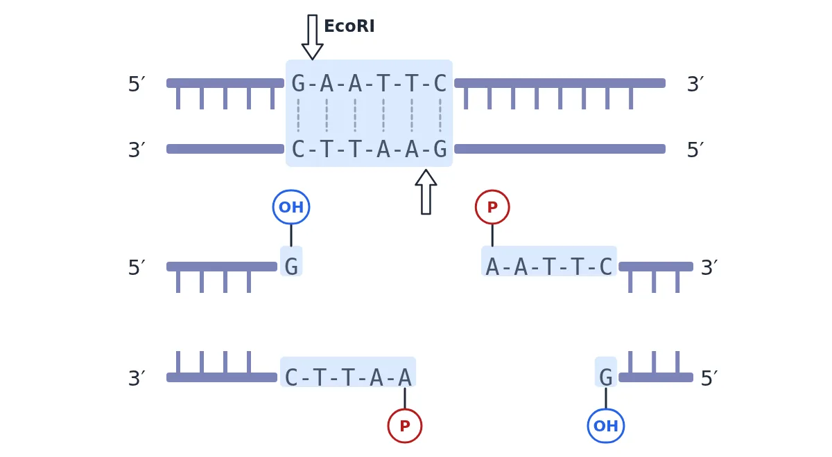
Note: the restriction enzyme breaks the bond between the sugar and the phosphate, i.e., the phosphodiester bond, in the sugar-phosphate backbone.
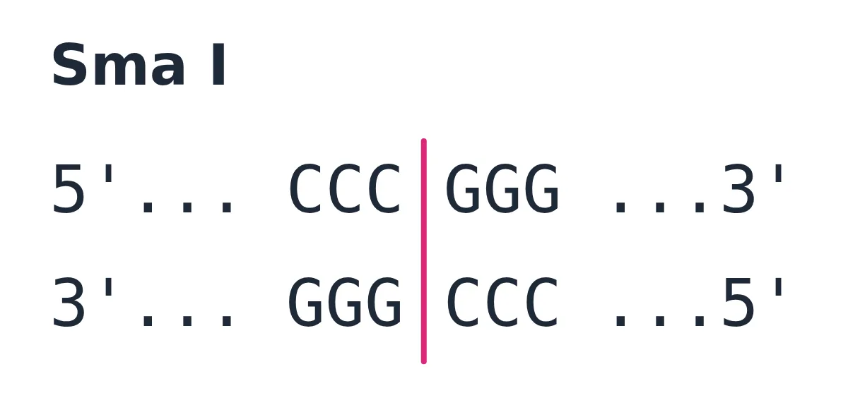
Sma I = Serratia marcescens, enzyme I. Sma I makes a flush (straight) cut through the middle of CCCGGG on both strands, so it forms blunt ends.
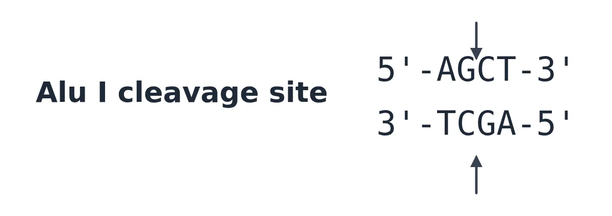
Alu I = Arthrobacter luteus, enzyme I. It cuts AG|CT and also forms blunt ends.
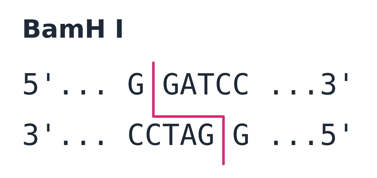
BamH I = Bacillus amyloliquefaciens strain H, enzyme I. It cuts G|GATCC and forms sticky ends.
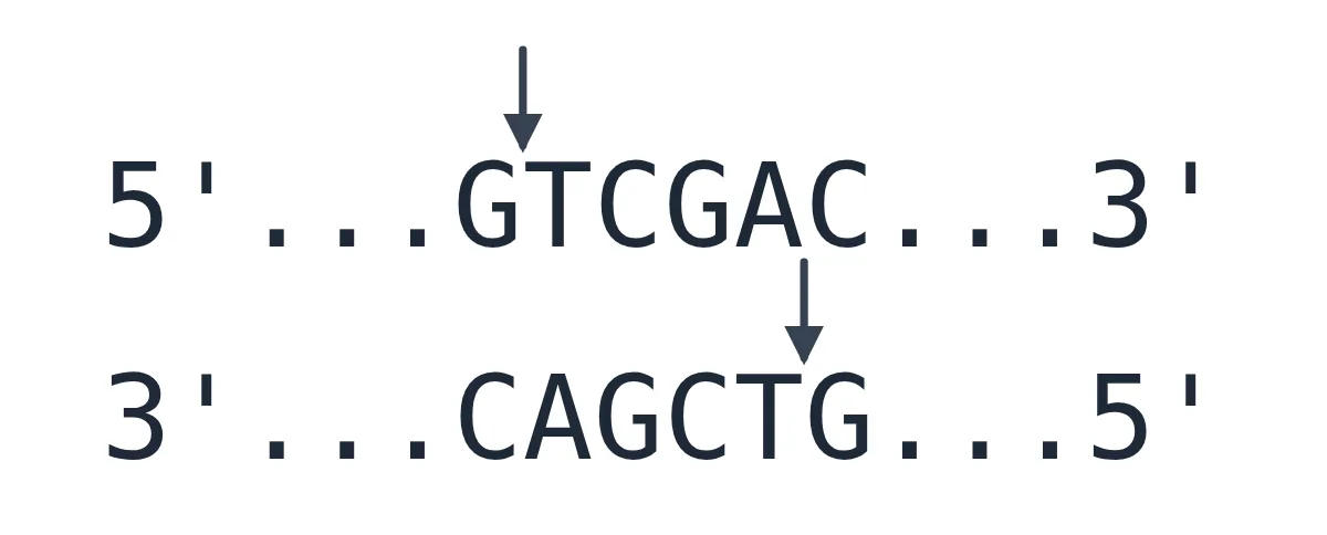
Sal I = Streptomyces albus, enzyme I. It cuts G|TCGAC and forms sticky ends.
Hind II = Haemophilus influenzae strain Rd, enzyme II.
Note: Hind II was the first restriction endonuclease whose functioning depended on a specific DNA nucleotide sequence. It was isolated and characterised in 1968, five years after the first restriction enzymes were isolated (1963). It was found that Hind II always cut DNA molecules at a particular point by recognising a specific sequence of six base pairs.
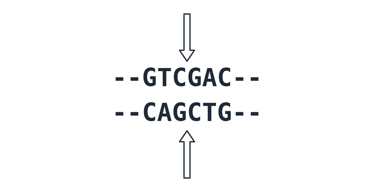
Sal I and Hind II: same site, different cut
Hind II recognises a family of 6 bp sequences, GTPy↓PuAC (Py = C or T, Pu = A or G). GTCGAC, the recognition sequence of Sal I, is one of them. So the two enzymes can recognise the same sequence (GTCGAC) but cut at different places: Sal I cuts G↓TCGAC (sticky ends), while Hind II cuts GTC↓GAC (blunt ends). In other words, two restriction enzymes can recognise the same sequence but cut it at different places. Note that Hind II is not the same as Hind III (A↓AGCTT, sticky ends), which comes from the same bacterial strain.
How often does a recognition site occur?
Note: if a restriction enzyme recognises a DNA sequence of, say, GATC (4 bases), then the expected frequency of this 4-base sequence in a random DNA strand is once in (4)4 = 4 × 4 × 4 × 4 = 256 bases (i.e., GATC would occur about once every 256 bases). Likewise, a 5-base recognition sequence would be present about once every 1024 bases:
(4)5 = 4 × 4 × 4 × 4 × 4 = 1024
Following the same working, a 6-base site such as GAATTC occurs about once every (4)6 = 4096 bases. Longer recognition sites therefore cut DNA less often and give bigger fragments.
Separating the cut fragments
Fragments made by restriction enzymes are separated by agarose gel electrophoresis: being negatively charged, DNA moves towards the anode, and smaller fragments move farther. The bands are stained with ethidium bromide, seen under UV light, cut out and eluted. See the full step, with a diagram, in the recombinant DNA technology article.
7 Cloning vectors
Plasmids and bacteriophages can replicate inside bacterial cells independently of the chromosomal DNA. Bacteriophages, because of their high number per cell, have very high copy numbers of their genome. Some plasmids have only one or two copies per cell, while others have 15 to 100 copies per cell, or even more. If we link an alien piece of DNA to such a plasmid or phage DNA, it is multiplied to the same copy number. Vectors used today are engineered so that foreign DNA can be linked easily and recombinants can be told apart from non-recombinants.
- Q. What are the conditions for any suitable vector?
Show answer
- It must have an origin of replication (ori).
- It should have selectable markers: genes on the basis of which we can select which cells have taken up the vector (transformants) and which have not (non-transformants). Antibiotic resistance genes are used as selectable markers.
- Cloning (recognition) sites: sites where the restriction enzyme can cut the vector. Preferably there should be only one site for the chosen enzyme, and it is useful if that site lies within a selectable marker gene, so that recombinants can be picked out by insertional inactivation.
- Q. Which of the following should be the features of a suitable vector?
- (a) The vector must contain an origin of replication so that it is independently able to replicate within the host.
- (b) The vector should incorporate a selectable marker, a gene whose product can identify the host cells containing the vector.
- (c) The vector must have one unique restriction enzyme recognition site, which can be used for cutting and introducing an insert.
- (d) The vector should be small in size, thereby facilitating entry/transfer into the host cell.
- (e) All of the above
Show answer
Ans: (e) All of the above.
(i) Origin of replication (ori)
This is the sequence from where replication starts. Any piece of DNA linked to it can be made to replicate within the host cells. The ori also controls the copy number of the linked DNA, so to get many copies of the target DNA, it should be cloned in a vector whose ori supports a high copy number.
(ii) Selectable marker
A selectable marker helps to identify and eliminate non-transformants and selectively permits the growth of transformants. (Transformation is the procedure by which a piece of DNA is introduced into a host bacterium.) Genes for resistance to antibiotics such as ampicillin, chloramphenicol, tetracycline or kanamycin are useful selectable markers for E. coli, because normal E. coli cells do not carry resistance against any of these antibiotics.
(iii) Cloning sites
To link the alien DNA, the vector should have very few, preferably single, recognition sites for the commonly used restriction enzymes. More than one site for the same enzyme would cut the vector into several fragments and complicate gene cloning.
Insertional inactivation in pBR322
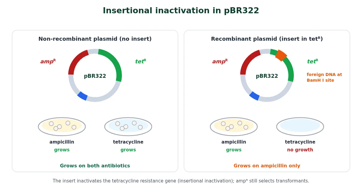
Foreign DNA is ligated at a restriction site inside one of the two antibiotic resistance genes. For example, foreign DNA can be ligated at the BamH I site of the tetracycline resistance gene (tetR) of pBR322. The recombinant plasmid loses tetracycline resistance but keeps ampicillin resistance. So the transformants are first grown on an ampicillin medium and then transferred to a tetracycline medium: recombinants grow on ampicillin but not on tetracycline, while non-recombinants grow on both. One resistance gene selects the transformants; the other is inactivated due to insertion of the alien DNA and helps to select the recombinants.
NCERT note for NEET
One line in NCERT 9.2.2 says the recombinants can be selected “by plating the transformants on tetracycline containing medium”. The lines that follow make the logic clear: transformants are selected on ampicillin, and recombinants are the ones that do not grow on tetracycline. Learn the logic.
Blue-white screening (chromogenic substrate)
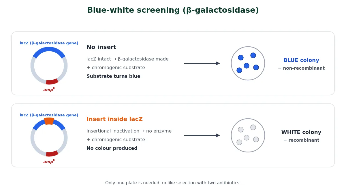
Selecting recombinants by inactivating an antibiotic resistance gene is cumbersome, because cells must be plated on two plates with different antibiotics. So alternative markers were developed that tell recombinants from non-recombinants by their ability to produce colour in the presence of a chromogenic substrate:
- The recombinant DNA is inserted within the coding sequence of the enzyme β-galactosidase (the lacZ gene).
- This inactivates the gene for the enzyme: insertional inactivation.
- If the plasmid has no insert, the enzyme is made and the colonies turn blue with the chromogenic substrate.
- If the insert is present, no enzyme is made and the colonies produce no colour (white). These are the recombinant colonies.
(iv) Vectors for cloning genes in plants and animals
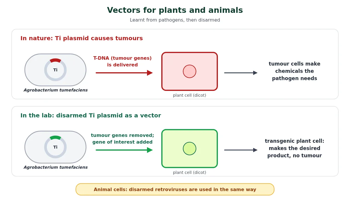
We learnt how to transfer genes into plants and animals from bacteria and viruses, which have always known how to deliver their genes into eukaryotic cells and make those cells do what the pathogen wants.
- Agrobacterium tumefaciens, a pathogen of several dicot plants, delivers a piece of DNA called T-DNA that transforms normal plant cells into a tumour and makes the tumour cells produce chemicals the pathogen needs.
- Retroviruses in animals can transform normal cells into cancerous cells.
- The tumour-inducing (Ti) plasmid of A. tumefaciens has been modified into a cloning vector that is no longer pathogenic to plants but can still deliver the genes we want into many plants. Retroviruses have likewise been disarmed and are used to deliver desirable genes into animal cells.
Once a gene or DNA fragment has been ligated into a suitable vector, it is transferred into a bacterial, plant or animal host, where it multiplies.
8 Types of vectors
1. Plasmid vectors
The term plasmid was coined by Joshua Lederberg in 1952. Plasmids are extra-chromosomal, self-replicating, circular, double-stranded DNA molecules. The number of copies per cell can be just 1–2, or 15–100 (NCERT), and some engineered high-copy vectors reach several hundred copies per cell. Natural plasmids need to be modified before they can be used as vectors. pBR322 was one of the first and most widely used artificial plasmid vectors of E. coli. Normal E. coli cells don’t carry resistance against any of the antibiotics ampicillin, tetracycline, chloramphenicol and kanamycin, which is why resistance genes for them work as markers.
pBR322
- p: plasmid
- B: Bolivar
- R: Rodriguez
- 322: the number used to distinguish it from other plasmid vectors
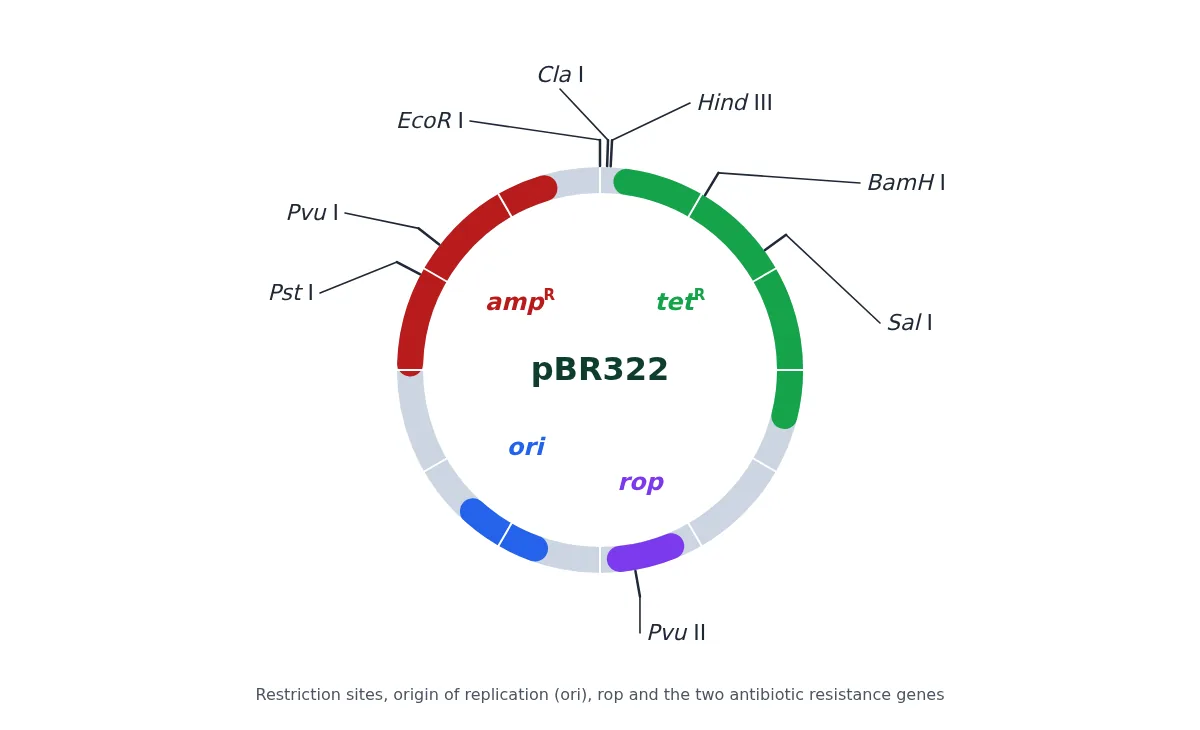
- Restriction sites: Pst I, Pvu I, EcoR I, Cla I, Hind III, BamH I, Sal I and Pvu II. BamH I and Sal I lie inside tetR; Pst I and Pvu I lie inside ampR.
- rop codes for the proteins involved in the replication of the plasmid (it helps to control the copy number).
- Selectable markers: the ampicillin resistance (ampR) and tetracycline resistance (tetR) genes.
- ori: the origin of replication.
pUC18/19
In this plasmid, the selectable marker is the ampicillin resistance gene, and the lacZ gene, which codes for the enzyme β-galactosidase, is used for screening. This lacZ region also contains a polylinker (multiple cloning site), so the insertion of any foreign DNA into any of its restriction sites results in an altered, non-functional enzyme. During screening of host cells containing the recombinant plasmid, the absence of β-galactosidase activity (white colonies) indicates plasmids containing the insert.
Shuttle vectors
Note: pBR322 and pUC vectors can replicate only in E. coli. Many of the vectors used in eukaryotic cells are constructed so that they can exist both in the eukaryotic cell and in E. coli. Such vectors are known as SHUTTLE VECTORS. These vectors contain two types of origin of replication and selectable marker genes: one set that functions in eukaryotic cells (e.g., yeast) and another in E. coli. An example of a shuttle vector is the yeast episomal plasmid YEp. In the case of plants, a naturally occurring plasmid of the bacterium Agrobacterium tumefaciens, called the Ti plasmid, has been suitably modified to function as a vector.
2. Bacteriophage-based vectors
In this category, there are two cloning vectors: lambda phage and M13 phage.
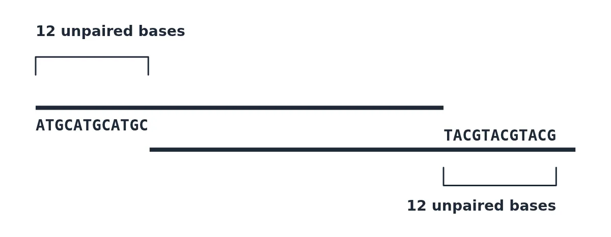
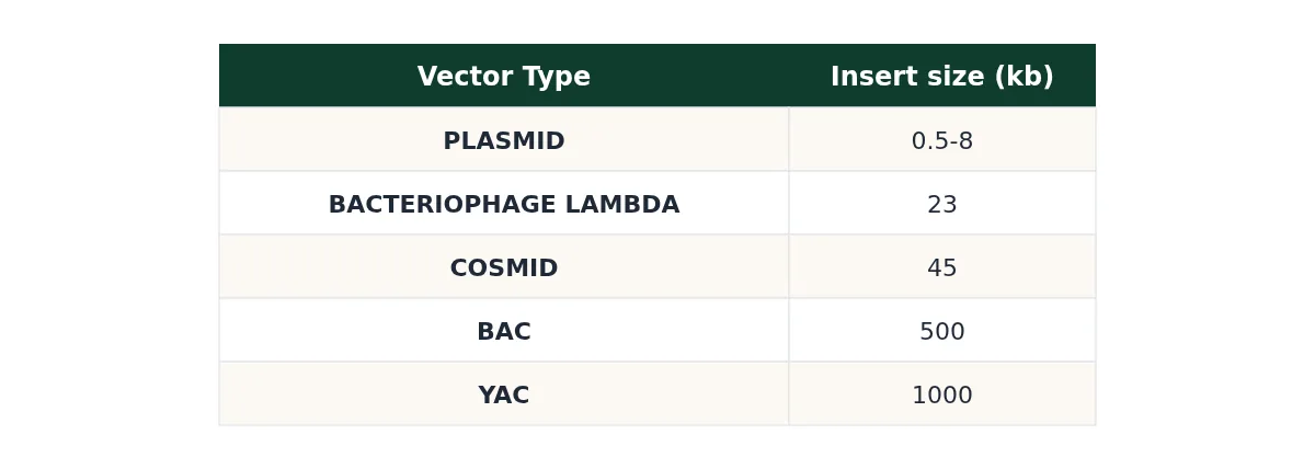
Bacteriophage lambda: it has a double-stranded, linear DNA genome of 48,502 bp, in which 12 bases at each end are unpaired and complementary. These cohesive end sites are called cos sites. An important feature of the lambda genome is that a large fragment in the central region of its genome is not essential for lytic infection of E. coli cells, and so this region can be replaced by foreign insert DNA. The linear phage DNA becomes circular because of the complementary end sequences. These phage vectors allow cloning of DNA fragments up to about 23 kb in size.
M13 phage: it infects E. coli cells that have a pilus (F pilus), which is present only in cells containing the F plasmid (called F+ cells).
- Q. Arrange vectors on the basis of their insert DNA size capacity.
- (a) Plasmid < bacteriophage lambda < cosmid < BAC < YAC
- (b) Plasmid < cosmid < bacteriophage lambda < BAC < YAC
- (c) Cosmid < plasmid < bacteriophage lambda < BAC < YAC
- (d) Cosmid < plasmid < bacteriophage lambda < YAC < BAC
Show answer
Ans: (a) Plasmid < bacteriophage lambda < cosmid < BAC < YAC.
9 Competent host (for transformation with recombinant DNA)
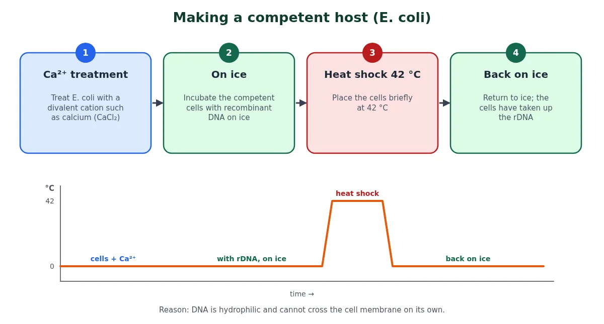
A competent host means the host cell must be capable of taking up the plasmid. For example, if normal host cells and plasmids are simply mixed together, the plasmid will not enter the host cell unless the host cell is given some specific treatment. In other words, a specific treatment makes the cell competent to take up the DNA.
Why is the treatment needed? DNA is a hydrophilic molecule, so it cannot pass through cell membranes on its own.
- The bacterial cells are treated with a specific concentration of a divalent cation, such as calcium (Ca2+). This increases the efficiency with which DNA enters the bacterium through pores in its cell wall.
- The competent cells are incubated with the recombinant DNA on ice.
- They are then placed briefly at 42 °C (heat shock).
- Finally they are put back on ice. This enables the bacteria to take up the recombinant DNA.
Other ways to introduce alien DNA
- Micro-injection: recombinant DNA is injected directly into the nucleus of an animal cell.
- Biolistics (gene gun): suitable for plants; cells are bombarded with high-velocity micro-particles of gold or tungsten coated with DNA.
- Disarmed pathogen vectors: when allowed to infect the cell, they transfer the recombinant DNA into the host (for example, the disarmed Ti plasmid or disarmed retroviruses).
Diagrams of micro-injection and the gene gun are given in the recombinant DNA technology article, along with how transformants and recombinants are selected.
So, in this article, I have finished (A) the principles of biotechnology and (B) the tools used in genetic engineering. In the next part, recombinant DNA technology, I finish (C) the process of genetic engineering.
10 Summary table: principles and tools at a glance
| Topic | Key points |
|---|---|
| Definition | EFB: integration of natural science and organisms, cells, parts thereof and molecular analogues for products and services |
| Core techniques | Genetic engineering (change the genetic material) and bioprocess engineering (sterile, large-scale culture) |
| First rDNA | Cohen and Boyer, 1972: antibiotic resistance gene + native plasmid of Salmonella typhimurium, cloned in E. coli |
| Key tools | Restriction enzymes, polymerases, ligases, vectors, host |
| Restriction enzymes | First sequence-specific RE: Hind II (6 bp); over 900 known; recognise palindromes; EcoRI = E. coli RY13, enzyme I |
| Vector features | ori (controls copy number), selectable marker, single cloning site |
| pBR322 | ampR (Pst I, Pvu I), tetR (BamH I, Sal I), ori, rop |
| Screening | Insertional inactivation; blue (no insert) vs white (recombinant) with β-galactosidase |
| Plant/animal vectors | Disarmed Ti plasmid of Agrobacterium tumefaciens; disarmed retroviruses |
| Competent host | Ca2+, ice, 42 °C heat shock, ice; also micro-injection, biolistics, disarmed pathogens |
Swipe the table sideways on a phone.
11 Common doubts about the principles of biotechnology
Do Sal I and Hind II recognise the same sequence?
Partly. Hind II recognises the family GTPy↓PuAC, and Sal I’s site GTCGAC belongs to that family, so both can recognise GTCGAC. They cut it differently: Sal I cuts G↓TCGAC and leaves sticky ends, while Hind II cuts GTC↓GAC and leaves blunt ends.
What is the difference between Hind II and Hind III?
Both come from Haemophilus influenzae strain Rd. Hind II was the first sequence-specific restriction enzyme and makes blunt ends at GTPy↓PuAC. Hind III recognises A↓AGCTT and makes sticky ends; it is one of the sites in pBR322.
Why does the R in EcoRI stand for RY13?
The letter after the genus and species shows the strain the enzyme came from. EcoRI was isolated from Escherichia coli strain RY13, and the Roman numeral I shows it was the first enzyme isolated from that strain.
Why is the cut vector treated with alkaline phosphatase?
Removing the 5′ phosphate groups stops the cut vector from joining back on itself (self-ligation). The vector can then close only when a foreign DNA fragment, which still has its phosphates, is ligated into it.
Why should a vector have only one recognition site for the enzyme used?
If the enzyme cut the vector at two or more places, the vector would fall apart into several fragments and the gene could not be inserted cleanly. A single site opens the circle at one point only.
Why is E. coli given a heat shock at 42 °C?
Calcium-treated cells are kept on ice with the recombinant DNA and then briefly warmed to 42 °C. This sudden change in temperature helps the DNA enter through pores in the cell wall, after which the cells are cooled on ice again.
Is lacZ a selectable marker?
In pUC vectors the selectable marker is the ampicillin resistance gene, which kills off non-transformants. The lacZ gene (β-galactosidase) is used for screening: blue colonies have no insert and white colonies are recombinants. NCERT calls it an alternative selectable marker.
12 Quick check
Tap each question to reveal the answer and check your grasp of the principles of biotechnology before you try the full MCQ set.
Test yourself
1. Name the two core techniques that enabled modern biotechnology.
Genetic engineering and bioprocess engineering.
2. Who made the first recombinant DNA, and in which year?
Stanley Cohen and Herbert Boyer, in 1972, using a native plasmid of Salmonella typhimurium.
3. Which was the first sequence-specific restriction endonuclease, and how long was its recognition sequence?
Hind II; it recognises a 6 bp sequence.
4. What do E, co, R and I stand for in EcoRI?
Escherichia (genus), coli (species), RY13 (strain) and the first enzyme isolated from that strain.
5. Which cofactor does a class II restriction enzyme need?
Only Mg²⁺ ions (no ATP).
6. How often is a 6 bp recognition site expected in random DNA?
Once every 4⁶ = 4096 bases.
7. In pBR322, foreign DNA is inserted at the BamH I site. Which antibiotic resistance is lost?
Tetracycline resistance (insertional inactivation of tetR).
8. In blue-white screening, which colonies are recombinant?
The white (colourless) colonies.
9. Which plasmid is used as a vector for plants?
The disarmed Ti plasmid of Agrobacterium tumefaciens.
10. Which divalent cation is used to make bacterial cells competent?
Calcium (Ca²⁺).
Related reading
- Next part: recombinant DNA technology (the seven steps, PCR and bioreactors)
- Gene cloning and its steps
- Practise: MCQs on biotechnology principles and processes
- All notes in Biotechnology: Principles and Processes
Notes by Competitors Point for NEET Biology, based on NCERT Class XII Chapter 9 (rationalised syllabus). All diagrams drawn for this page.